General Dentistry
July 21, 2026
Tooth Formation Stages: How Teeth Develop From Dental Lamina to Eruption
 Dr. Rifat Alsaman
Medically reviewed by Dr. Rifat— Last updated July 2026
Introduction
Tooth formation stages describe how a tooth develops long before it ever appears in the mouth. Months before a baby tooth erupts, hidden work is already underway inside the jaw. A simple thickening of tissue slowly transforms into a hard, mineralized structure, through a precise, step-by-step biological sequence.
Understanding this process matters beyond dental school textbooks. Parents can use it to recognize why certain enamel defects show up later. Clinicians can pinpoint exactly when development went off track. Patients gain a clearer picture of conditions like fluorosis and amelogenesis imperfecta, and why they happen at all.
In this guide, we walk through each of the tooth formation stages in order, from the dental lamina to final enamel maturation. Then we follow the timeline through to the actual eruption. Along the way, we answer the specific questions people search for most. Which stage produces the dental lamina? What does the bell stage actually look like? When does fluorosis occur, and why? We also share clinical notes from our team at Vitrin Clinic. We evaluate enamel and dentin conditions in both children and adults every week.
Quick Answer
The stages of tooth formation are the sequence of biological steps that turn embryonic tissue into a fully mineralized tooth. There are six stages in total: initiation, bud, cap, bell, apposition, and maturation. Each stage produces a specific structure: the dental lamina, the tooth bud, the enamel organ, and eventually the hardened crown. The process starts as early as the sixth week in utero. It continues for months afterward.
Stage | What Happens | Approx. Timing |
Dental Lamina (Initiation) | Initiation of tooth development | 6th week in utero |
Bud Stage | Tooth germ formation begins | 8th week |
Cap Stage | Enamel organ forms, differentiation starts | 9th–10th week |
Bell Stage | Cell differentiation, crown shape forms | 11th–12th week |
Apposition & Maturation | Enamel, dentin, and cementum laid down layer by layer | 4th month onward |
What Is Tooth Formation (Odontogenesis)?
Odontogenesis is the scientific term for the tooth formation stages as a whole the process of tooth development. It describes the full journey a tooth takes, from a small cluster of embryonic cells to a hardened, decades-lasting structure. This journey starts early. Odontogenesis begins around the sixth week of gestation. That's when the ectoderm lining the future mouth thickens to form the earliest tooth-forming tissue.
That early timing is more than trivia. MRI research tracking fetal development found something striking. All ten primary tooth buds were identifiable in only 5.4% of fetuses scanned between weeks 18 and 21. That figure rose to 75.5% by weeks 26–29, and reached 90.6% by week 34 or later. In other words, the tooth formation stages aren't a single event. They unfold gradually, over months, well before a baby ever smiles.
This matters for a simple reason. A defect that shows up on a tooth years later usually has its roots in one specific window of development. Knowing which window helps explain why it happened, not just that it happened.
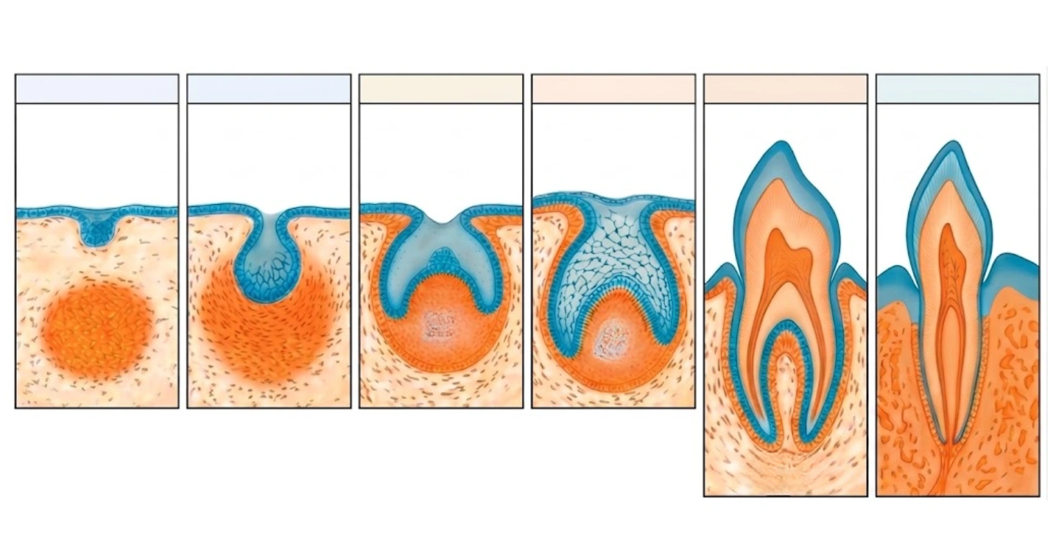
Tooth Formation Stages: Stage-by-Stage Overview
Every tooth develops through six distinct tooth formation stages. Each one builds directly on the last:
Initiation – the dental lamina forms
Bud Stage – the first tooth germ appears
Cap Stage – the enamel organ takes shape
Bell Stage – cells differentiate and the crown is patterned
Apposition – enamel, dentin, and cementum are laid down layer by layer
Maturation – the tooth's hard tissues fully mineralize
A disruption at any single point in these stages can leave a visible mark on the tooth. Sometimes it doesn't show up until years after the tooth erupts. The sections below walk through each stage in order.
Stage 1 — Initiation and the Dental Lamina
Initiation is the first of the tooth formation stages. The dental lamina forms during this stage. Around week six, the oral epithelium thickens along the future jaws. It forms a horseshoe-shaped band called the dental lamina. This band becomes the launching point for every tooth bud that follows, both primary and permanent.
Histologically, this stage is simple but foundational. It involves localized epithelial thickening, supported by an intact basement membrane. Beneath it, neural-crest-derived mesenchymal cells begin to condense in response to epithelial signals. This early cellular conversation sets the pattern for everything that comes next.
Clinic Note: In our observation at Vitrin Clinic, irregularities that begin this early are rarely visible until much later. This is one reason we recommend early developmental screening, along with sound prenatal nutrition counseling for expectant parents.
Stage 2 — Bud Stage
The bud stage is the second of the tooth formation stages. By week eight, the dental lamina grows downward into the mesenchyme at ten points per arch. Each of these points becomes a tooth bud, corresponding to the ten primary teeth in that arch. The bud itself is a rounded mass of epithelial cells, surrounded by mesenchyme that continues to condense around it.
This stage is also where things can go visibly wrong. Hypodontia, or missing tooth buds, affects roughly 1% of primary teeth and 6.4% of permanent teeth in the general population. On the opposite end, supernumerary (extra) teeth occur when this stage overproduces buds. Worldwide prevalence estimates range from about 0.1% to 3.8% of the population, depending on ethnicity and dentition type.
Stage 3 — Cap Stage
The cap stage is the third of the tooth formation stages. Around weeks nine to ten, the tooth bud folds inward and takes on a cap-like shape. This marks the start of the enamel organ, the dental papilla, and the dental sac. These three structures will eventually become enamel, dentin and pulp, and the supporting periodontal tissues.
This is also where the layer-by-layer construction of the tooth begins to take shape architecturally. Actual hard-tissue deposition won't start for a few more weeks. Think of the cap stage as the blueprint. It draws the plan; apposition later fills it in.
Stage 4 — Bell Stage (Labeled Diagram)
The bell stage is the fourth of the tooth formation stages. By weeks eleven to twelve, the enamel organ deepens into a bell shape. Four distinct cell layers become identifiable at this point:
Outer enamel epithelium – a protective outer layer
Stellate reticulum – a star-shaped network that nourishes the developing enamel
Stratum intermedium – supports enamel-forming activity
Inner enamel epithelium – cells that will become ameloblasts, the enamel producers
Beneath these layers, the dental papilla differentiates into odontoblasts and the future pulp. The dental sac surrounds the whole structure. Over time, it forms cementum, the periodontal ligament, and alveolar bone.
Early vs. Late Bell Stage
The bell stage has two distinct phases. In the early bell stage, cells are still specializing — a process called histodifferentiation. In the late bell stage, the crown's actual shape is finalized, called morphodifferentiation. This is where the specific cusp pattern for each tooth type gets locked in.
Stage 5 — Apposition and the Layer-by-Layer Formation of Tooth Structure
Apposition is the fifth of the tooth formation stages, and it's where planning turns into construction. Ameloblasts begin secreting an enamel matrix. Odontoblasts secrete dentin matrix. Cementoblasts lay down cementum along the developing root. This happens in a specific order:
Dentin is deposited first, just beneath the future enamel layer
Enamel matrix follows, secreted outward in successive layers
Cementum is deposited last, along the root surface
This layered process ultimately determines enamel thickness and dentin strength. Both factors shape a tooth's long-term resistance to decay and sensitivity.
Clinic Note: At Vitrin Clinic, we regularly see how variations formed during this stage affect a patient's restorative needs decades later. Thinner or irregular enamel often calls for more conservative bonding rather than aggressive prep work. Our diagnostic imaging can flag this well before it becomes painful. See our restorative diagnostic services →
Stage 6 — Maturation Stage
Maturation is the sixth and final of the tooth formation stages. Once the organic scaffold of enamel and dentin is in place, maturation begins. Ameloblasts shift roles here. Instead of producing matrix, they actively transport minerals into the enamel, hardening it into its final, dense form.
It's worth being precise about the difference between apposition and maturation, since the two are often confused. Apposition lays down the soft organic framework. Maturation hardens that framework into its final mineralized state. This distinction isn't just academic. Different developmental conditions target one stage specifically and not the other which is exactly what separates amelogenesis imperfecta from fluorosis, covered later in this guide.
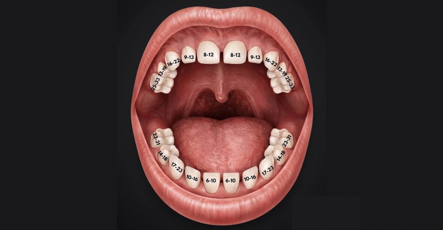
Tooth Eruption Timeline: When Do Teeth Actually Appear?
The six tooth formation stages we just covered explain how a tooth is built. Eruption explains when it shows up in the mouth and that's usually what parents actually want to know. Once apposition and maturation are far enough along, the tooth begins its slow migration through bone and gum tissue. This section gives a quick overview by age. Each tooth type below has its own dedicated guide for a deeper breakdown.
Primary (Baby) Teeth Eruption
The tooth formation stages we covered above explain how each of these teeth was built before it ever appeared.
Tooth | Typical Eruption Age |
Central incisors | 6–10 months |
Lateral incisors | 9–16 months |
First molars | 13–19 months |
Canines | 16–23 months |
Second molars | 23–33 months |
According to the American Dental Association, all 20 primary teeth are usually in place by around age 2½ to 3. A variation of four to five months earlier or later than these averages is still considered normal.
Permanent (Adult) Teeth Eruption
Tooth | Typical Eruption Age |
First molars ("6-year molars") | 6–7 years |
Central incisors | 6–7 years |
Lateral incisors | 7–8 years |
Canines | 9–12 years |
First premolars | 10–11 years |
Second premolars | 10–12 years |
Second molars | 11–13 years |
Third molars (wisdom teeth) | 17–21 years |
By age 21, all 32 permanent teeth have usually erupted, wisdom teeth included. Permanent tooth eruption can vary by up to 12 to 18 months in either direction without being a cause for concern.
Clinic Note: We get asked constantly whether a specific tooth is "late." In most cases, it's within normal range. But persistent delays especially past 18 months beyond the average for a given tooth are worth a quick evaluation. It's worth ruling out an underlying issue at the bud or cap stage.
This is a general overview. For a full breakdown of each tooth type, timing variations, and what to do about early or late eruption, see our dedicated guides on incisors, canines, premolars, primary vs. permanent teeth, delayed baby tooth eruption, and wisdom teeth.
What Is the Final Stage of Tooth Formation?
Among all the tooth formation stages, this is the one people ask about most. The answer depends on how you define "formation." At the histological level, maturation is the final stage of tooth formation. This is when enamel reaches its full mineral density and the tooth's hard tissues are essentially complete.
But many people mean something broader when they ask this question. They're really asking when the tooth is finished, in the everyday sense and that points to eruption, not maturation. Eruption is the clinical process of a tooth moving from its position in the jaw into the mouth. It's distinct from the microscopic process of tissue formation itself.
So both answers are correct, depending on intent. If you're asking from a developmental-biology standpoint, maturation is the final stage. If you're asking "when do I actually see the tooth," eruption is the final visible milestone. It can continue for years after the crown itself is fully formed, since root development often isn't complete until well after a tooth emerges.
When Tooth Development Goes Wrong: Clinical Correlations
Understanding these tooth formation stages becomes especially useful for explaining two conditions we see often at Vitrin Clinic: amelogenesis imperfecta and fluorosis. Both affect enamel. Both trace back to a specific point in development.
At Which Stage Does Amelogenesis Imperfecta Affect Tooth Formation?
Amelogenesis imperfecta primarily affects the apposition and maturation stages. It's caused by genetic mutations that disrupt ameloblast function, the cells responsible for building and hardening enamel. Depending on which stage is disrupted, it presents in three main forms.
Hypoplastic type – reduced enamel matrix during apposition, so too little enamel is laid down
Hypomaturation type – enamel forms in normal quantity but fails to harden properly
Hypocalcified type – enamel is poorly mineralized despite normal thickness
Prevalence varies widely by population from as common as 1 in 700 people in some regions, to as rare as 1 in 14,000 in others. That wide range shows how disrupting a single developmental stage can reshape an entire dentition, even though the condition stays rare overall.
Clinic Note: At Vitrin Clinic, we treat amelogenesis imperfecta with a mix of protective bonding, veneers, or full-coverage crowns. The approach depends on severity and the patient's age. Early diagnosis, often possible as soon as primary teeth erupt, gives us far more conservative options than waiting until adulthood.
During Which Stage of Tooth Formation Does Fluorosis Occur?
Dental fluorosis occurs during the maturation stage, when ameloblasts are actively mineralizing enamel. Excessive fluoride exposure during this window disrupts mineralization. The result is visible white streaks or mottling in milder cases, and brown staining or pitting in more severe ones.
This isn't a rare condition. CDC survey data has shown dental fluorosis affecting 65% or more of U.S. adolescents to some degree, though the large majority of these cases are mild or very mild rather than severe. Because fluorosis is tied so specifically to the maturation stage, timing matters more than total lifetime exposure. Risk is highest from infancy through around age eight, while permanent teeth are still maturing.
Clinic Note: We counsel parents on age-appropriate fluoride toothpaste amounts a rice-grain smear for toddlers, growing to a pea-sized amount later. Fluorosis is preventable with the right dosing during early childhood, well before permanent teeth erupt.
When to See a Dentist About Tooth Development Concerns
Not every irregularity across the tooth formation stages needs urgent attention, but some signs are worth having checked rather than watching and waiting. Unusual discoloration, pitted or rough enamel, or delayed eruption are all reasonable reasons to book an evaluation. Most developmental enamel conditions are far easier to manage when caught early — before the affected teeth face years of wear and decay risk.
Timing matters here. A tooth that looks slightly off at age three is much easier to treat conservatively than the same tooth left unaddressed until adolescence. If something looks unusual, it's worth asking about — even if it turns out to be within normal variation.
Concerned about a child's tooth development or possible enamel defects? Book a consultation at Vitrin Clinic — our team can evaluate exactly which developmental stage may have been affected and recommend the most conservative treatment path available.
References
Nanci, A. Ten Cate's Oral Histology: Development, Structure, and Function. Elsevier.
Kondo, S., et al. "Comparison of the Visibility of Fetal Tooth Buds on 1.5 and 3 Tesla MRI." PMC, National Center for Biotechnology Information. ncbi.nlm.nih.gov/pmc/articles/PMC7693030
MedlinePlus Genetics. "Amelogenesis Imperfecta." U.S. National Library of Medicine. medlineplus.gov/genetics/condition/amelogenesis-imperfecta
"Patient-Reported Outcome Measures in Individuals with Amelogenesis Imperfecta: A Systematic Review." PMC. pmc.ncbi.nlm.nih.gov/articles/PMC9750902
"Supernumerary Teeth: A Pictorial Review and Revised Classification." PMC. pmc.ncbi.nlm.nih.gov/articles/PMC11938152
Centers for Disease Control and Prevention (CDC), National Center for Health Statistics. "Prevalence and Severity of Dental Fluorosis in the United States." NCHS Data Brief No. 53. cdc.gov/nchs/data/databriefs/db53.pdf
FAQs
Dr. Rifat Alsaman has more than 5 years of clinical experience in dentistry and currently serves as the Head of the Medical Team at Vitrin Clinic. He is dedicated to providing exceptional patient care, overseeing treatment planning, and ensuring the highest clinical standards across the team. His expertise, attention to detail, and commitment to continuous professional development have helped countless patients achieve healthier, more confident smiles.


.webp&w=3840&q=75)

