Orthodontics
September 21, 2026
Impacted Canine Tooth: Symptoms, Causes, Treatment, and Surgery
 Dr. Rifat Alsaman
Impacted canine tooth problems occur when a canine stays hidden in the gum or bone instead of erupting. The upper canine matters for chewing, bite guidance, facial aesthetics, and the alignment of neighboring teeth. An impacted canine may cause no obvious symptoms, so early assessment with a clinical examination and dental imaging is important. Treatment depends on the tooth's position, angle, space, age, root development, and surrounding teeth. Options include monitoring, space creation, impacted canine exposure, surgical-orthodontic care, and removal when preservation is unpredictable.
Impacted Canine Tooth: What Does It Mean?
Impacted canine tooth means the canine is present but cannot erupt into its normal place. Normally, the upper canine erupts around ages eleven to thirteen and guides the bite. When its path is blocked or badly delayed, the tooth stays trapped. A partially erupted canine has broken through the gum, while a fully impacted canine stays covered by bone or tissue. This matters beyond appearance because the trapped tooth can push against neighboring roots, disturb alignment, and affect bite function.
Where Can an Impacted Canine Develop?
An impacted canine tooth most often develops on the palatal side, toward the roof of the mouth. Buccal or labial impaction places the tooth toward the lips and cheeks, and it is usually easier to reach. Less common positions include horizontal canines and abnormal eruption paths that cross the midline. Location strongly influences planning, because it determines the surgical approach, the direction of orthodontic pulling, and the risk to neighboring roots. Accurate imaging therefore comes before any decision.
How Is an Impacted Canine Different From a Missing Canine?
A canine that cannot be seen is not always absent. In an impacted case, the tooth is present but trapped in the gum or bone. A congenitally missing canine never developed at all. Dental imaging separates the two, because a panoramic X-ray can reveal a hidden crown. When the picture is unclear, three-dimensional scans can locate the unerupted tooth precisely. This distinction matters, since a hidden canine may still be saved, while a missing one needs a completely different plan.
Discover the connection between congenital adrenal hyperplasia and oral health
Signs and Symptoms of an Impacted Canine
Many patients notice nothing at all. Still, several signs can point toward an impacted canine tooth. The permanent canine may erupt later than the opposite side, and the baby canine may stay in place. Some patients see a gap, crowding, or shifted teeth near the canine area. The gum or dental arch may look uneven, and mild tenderness can occur. However, symptoms alone cannot show the tooth's exact position or condition, so imaging remains essential for a reliable diagnosis.
Dental Problems That May Develop Around an Impacted Canine
An untreated impacted canine tooth can affect neighboring teeth. It may cause root resorption of the lateral incisor, which weakens that tooth. Adjacent teeth can drift or tilt out of position. In some cases, a cyst forms around the unerupted crown. Crowding and bite problems may follow, and a partly erupted tooth can trap plaque and irritate the gum. Missing or displaced canine eruption also creates aesthetic concerns, particularly in the smile line. These complications explain why early assessment matters.
Tips for Patients
Do Not Ignore a Retained Baby Canine: A baby canine that stays long after its usual age deserves a dental check.
Ask Where the Permanent Canine Is Located: Request a clear explanation of the tooth's position from your imaging.
Understand Whether Orthodontics Is Part of the Plan: Most preserved canines need braces to move into place.
Follow Post-Surgical Instructions Carefully: Good hygiene and attendance at follow-ups protect healing.
Do Not Compare Treatment Plans Based on Price Alone: Compare the imaging, team, and full scope of care.
Note: All images used are for editorial and illustrative purposes only and may not originate from the original news provider or associated company.
Find out why your mouth may suddenly produce so much saliva
What Causes an Impacted Canine Tooth?
The cause is often multifactorial rather than a single issue. Crowding, abnormal eruption paths, retained baby teeth, developmental factors, and genetics can all contribute. The upper canine has the longest eruption route, which makes it vulnerable to deflection. Understanding the cause helps the dental team choose between space creation, exposure, or removal. Each patient's combination of factors differs, so two people with the same diagnosis can need different treatment. A careful history and examination show which factors apply.
Lack of Space in the Dental Arch
Crowding can block the canine's path. When the jaw is too small for all the teeth, the canine may be deflected or trapped above its neighbors. Enough orthodontic space strongly supports successful eruption, while a narrow arch often does the opposite. That is why space creation is frequently the first step of treatment, and it may involve braces, expanders, or selected extractions. Without adequate room, even a well-positioned canine may fail to reach the arch. Space planning therefore begins early.
Abnormal Tooth Position or Eruption Path
Sometimes the canine simply forms at an unusual angle. Instead of moving downward, it may drift toward the palate or lie horizontally. The tooth can then become trapped in dense bone or thick soft tissue. It may also press against the root of the lateral incisor, which can alter its path further. The more abnormal the angle and depth, the harder it becomes to guide the tooth naturally into the arch. Three-dimensional imaging clarifies how severe the deviation is.
Retained Baby Teeth
A retained primary canine is often linked to delayed eruption of the permanent canine. The baby tooth may occupy the space, or it may signal that the permanent tooth is off course. Normally, the root of the baby canine dissolves as the permanent tooth approaches. When that does not happen, the dentist should investigate. Early removal of a retained baby canine, when clinically indicated, can sometimes help the permanent tooth find its path. Timing matters, so assessment should not wait.
Other Factors
Several other factors contribute. Developmental variations can change how the tooth bud forms, and genetics or family history may raise the likelihood. Obstacles in the eruption path, such as extra teeth or growths, can block movement. Abnormalities of neighboring teeth, including small or peg-shaped lateral incisors, are also associated with canine problems. Most cases involve several causes at once, so individual evaluation is essential. No single explanation fits every patient, and family history alone does not predict the outcome.
Explore pneumococcal disease and oral health
How Is an Impacted Canine Diagnosed?
Diagnosis combines a clinical examination with imaging. The dentist checks the arch, feels the gum where the canine should appear, and looks for retained baby teeth. Available space and comparison with the opposite canine complete the clinical picture. Panoramic X-rays then locate the tooth, and three-dimensional imaging is added when more detail is needed. Together, these findings show the tooth's position, depth, and relationship to neighboring roots, which guides every later treatment decision. Accurate diagnosis prevents guesswork and unnecessary procedures.
Clinical Dental Examination
The examination begins with the dental arch, checking symmetry and alignment. The dentist gently palpates the gum where the canine should erupt. A bulge on the palate or cheek side can suggest its location. A retained primary tooth is noted, and crowding and available space are measured. Finally, the dentist compares the affected side with the opposite canine. A difference in eruption timing or position is often the first clue. This step is quick, painless, and highly informative.
Dental X-Rays and 3D Imaging
Imaging turns suspicion into a clear diagnosis. Two-dimensional images, such as panoramic X-rays, give a broad overview of the jaws and unerupted teeth. Three-dimensional imaging adds depth, showing exactly where the canine lies and how close it sits to neighboring roots. The dentist chooses the type of imaging according to clinical need, not routine. Together, these tools support safe surgical planning and help predict whether the canine can be guided into the arch. Each scan is justified individually.
Panoramic Imaging
A panoramic X-ray captures both jaws in one image. It helps locate the canine and shows whether the tooth is tilted, high, or close to the midline. It also reveals neighboring structures, including the lateral incisor roots, retained baby teeth, and any obvious cysts. Panoramic imaging is fast and widely available, so it is often the first scan. However, it flattens anatomy into two dimensions, so it cannot always show depth precisely. Further imaging may then be considered.
CBCT and 3D Planning
CBCT produces a three-dimensional image of the teeth and jaw. It may be indicated when panoramic images do not clearly show the canine's position. CBCT can clarify whether the crown lies toward the palate or cheek, and how close it is to adjacent roots. It also helps identify resorption. However, CBCT is not necessary for every patient. The dentist recommends it only when the added information could change the treatment plan. This keeps radiation exposure appropriate and the plan precise.
Why the Canine's Position Matters
Position shapes almost every decision. The angle, the depth in the bone, the stage of root development, and the closeness to neighboring roots all influence what is realistic. A shallow, upright canine may be guided into place with relative ease. A deep, horizontal canine near a lateral incisor root carries more risk and may need a different approach. Careful assessment therefore protects the neighboring teeth and improves the chances of a predictable result. Position also affects treatment length.
Impacted Canine Tooth Treatment Options
Treatment is individualized, and no single protocol suits every patient. Options range from conservative to more invasive. They include monitoring, orthodontic space creation, exposure with guided eruption, combined surgical-orthodontic care, and removal followed by replacement. Impacted canine tooth treatment depends on the tooth's position, the patient's age, and the health of neighboring teeth. The following sections explain each approach so patients can discuss their choices with confidence. The best plan balances preservation of the natural tooth against the risks and predictability of treatment.
Monitoring an Impacted Canine
Observation may be appropriate when the impacted canine tooth is not harming neighboring teeth, or when a young patient still has eruption potential. It relies on periodic clinical examinations and follow-up radiographs. Monitoring is not the same as ignoring the condition. It is an active plan with defined review points. If the canine moves in the wrong direction or a neighboring root shows damage, the dentist can change course promptly. Regular visits keep every option open. Patients should attend every scheduled review.
Orthodontic Treatment for an Impacted Canine
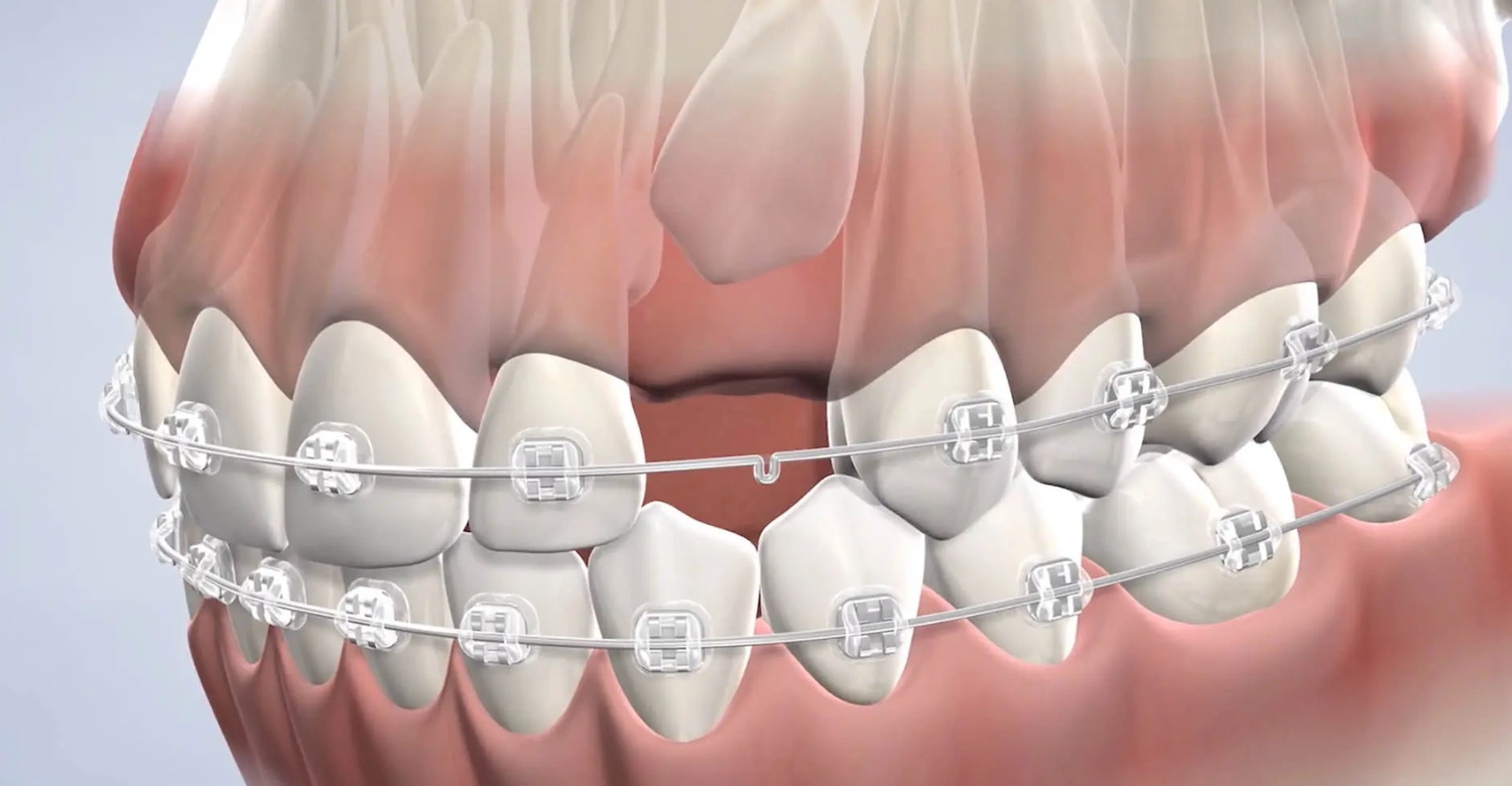
Braces play two roles. First, they create space in the arch, and second, they guide the canine into position. Impacted canine braces are often worn for a long period because the tooth may need to travel a considerable distance. Space creation is essential before traction begins. However, braces alone may not be enough when the canine is deeply embedded. The tooth must first be uncovered surgically. Close teamwork between surgeon and orthodontist keeps treatment efficient.
Impacted Canine Exposure
Impacted canine exposure is a surgical procedure that uncovers the tooth so an orthodontist can guide it into position. The sequence is straightforward. First, orthodontic space is created or confirmed. Next, the surgeon exposes the canine and, when appropriate, attaches an orthodontic bracket or chain. Then the orthodontist gradually guides the tooth into the arch. Healing and movement both require time, so patience and regular appointments are important. Movement often takes many months, depending on the tooth's distance and angle.
Surgical-Orthodontic Treatment
A combined approach is used when the canine cannot erupt on its own but can still be saved. The oral surgeon uncovers the tooth, and the orthodontist moves it using controlled force. Close coordination between the two is essential. Success depends on the canine's position, root development, available space, and the patient's age and cooperation. Deeply displaced teeth or teeth near vital roots have a lower chance of success, which the team explains before treatment begins. Realistic expectations matter.
Impacted Canine Tooth Removal
Impacted canine tooth removal may be considered when preserving the tooth is not predictable or beneficial. Examples include severe displacement, an unfavorable position, associated pathology such as a cyst, or damage to adjacent roots. Age and health can also influence the decision. Extraction is not automatically the first choice, because a natural canine is valuable. The dentist weighs the risks of moving the tooth against the benefits of removing it, then explains the reasoning clearly. Patients should feel free to ask questions.
Impacted Canine Extraction and Replacement Options
Impacted canine extraction leaves a space that needs a plan. In selected cases, orthodontics can close the space by moving neighboring teeth forward. Otherwise, a prosthetic replacement, such as a bridge, may be used. A dental implant is an option once growth and bone development are complete. Replacement planning should ideally happen before extraction, because the choice affects bone preservation, aesthetics, and orthodontic sequencing. Discussing the full pathway early prevents surprises later. Each option has different timelines, costs, and maintenance needs.
Discover more about fixed dentures cost in Turkey
Impacted Canine Tooth Surgery: What Should Patients Expect?
Patients can expect a structured process. It starts with examination, imaging, and planning, continues with a carefully chosen procedure, and ends with follow-up care. Impacted canine tooth surgery is usually performed under local anesthesia, with sedation considered when appropriate. The exact experience depends on the tooth's location and whether the goal is exposure or removal. Knowing each stage in advance reduces anxiety and helps patients prepare for recovery and orthodontic visits. The next sections walk through each stage.
Before Surgery
Before surgery, the dentist performs a clinical examination and reviews the imaging. A treatment plan is then prepared, including whether the canine will be exposed or removed. Anesthesia or sedation options are discussed when clinically appropriate. When braces are involved, the surgical and orthodontic teams coordinate so the timing fits together. Patients should also share their medical history and medications, and ask any questions before the appointment. Clear preparation makes the procedure smoother. Fasting instructions are given if sedation is planned.
During Surgery
The purpose of surgery is either to expose the canine or to remove it. The surgeon numbs the area, then gently reaches the tooth through the gum and, if needed, the overlying bone. When exposure is planned, a bracket may be attached. The exact technique depends on the canine's location and surrounding anatomy. Palatal and buccal cases differ, and each is planned individually. Most patients remain comfortable throughout, and the procedure length varies with complexity. The team explains each step beforehand.
After Surgery
Some swelling and tenderness are expected for a few days. Patients receive oral hygiene instructions, including gentle cleaning and any prescribed rinses. Follow-up appointments check healing. When exposure is performed, orthodontic activation begins afterward to move the tooth. Contact the dental team if you notice heavy bleeding, worsening pain, fever, spreading swelling, or a loose bracket. Following the aftercare plan carefully protects healing and supports a better final result. Soft foods and rest usually help during the first days.
Learn how eating before bed can affect oral health
Impacted Canine Tooth Surgery Cost: What Determines the Price?
There is no single fixed impacted canine tooth surgery cost. The price depends on the depth and complexity of the impaction, the canine's position and angulation, and the imaging required. Surgical complexity, anesthesia or sedation, and the number of appointments also matter. Cost also differs between exposure and extraction, and when orthodontic treatment is needed. The cost of surgery is separate from the overall cost of surgical-orthodontic treatment. Patients should request an individualized plan rather than comparing surgery prices alone.
How Long Does Impacted Canine Treatment Take?
Treatment duration varies significantly. Patient age and dental development, the canine's depth and angulation, available space, root development, and response to orthodontic movement all affect timing. Impacted canine braces may remain necessary after the canine is exposed, sometimes for many months. Surgical exposure itself is usually only one stage of the process, not the entire treatment. A personalized assessment provides the most realistic timeline for each patient. Younger patients often respond faster, while deeper canines usually need more time.
Find out about leptomeningeal disease and oral health
Benefits and Expected Results of Treating an Impacted Canine
Treating an impacted canine tooth can improve function, aesthetics, and the health of neighboring teeth. A successfully guided canine restores alignment, supports the bite, and completes the smile. Benefits depend on the tooth's position, treatment timing, and patient cooperation. Although treatment cannot guarantee that every complication will be prevented, it often creates a more stable and balanced result. The following sections describe functional gains, aesthetic gains, protection for adjacent teeth, and what to expect at each stage.
Functional Benefits
A canine in the arch improves dental alignment and supports proper chewing. The canine also provides guidance during side-to-side movements, protecting the other teeth from excessive force. Better canine guidance and bite function contribute to more stable arch relationships. Over time, a balanced bite reduces uneven wear and strain. These functional gains are among the strongest reasons to preserve a healthy natural canine whenever its position and prognosis make treatment realistic. Function and aesthetics improve together.
Aesthetic Benefits
Aesthetic gains are often noticeable. Treatment restores a complete-looking smile and improves symmetry between the right and left sides. The canine takes its natural position in the arch, supporting the shape of the smile. Because canines frame the front teeth, their position affects how balanced the smile appears. Patients often feel more confident once the gap or crowding has been corrected. Cosmetic goals should always remain realistic and respect the biological limits of the tooth. Good planning protects both beauty and health.
Protecting Neighboring Teeth
Appropriate treatment may help address risks linked to a displaced canine, such as pressure on adjacent roots. Guiding or removing the tooth at the right time can limit further resorption and crowding. However, treatment does not guarantee prevention of every complication. Some changes may already exist when the canine is found. Regular follow-up and imaging help the dental team monitor neighboring teeth and act early if problems appear. Protecting healthy teeth is a central goal of every plan.
Before-and-After Expectations
Before treatment, patients may have a missing or unerupted canine, crowding, or displaced teeth. During treatment, the canine moves gradually under orthodontic force, so changes are slow and steady. After successful treatment, the canine sits within the arch and the bite functions properly. Final results depend on anatomy, treatment timing, and patient cooperation. Wearing braces as directed and keeping every appointment greatly improve the outcome. Photographs and scans taken at each stage help track progress.
Clinical Note
Dr. Rifat Alsaman, Head of the Medical Team at Vitrin Clinic and Cosmetic Dentist, stresses that the key question is where a canine sits and what surrounds it. Two patients may need completely different plans. A favorable canine may be guided into the arch after exposure, while a severely displaced one may need extraction and replacement. CBCT may be recommended when two-dimensional images fall short. Decisions should rest on clinical findings and imaging, not age or appearance alone.
Explore the role of polyphenols in oral health
Preventing Complications From an Impacted Canine
An impacted canine tooth cannot always be prevented, but early attention helps. Regular dental examinations during childhood and adolescence allow the dentist to monitor eruption patterns. Retained baby teeth should be addressed when clinically indicated. If the eruption seems delayed, an early orthodontic assessment is wise. Neighboring teeth should also be checked for displacement or root resorption. Early assessment can reveal treatment options before complications become more advanced, which often makes care simpler and more predictable. Parents should not wait for obvious problems.
What We Notice Clinically
Dr. Rifat Alsaman emphasizes that an impacted canine is a three-dimensional problem, not simply a missing tooth. He considers the canine's position, neighboring roots, available space, periodontal condition, and overall bite before recommending treatment. Preserving a natural canine is reasonable when its position and prognosis make orthodontic guidance realistic. He does not apply one protocol to every case, and he stresses individualized imaging and multidisciplinary planning. Cosmetic expectations should never override the tooth's biological limits.
Discover the causes and effects of tooth erosion
Why Choose Vitrin Clinic for Impacted Canine Treatment?
Vitrin Clinic takes an individualized approach to complex cases, including each impacted canine tooth. Every plan starts with a clinical examination and appropriate diagnostic imaging, including CBCT and 3D planning when clinically indicated. Digital dental technology supports accurate decisions, while surgical and orthodontic considerations are coordinated in one plan. The team discusses preservation versus extraction openly, based on your anatomy and prognosis. Patients receive support during diagnosis, planning, procedures, and follow-up. Recommendations follow individual clinical findings, never a one-size-fits-all protocol.
References:
FAQs
Dr. Rifat Alsaman has more than 5 years of clinical experience in dentistry and currently serves as the Head of the Medical Team at Vitrin Clinic. He is dedicated to providing exceptional patient care, overseeing treatment planning, and ensuring the highest clinical standards across the team. His expertise, attention to detail, and commitment to continuous professional development have helped countless patients achieve healthier, more confident smiles.


.webp&w=3840&q=75)

