General Dentistry
September 28, 2026
What Is Enamel Hypoplasia and How Is It Treated?
 Dr. Rifat Alsaman
Enamel is the hard outer layer that protects each tooth from daily chewing forces. Developmental problems can change its thickness, strength, shape, or appearance. These changes can affect baby teeth, permanent teeth, or both. Enamel hypoplasia describes a reduction in the amount or thickness of enamel formed during tooth development. Enamel hypoplasia can appear as pits, grooves, or areas where enamel is partly missing. The condition may have inherited, systemic, nutritional, prenatal, or local causes. Treatment depends on the affected tooth, defect severity, symptoms, age, and future dental needs. A dentist first identifies the pattern and likely cause. Preventive care may help mild enamel hypoplasia defects remain stable. Bonding, restorations, or crowns may protect teeth with greater structural damage. Vitrin Clinic evaluates each tooth individually before recommending treatment. This approach helps balance tooth preservation, function, and appearance.
What Is Enamel Hypoplasia?
This condition is a developmental defect involving reduced enamel formation. The enamel may be thinner than expected in specific areas. It can affect one tooth, several teeth, or broader areas of the dentition. Clinical patterns of enamel hypoplasia can include pits, grooves, furrows, or missing enamel. The surface defect develops while the tooth is forming, rather than after eruption. This difference separates developmental defects from many acquired forms of tooth wear. The timing of the disturbance can influence which tooth surfaces become affected by enamel hypoplasia. A dentist considers the location, symmetry, severity, and distribution during examination. The clinical appearance does not always reveal the original cause. Dental history and developmental timing can therefore provide important supporting information. Research classifies enamel hypoplasia separately from enamel opacities and other developmental abnormalities.
How Does Enamel Hypoplasia Affect Tooth Enamel?
Tooth enamel develops through coordinated formation and maturation stages. A disturbance during matrix formation can reduce the amount of enamel produced. The resulting surface may become thinner, irregular, or locally absent. Unlike normal enamel, these areas may provide less protection for underlying tooth tissues. Pits and grooves can also create spaces where plaque and food debris remain. This may complicate cleaning, especially around posterior teeth. Clinical severity varies widely between individuals and individual teeth. Some defects are small and mainly cosmetic. Others create structural weakness that contributes to sensitivity, wear, or decay. The defect itself does not mean the entire tooth lacks enamel. Many affected teeth retain normal enamel around the abnormal area.
What Does Thin or Underdeveloped Enamel Look Like?
Thin enamel may look visibly uneven when examined under good lighting. Some teeth show shallow surface depressions or linear grooves. Others may have broader regions with visibly reduced enamel thickness. Color differences can occur when underlying tooth tissues become more noticeable. The appearance varies according to defect depth, location, and developmental timing. Some changes are obvious after an eruption, while others are subtle. Plaque, saliva, and lighting can sometimes make visual assessment more difficult. Dentists therefore clean and dry the tooth before detailed examination. Magnification may also help identify small surface irregularities. A developmental defect should not automatically be interpreted as decay. Caries can also alter enamel appearance after eruption. Clinical history and surface characteristics help separate these possibilities.
Pits, Grooves, and Areas of Missing Enamel
Pits are small depressions within the tooth surface. Grooves are longer linear defects that may follow a developmental pattern. More extensive defects can leave areas with very little enamel. These findings are classic examples of reduced enamel quantity. Modified developmental enamel defect classifications specifically describe pits, fissures, grooves, and missing enamel. Their size and distribution can help dentists document severity. A localized defect may affect only one tooth surface. A broader developmental disturbance can affect several teeth. Deep surface defects may retain plaque more easily than smooth enamel. This can increase cleaning challenges around the affected area. Restorative protection may be considered when structural loss compromises function. The exact treatment depends on tooth condition and patient-specific risk factors.
White, Yellow, or Brown Changes in Tooth Color
Color changes do not always indicate reduced enamel thickness. White areas can represent developmental opacities with normal enamel thickness. Yellow or brown changes may reflect staining or deeper structural differences. Some defects can combine altered color with areas of enamel loss. This makes visual diagnosis more complicated. Dentists examine both color and surface continuity during assessment. A smooth surface may suggest an opacity rather than a true loss of enamel. A disrupted surface with pits or missing tissue suggests a quantitative developmental defect. Fluorosis can also cause enamel discoloration and requires separate evaluation. Color alone cannot reliably establish the underlying diagnosis. Photographs can help monitor changes, especially when appearance is a major concern. Treatment should address the underlying defect rather than color alone.
Is Enamel Hypoplasia Present From Birth?
The defect begins while the tooth is developing before eruption. Therefore, the abnormal enamel is already formed before the tooth enters the mouth. However, it is not necessarily visible immediately after birth. Many teeth affected during early development erupt months or years later. The timing depends on the particular tooth and its developmental stage. Prenatal and early-life factors can influence enamel formation. Prematurity and very low birth weight have been associated with developmental enamel defects. Research suggests these associations are stronger in primary teeth. Evidence also identifies multiple possible contributing pathways, including nutritional and systemic disturbances. Importantly, the presence of a defect does not prove one specific childhood event caused it. Dentists use the tooth pattern and history to develop a more informed explanation.
Learn How to Stop Acid Erosion on Teeth
What Are the Symptoms of Enamel Hypoplasia?
Symptoms depend on the amount of enamel formed and the tooth location. Some patients notice only visible surface irregularities. Others develop sensitivity when exposed to temperature, sweetness, or acidic foods. Rough surfaces may feel different when the tongue passes across the tooth. Deeper defects can contribute to chipping, accelerated wear, or plaque retention. Children may complain during brushing when affected areas are particularly sensitive. Symptoms are not always present, especially with small defects. Appearance may be the only noticeable concern in mild cases. Dentists assess symptoms alongside enamel thickness and surface structure. This helps determine whether monitoring is sufficient. More extensive defects may need preventive or restorative protection. Early assessment can help reduce avoidable complications.
Common Enamel Hypoplasia Symptoms
Common clinical features include reduced enamel thickness and irregular tooth surfaces. Pits and grooves may be visible on the crown. Some teeth show missing enamel across a larger surface. White, yellow, or brown changes may accompany structural abnormalities. Sensitivity can occur when thinner enamel provides less protection for underlying tissues. Weak areas may chip during normal chewing forces. Rough surfaces can collect plaque and food more easily. Symptoms often vary between teeth in the same patient. Some affected teeth may remain comfortable for years. Others may become symptomatic as wear or decay develops. Children can be particularly affected because primary enamel is vulnerable to environmental disturbances. A structured examination helps distinguish developmental findings from acquired problems.
Thin or Uneven Tooth Enamel
Thin enamel can create an uneven surface across part of the crown. The defect may appear as a shallow depression or reduced enamel band. Some affected surfaces feel rough compared with nearby healthy enamel. Uneven thickness can also expose underlying dentin more easily when enamel is severely reduced. This may contribute to temperature sensitivity in some patients. However, sensitivity varies considerably between cases. Small defects may cause no discomfort at all. Dentists assess the remaining tooth structure before recommending protective treatment. A conservative approach may be appropriate when the tooth remains strong. More extensive loss may require a restoration to protect vulnerable areas. Vitrin Clinic considers defect depth and tooth function during individualized assessment. This helps avoid unnecessary removal of healthy tooth structure.
Pits and Grooves in Teeth
Pits and grooves represent characteristic surface defects caused by incomplete enamel formation. Their depth can range from barely visible depressions to pronounced structural defects. Deep grooves may retain plaque and food despite regular brushing. This can make affected teeth harder to clean effectively. Posterior teeth can be especially challenging because chewing surfaces naturally contain anatomical grooves. A dentist may recommend sealants when suitable for the tooth and defect. Small restorative materials can also protect localized structural weaknesses. The treatment decision depends on depth, cleanliness, caries risk, and remaining enamel. Regular professional monitoring helps identify changes before damage becomes extensive. Parents should mention unusual pits or grooves during routine pediatric visits. Early preventive treatment can reduce the need for larger restorations later.
White Spots or Discolored Areas
White spots can occur with several different enamel conditions. They may represent developmental opacities rather than reduced enamel thickness. Some defects become yellow or brown because the surface retains stains more easily. Color can also change when enamel is thinner and underlying dentin becomes more visible. A dentist examines surface texture, margins, distribution, and tooth history. This helps distinguish developmental defects from decay and fluorosis. Symmetry across multiple teeth may provide useful diagnostic clues. Isolated changes can suggest a localized developmental event. Photographs can help monitor visible changes over time. Cosmetic treatment is not always necessary when the tooth remains structurally healthy. Vitrin Clinic assesses both appearance and tooth integrity before discussing aesthetic options.
Tooth Sensitivity to Hot, Cold, or Sweet Foods
Sensitivity can occur when enamel is markedly thin or absent. Reduced enamel protection may allow external stimuli to reach dentin more readily. Patients may notice brief discomfort from cold drinks, hot foods, or sweets. Acidic foods may also trigger irritation in vulnerable areas. Sensitivity does not confirm a developmental enamel defect by itself. Cavities, cracks, worn restorations, and gum recession can cause similar symptoms. Dentists therefore investigate the exact source before recommending treatment. A desensitizing toothpaste may help some patients with exposed or sensitive dentin. Fluoride therapy may also be considered based on individual risk. Persistent pain requires professional evaluation rather than repeated self-treatment. Treatment should address the underlying cause while protecting remaining tooth structure.
Chipping, Wear, or Rough Tooth Surfaces
Reduced enamel thickness can make affected surfaces more vulnerable to mechanical damage. Repeated chewing forces may enlarge existing defects over time. Rough edges can develop when thin enamel chips away. Wear may become more noticeable on teeth exposed to heavy biting forces. Bruxism can increase mechanical stress on compromised surfaces. Acid exposure can also weaken the overall dental environment. However, post-eruptive wear should be distinguished from developmental enamel defects. A defect forms during tooth development, while attrition develops after eruption. Dentists examine the shape and distribution of the affected area. They also consider diet, oral habits, and bite forces. Protective restorations may be useful when structural loss threatens tooth function. Conservative treatment remains preferable when sufficient healthy structure remains.
Can Enamel Hypoplasia Cause Tooth Pain?
Enamel hypoplasia itself does not always cause pain. Pain becomes more likely when structural defects expose or irritate deeper tooth tissues. Sensitivity may occur when thin enamel provides less protection. Pain can also result when cavities develop inside irregular surfaces. Deep cracks or substantial enamel loss can produce additional discomfort. Children may describe sensitivity during eating, brushing, or drinking. The pattern of pain matters during dental diagnosis. Brief temperature sensitivity differs from spontaneous or lingering pain. Persistent pain may indicate decay, pulpal inflammation, or another dental problem. Therefore, pain should not automatically be attributed to the developmental defect. A dentist examines the tooth and surrounding tissues before selecting treatment. Early protection can sometimes prevent minor sensitivity from becoming a larger problem.
When Can Symptoms Become More Noticeable?
Symptoms may become more noticeable after teeth erupt into the mouth. Chewing, brushing, dietary acids, and plaque exposure can gradually stress vulnerable surfaces. A defect that was initially painless may later become sensitive. Caries can also develop within or around irregular enamel. Research links developmental enamel defects with increased caries risk in children. One longitudinal study found higher caries risk in primary teeth with hypoplasia. A meta-analysis also reported an association between developmental enamel defects and early childhood caries. These findings support closer preventive monitoring for affected children. Vitrin Clinic recommends evaluating changing symptoms rather than waiting for significant structural damage. Individual risk factors can determine how frequently follow-up is appropriate.
What Causes Enamel Hypoplasia?
The underlying causes are diverse because enamel develops through a sensitive biological process. Disturbances can occur before birth, during infancy, or during early childhood. Genetic changes can directly affect enamel formation and maturation. Systemic illnesses may interfere with normal tooth development during critical periods. Nutritional deficiencies can also contribute when they impair normal tissue development. Prematurity and low birth weight have been associated with developmental defects. Local trauma or infection involving primary teeth may affect developing permanent successors. Fluoride exposure during enamel development can produce fluorosis, which requires separate diagnosis. Importantly, many cases have no single clearly identifiable cause. A dentist focuses on distribution, chronology, family history, and clinical appearance. This approach avoids assuming a specific cause without supporting evidence.
Enamel Development and Disturbances During Tooth Formation
Enamel forms through specialized cells called ameloblasts. These cells first produce an enamel matrix and later support its maturation. A disturbance during formation can reduce the amount of enamel produced. A disturbance during maturation can instead alter mineral quality. These mechanisms create different clinical patterns. Hypoplasia reflects a quantitative deficiency in enamel thickness. Hypomineralization generally affects enamel quality or translucency with normal thickness. The distinction matters because the treatment and likely causes may differ. Developmental timing also affects which portion of the crown becomes abnormal. Dentists can sometimes infer when a disturbance occurred from the defect's position. However, exact timing is not always possible from clinical appearance alone.
Genetic Causes of Enamel Hypoplasia
Inherited enamel abnormalities can affect enamel structure throughout the dentition. Genetic conditions may produce generalized defects affecting many teeth. The pattern can involve both primary and permanent teeth. Family history becomes particularly important when defects appear widespread or symmetrical. Amelogenesis imperfecta is one example of an inherited enamel disorder. It includes several genetic forms with different structural patterns. Modern diagnostic approaches may combine clinical findings, family history, radiographs, and genetic testing. Not every generalized enamel defect has a confirmed genetic cause. Acquired developmental disturbances can produce overlapping appearances. Clinicians therefore avoid assigning an inherited diagnosis based only on visible enamel changes. Referral may be appropriate when the pattern strongly suggests a hereditary condition.
Amelogenesis Imperfecta and Inherited Enamel Defects
Amelogenesis imperfecta is a group of inherited disorders affecting enamel formation. Different genetic changes can produce hypoplastic, hypomature, or hypocalcified patterns. Teeth may appear unusually thin, rough, discolored, or fragile. The condition commonly affects multiple teeth rather than a single isolated tooth. Symmetry and involvement of both dentitions can provide useful diagnostic clues. Family history may reveal similarly affected relatives. Some cases occur without an obvious family history because of new genetic changes. A definitive diagnosis may require specialist assessment. Genetic testing can support diagnosis when the clinical picture is uncertain. Treatment focuses on protecting teeth, restoring function, and addressing appearance when appropriate. Management may involve pediatric dentistry, restorative dentistry, orthodontics, and prosthodontics.
Nutritional and Systemic Factors
Normal enamel development requires adequate biological support during tooth formation. Systemic disturbances can interfere with developing enamel during vulnerable periods. Nutritional problems may contribute when deficiencies affect tissue development. Childhood illnesses have also been associated with developmental enamel defects in some studies. These associations do not prove that one illness caused an individual tooth defect. Researchers have identified multiple possible prenatal and postnatal influences. Prematurity is one documented factor linked with developmental enamel changes. Very low birth weight has also been associated with higher risks in primary teeth. Evidence remains heterogeneous for many individual systemic factors. A dentist therefore reviews the broader developmental history rather than searching for one isolated explanation.
Nutritional Deficiencies and Enamel Development
Nutrients support normal growth and mineralization of developing dental tissues. Significant nutritional disturbances during tooth formation may therefore affect enamel development. Published reviews identify nutritional disorders among possible acquired influences. However, a visible defect cannot establish that a specific deficiency caused it. Many developmental factors can occur together during infancy and childhood. Nutritional history should therefore be interpreted alongside medical and dental records. Severe or prolonged nutritional problems may be more relevant than short-term dietary variation. Parents should not start high-dose supplements solely because a child has an enamel defect. Appropriate dietary evaluation should come from a qualified healthcare professional. Dental care should focus on protecting the affected tooth regardless of whether the original cause is known.
Illnesses or Medical Conditions During Tooth Development
Some illnesses can coincide with disturbances in developing teeth. Fever, infection, metabolic problems, and other systemic stressors have been discussed as possible influences. The relationship depends on disease timing and the tooth's developmental stage. A dentist may ask about significant childhood illnesses and hospitalizations. The pattern across teeth can help determine whether a systemic event is plausible. Multiple teeth affected during a similar developmental period can provide useful clues. However, these findings cannot identify a specific illness with certainty. Modern reviews emphasize the multifactorial nature of developmental enamel defects. Prematurity, low birth weight, and early-life health challenges have received particular research attention. Good clinical documentation helps distinguish suspected associations from established causes.
Prenatal and Early-Life Factors
Tooth development begins before eruption and continues through several carefully timed stages. Prenatal health can therefore influence the environment surrounding developing teeth. Early-life conditions may also affect enamel formation after birth. Prematurity has been associated with increased developmental enamel defects, particularly in primary teeth. One systematic review found an association across several studies. More recent evidence continues to identify prematurity and very low birth weight as areas of interest. However, newer reviews describe important limitations and remaining knowledge gaps. These findings support careful history-taking rather than automatic attribution. Vitrin Clinic considers developmental history alongside the observed tooth pattern. This helps create a more individualized explanation and treatment plan.
Local Causes Affecting Individual Teeth
A localized developmental disturbance may affect one tooth while nearby teeth remain normal. This pattern can point toward a local event rather than a generalized condition. Trauma or infection involving a primary tooth can disturb the developing permanent successor. The resulting defect may appear on a specific permanent tooth surface. Localized changes can therefore provide useful clues about developmental timing. Dentists examine neighboring teeth to identify whether the problem is isolated or widespread. A single affected tooth does not automatically indicate a genetic or systemic disorder. Treatment is usually directed toward protecting the individual tooth. Small defects may need only preventive monitoring. Larger structural defects may require bonding, sealing, or another restoration.
Trauma or Infection of a Primary Tooth
An injured or infected primary tooth can sometimes affect the permanent tooth developing beneath it. This is a recognized local pathway for developmental enamel defects. The resulting abnormality may appear on the crown of the permanent successor. The pattern can be localized rather than generalized across the mouth. Clinical history is especially useful when one permanent tooth shows an unusual defect. Parents may remember a previous injury, infection, or treatment involving the baby tooth. However, absence of a remembered event does not exclude other causes. The dentist evaluates the tooth pattern before assigning a likely explanation. Treatment depends on the amount of enamel affected and the health of the permanent tooth.
Can Enamel Hypoplasia Develop From Fluoride Exposure?
Excess fluoride exposure during tooth development can cause dental fluorosis. Dental fluorosis is a developmental condition that mainly changes enamel appearance. It can produce white flecks, spots, lines, or more extensive discoloration. Unlike a localized developmental defect, fluorosis often follows a diffuse or symmetrical distribution. The enamel may retain its normal thickness in mild cases. Severe fluorosis can produce surface changes and structural defects. Fluoride exposure matters only while developing teeth are forming beneath the gums. The CDC states that only young children can develop dental fluorosis. By about age eight, permanent tooth enamel is generally fully formed. Correct diagnosis therefore requires attention to tooth distribution and exposure history. Parents should avoid changing fluoride use without professional guidance. Fluoride remains an important preventive tool against dental cavities.
Understand the link in Sinonasal Disease and Dental Health
Enamel Hypoplasia in Children: What Should Parents Know?
Children can develop developmental enamel defects while their teeth are forming. These abnormalities may involve baby teeth, permanent teeth, or both. Parents may first notice unusual color, pits, grooves, or rough surfaces. Some children also experience sensitivity during brushing or eating. Early examination helps determine whether the finding represents a developmental defect or another condition. A dentist may also identify areas that could trap plaque more easily. Children with vulnerable enamel can benefit from individualized preventive care. This may include fluoride applications, sealants, dietary guidance, and closer monitoring. The appropriate approach depends on age, tooth development, caries risk, and defect severity. Pediatric dental evaluation can also identify unrelated problems that look similar. Early recognition is especially valuable when structural protection may prevent later restorative treatment.
Why Enamel Development Matters During Childhood
Enamel protects developing teeth against mechanical forces, acids, and bacterial challenges. During childhood, permanent teeth continue forming beneath the gums for several years. Disturbances during this period can leave localized or generalized developmental changes. The timing of the disturbance can influence which part of the crown becomes affected. Primary teeth also develop before eruption and remain important for chewing and speech. Premature tooth loss can create additional orthodontic and functional concerns. Developmental enamel defects can also increase susceptibility to caries and sensitivity. Research identifies enamel defects as relevant risk factors for childhood oral disease. Parents should therefore treat unusual enamel findings as a reason for assessment. They should not assume that discoloration is simply cosmetic. Vitrin Clinic considers the child's developmental stage when evaluating treatment options.
Enamel Hypoplasia in Baby Teeth vs Permanent Teeth
Developmental defects can affect both primary and permanent teeth. Primary teeth may be affected by prenatal events and early-life disturbances. Permanent teeth may reflect later childhood events during their formation. The location of a defect can therefore provide clues about when development was interrupted. A defect in one permanent tooth may suggest a local event. Several teeth affected across similar developmental periods may suggest a broader disturbance. Primary and permanent teeth also differ in size, structure, and expected lifespan. These factors influence treatment decisions. A small defect in a baby tooth may require monitoring rather than extensive restoration. A similar defect in a permanent tooth may require longer-term protection. The dentist should consider expected eruption, growth, function, and future restorative needs.
How Can Parents Recognize Enamel Defects?
Parents may notice unusual pits, grooves, discoloration, or uneven tooth surfaces. Some defects become easier to see when teeth are clean and dry. White areas may be confused with early decay or fluorosis. Yellow or brown areas can also have several possible explanations. Children may complain about cold drinks or discomfort during brushing. Rough surfaces may feel different when the tongue touches them. Chipping around a developmental defect can also become noticeable. Parents should avoid trying to diagnose the cause from appearance alone. Dentists compare affected teeth with neighboring teeth and examine the distribution. They may also ask about early illnesses, trauma, medications, nutrition, and family history. Photographs can help document changes between appointments. Early assessment allows preventive measures before structural damage progresses.
Does Enamel Hypoplasia Increase the Risk of Cavities in Children?
Developmental enamel defects can increase susceptibility to dental caries. Reduced enamel thickness can expose deeper tooth structures more readily. Pits and grooves can also create plaque-retentive areas. Research has reported associations between developmental enamel defects and childhood caries. A systematic review found evidence linking developmental defects with increased early childhood caries risk. This association does not mean every affected child will develop cavities. Caries risk also depends on diet, fluoride exposure, oral hygiene, saliva, and bacterial activity. Preventive care can therefore be particularly valuable for vulnerable teeth. Dentists may recommend fluoride treatment or sealants where clinically appropriate. Parents should also monitor frequent snacking and sugary drinks. Regular dental visits help identify changes before cavities become extensive.
When Should a Child With Thin Enamel See a Pediatric Dentist?
A child should receive professional assessment when teeth appear unusually thin or structurally irregular. Evaluation is especially useful when multiple teeth show similar abnormalities. Sensitivity, chipping, food trapping, or visible cavities also warrant attention. A pediatric dentist can assess eruption patterns and developmental timing. The clinician can compare primary and permanent teeth during different growth stages. Early assessment does not automatically mean invasive treatment is required. Many children can be managed with preventive care and observation. More substantial defects may require sealants, bonding, or other protective restorations. Persistent pain requires prompt assessment because it can indicate decay or pulpal involvement. Children with generalized defects may benefit from multidisciplinary evaluation. Treatment should preserve tooth structure whenever a conservative option remains suitable.
Enamel Hypoplasia vs Other Tooth Enamel Defects
Several dental conditions can produce similar visual changes. Developmental defects include quantitative and qualitative enamel abnormalities. Quantitative defects involve reduced enamel amount or thickness. Qualitative defects primarily affect enamel translucency, color, or mineral quality. Fluorosis can create diffuse changes across multiple teeth. Amelogenesis imperfecta can produce generalized inherited enamel abnormalities. Tooth decay develops after eruption and follows different biological processes. Erosion results from chemical dissolution and typically shows patterns related to acid exposure. Correct diagnosis matters because treatment depends on the underlying condition. A dentist evaluates surface texture, distribution, symmetry, timing, and history. Recent expert guidance also emphasizes family history and systemic history when distinguishing developmental anomalies.
Enamel Hypoplasia vs Enamel Hypomineralization
Hypoplasia and hypomineralization describe different developmental problems. Hypoplasia primarily involves reduced enamel quantity or thickness. Hypomineralization primarily involves altered enamel quality or mineralization. A hypoplastic area may appear as a pit, groove, or missing enamel. A hypomineralized area often appears opaque, chalky, or discolored. The two conditions can occasionally occur together. They can also become difficult to distinguish after post-eruptive enamel breakdown. Affected teeth may develop sensitivity, surface loss, or caries. Dentists therefore assess the original surface morphology whenever possible. The border between normal and abnormal enamel can provide useful diagnostic information. Treatment should reflect the structural and mineral characteristics of the tooth. A simplified visual diagnosis can lead to an inappropriate restorative approach.
Enamel Hypoplasia vs Amelogenesis Imperfecta
Amelogenesis imperfecta is an inherited group of enamel-development disorders. It can affect many teeth across primary and permanent dentitions. Generalized and symmetrical involvement can raise suspicion of an inherited disorder. Family history can provide additional evidence. Different genetic forms can produce thin, rough, soft, or discolored enamel. A localized developmental defect is usually less suggestive of generalized hereditary disease. However, clinical appearances can overlap between conditions. A recent 2026 review highlights the value of distribution, symmetry, dentition involvement, family history, radiographic findings, and genetic testing. Genetic evaluation is not necessary for every patient with an enamel abnormality. It becomes more relevant when the clinical pattern suggests a hereditary condition. Specialist referral can help establish an accurate diagnosis and long-term treatment strategy.
Enamel Hypoplasia vs Dental Fluorosis
Dental fluorosis results from excessive fluoride exposure during tooth development. It commonly produces diffuse white flecks, spots, or lines. The CDC notes that only young children can develop fluorosis. Developmental defects involving reduced enamel thickness often appear as pits, grooves, or localized missing enamel. Distribution can help differentiate these conditions. Fluorosis is frequently more generalized and symmetrical. A single isolated defective tooth is less typical of generalized fluorosis. Exposure history can provide another useful diagnostic clue. Dentists also consider the child's age during tooth formation. Mild fluorosis usually affects appearance rather than causing pain. More extensive enamel abnormalities require careful evaluation because several conditions can overlap visually. Treatment depends on both structural integrity and aesthetic concerns.
Enamel Hypoplasia vs Tooth Decay
Tooth decay is an acquired process that develops after eruption. Developmental enamel defects form before the tooth enters the mouth. Decay commonly progresses through plaque-associated acid activity. It can create white, brown, or cavitated areas depending on its stage. Developmental defects may instead show smooth borders, pits, grooves, or consistent patterns. A defect present immediately after eruption can provide an important diagnostic clue. Caries can later develop within an existing developmental defect. This creates additional diagnostic complexity. Dentists assess plaque accumulation, lesion activity, surface texture, and location. Radiographs may be useful when decay extends into areas that cannot be evaluated visually. Correct differentiation prevents unnecessary removal of structurally sound tissue.
Enamel Hypoplasia vs Dental Erosion
Dental erosion involves chemical loss of tooth structure without bacterial activity being the primary cause. Common acid sources include dietary acids and gastric acid exposure. Erosion develops after eruption and often produces smooth, softened, or cupped surfaces. Developmental defects originate while enamel is forming. The distinction can sometimes be difficult when damaged enamel loses its original characteristics. Dentists consider the patient's age, diet, reflux history, oral habits, and distribution of surface loss. Wear from tooth contact can also coexist with erosion. Developmental enamel abnormalities may increase vulnerability to later erosive damage. Research identifies associations between developmental enamel defects and erosion-related outcomes. Preventive advice should therefore address both structural vulnerability and avoidable acid exposure. Treatment should protect remaining tooth tissue wherever possible.
How Can a Dentist Tell These Conditions Apart?
Diagnosis begins with careful clinical examination. The dentist evaluates thickness, color, surface texture, borders, location, and distribution. Timing is also important because developmental defects form before eruption. The clinician may ask about childhood illness, trauma, nutrition, medications, fluoride exposure, and family history. The number of affected teeth can provide valuable clues. Symmetrical abnormalities may suggest generalized developmental or inherited conditions. An isolated tooth may suggest a localized developmental event. Radiographs can provide additional information when hidden decay or structural changes are suspected. Genetic evaluation may be considered when an inherited disorder is possible. Recent expert consensus recommends integrating clinical findings with dental, systemic, family, and residency history. A precise diagnosis helps determine whether prevention, restoration, monitoring, or specialist referral is appropriate.
Check the effects of Secondhand Smoke and Tooth Enamel Development
Can Enamel Hypoplasia Cause Tooth Decay and Sensitivity?
Thin or structurally defective enamel can leave teeth more vulnerable to oral challenges. Reduced thickness can provide less protection for underlying dentin. Pits and grooves can also make plaque removal harder. These factors may contribute to sensitivity and carry susceptibility. Developmental enamel defects are recognized risk factors for dental caries and hypersensitivity. However, not every patient experiences these complications. Risk depends on the extent and location of the defect. Diet, fluoride exposure, oral hygiene, saliva, and caries history also matter. Sensitive teeth may benefit from preventive fluoride or desensitizing measures. Cavitated areas require direct dental assessment. Vitrin Clinic evaluates both structural integrity and symptoms before recommending restorative treatment. Protecting healthy tooth tissue remains an important treatment principle.
Enamel Hypoplasia and Tooth Decay
A developmental enamel defect can create a surface that is easier to retain plaque. Deep pits and grooves may reduce the effectiveness of routine brushing. Caries can then develop within vulnerable areas. Research has associated developmental enamel defects with increased caries risk. The presence of a defect does not mean decay is already present. Dentists look for active lesions, cavitation, plaque retention, and changes over time. Preventive treatment may reduce the likelihood of progression. Fluoride exposure can strengthen resistance against acid-related mineral loss. Sealants may help protect suitable pits and grooves. Children with higher caries risk may need closer recall intervals. A structured prevention plan can be more conservative than waiting for visible cavities.
Enamel Hypoplasia and Tooth Sensitivity
Sensitivity can occur when reduced enamel exposes dentin or creates thinner protective coverage. Cold drinks often trigger short, sharp discomfort. Sweet or acidic foods can also provoke symptoms in vulnerable areas. Sensitivity should still be investigated because several other conditions can cause similar symptoms. Cavities, cracks, recession, and worn restorations can produce comparable complaints. Dentists assess whether the pain is brief, prolonged, spontaneous, or associated with biting. The pattern can help determine whether deeper dental tissues may be involved. Desensitizing toothpaste may reduce symptoms for some patients. Fluoride-based treatments can also support preventive management. Persistent or worsening pain should not be managed solely with home products. Professional diagnosis remains important when symptoms continue.
Why Thin Enamel Can Leave Teeth More Vulnerable
Enamel provides a protective barrier over the dentin underneath. When less enamel is present, external stimuli can affect underlying tissues more easily. Structural irregularities can also create additional retention sites for plaque. Repeated chewing forces can stress already compromised areas. Acid exposure can further challenge vulnerable surfaces after an eruption. These combined factors can increase the likelihood of sensitivity, wear, and caries. The actual risk varies substantially between patients. Some defects remain stable with minimal symptoms. Others can become clinically significant when combined with poor plaque control or frequent acid exposure. Preventive strategies should therefore be individualized. The aim is to reduce additional stress while preserving as much natural tooth structure as possible.
Can Pits and Grooves Make Plaque Control More Difficult?
Deep pits and grooves can create sheltered areas where plaque remains after brushing. Standard toothbrush bristles may not fully reach narrow or irregular surfaces. Food particles can also accumulate in deeper developmental depressions. This does not guarantee decay, but it can increase local plaque-retention challenges. Dentists may recommend sealants when the anatomy and tooth condition are suitable. Fluoride can provide additional protection against demineralization. Children may need practical brushing support because their manual dexterity is still developing. Electric toothbrushes can sometimes improve plaque removal when used correctly. Interdental cleaning may be relevant when defects extend between teeth. Regular professional examinations help determine whether preventive changes are working.
What Complications Can Occur Without Appropriate Protection?
Unprotected defects may remain stable or gradually contribute to additional dental problems. Possible complications include sensitivity, plaque retention, caries, chipping, and surface wear. Extensive structural loss can make restoration more difficult later. Repeated damage may require progressively larger restorative procedures. Caries can also extend into deeper tooth structures if not detected early. Developmental enamel defects have been associated with functional and aesthetic concerns. The presence of a defect does not predict a specific outcome for every patient. Risk depends on tooth location, severity, oral hygiene, diet, and individual susceptibility. Preventive monitoring can identify changes before they require extensive intervention. Vitrin Clinic evaluates structural stability alongside the patient's symptoms and aesthetic concerns.
How Is Enamel Hypoplasia Diagnosed?
Diagnosis is primarily clinical and depends on recognizing developmental enamel patterns. Dentists examine surface thickness, texture, color, pits, grooves, and areas of missing tissue. They also determine whether the finding affects one tooth or several teeth. Dental history can help establish when the defect may have developed. Medical and family histories can provide additional clues about possible systemic or inherited causes. Radiographs are sometimes used to assess hidden decay or tooth structure. Genetic evaluation may be considered when an inherited disorder is suspected. Expert consensus supports combining clinical characteristics with broader health and family information. Diagnosis should also exclude acquired conditions such as erosion, decay, and tooth wear. Accurate identification is important before choosing restorative or cosmetic treatment.
What Happens During a Dental Examination?
The dentist begins by inspecting the affected teeth under appropriate lighting. Teeth may be cleaned and dried to make surface changes easier to evaluate. The clinician checks enamel thickness, color, texture, and structural integrity. Neighboring teeth are compared for similar or different findings. The dentist may also assess plaque accumulation around pits and grooves. A detailed history can identify previous trauma, infections, illness, medication use, or nutritional concerns. Family history may be relevant when multiple teeth appear similarly affected. Photographs can document appearance and support future comparisons. Additional tests depend on the clinical findings. Vitrin Clinic uses individualized assessment rather than relying on age or appearance alone. The final diagnosis guides whether monitoring, prevention, restoration, or referral is appropriate.
Visual Examination of Enamel Thickness, Color, and Surface Defects
Visual examination provides the initial evidence for a developmental enamel abnormality. The dentist examines thickness, continuity, surface texture, and color. Pits and grooves can indicate reduced enamel formation. Smooth white areas may instead represent enamel opacities. Yellow or brown changes may have multiple possible causes. The clinician also studies whether the changes follow a particular distribution. Multiple teeth affected in a similar pattern can suggest a broader developmental disturbance. One isolated tooth can suggest a local event. Drying the enamel often makes subtle defects easier to see. Intraoral photographs can improve documentation during follow-up. Visual examination alone may not explain every abnormality. Additional history or imaging can help resolve uncertain cases.
Dental History and Timing of Tooth Development
Dental history can help determine when an abnormality develops. The dentist may ask when the tooth erupted and when the defect was first noticed. The location of a defect can also provide developmental timing clues. A disturbance during crown formation may create a defect at a specific crown level. Previous dental treatment can provide additional information. Trauma to primary teeth is especially relevant when one permanent successor is affected. Records from earlier dental visits may show whether a defect was present before decay developed. This distinction can prevent misclassification of a developmental defect as an acquired lesion. Dentists interpret timing alongside clinical appearance rather than using chronology alone.
Medical, Nutritional, and Family History
Medical history can reveal significant childhood illnesses or developmental challenges. Nutritional history may identify prolonged dietary problems or documented deficiencies. Family history becomes more important when several relatives have similar enamel abnormalities. The dentist may also ask about fluoride exposure and residential history. This can help distinguish diffuse fluorosis from other developmental patterns. Modern diagnostic recommendations emphasize combining these histories with clinical examination. The information does not necessarily identify one definitive cause. Many enamel abnormalities are multifactorial or remain unexplained. Good history-taking instead helps narrow the differential diagnosis. Patients should provide previous dental records when available. Parents can also document relevant childhood medical events before a pediatric dental consultation.
When Are Dental X-Rays or Other Imaging Useful?
Imaging is not required for every visible enamel defect. It becomes useful when the dentist needs information that cannot be obtained clinically. Radiographs can help identify hidden caries, pulpal involvement, or other structural abnormalities. They can also support assessment of developing permanent teeth. Advanced imaging such as CBCT is generally reserved for situations where three-dimensional information could change management. The benefit should justify the additional radiation exposure. Imaging cannot independently establish every developmental enamel diagnosis. Clinical examination and history remain essential. Vitrin Clinic may use digital dental imaging when clinically appropriate. The goal is to answer a specific diagnostic question rather than perform unnecessary scans.
How Does a Dentist Identify the Underlying Cause?
Dentists usually consider several possible explanations rather than one cause immediately. They examine the distribution, symmetry, severity, and surface characteristics of the defect. The tooth's development and eruption history are also considered. Medical history can reveal systemic events during relevant developmental periods. Family history may increase suspicion of an inherited enamel disorder. A localized defect may suggest previous trauma or infection involving a primary tooth. Diffuse changes may suggest environmental or systemic influences. Fluoride exposure history can help when fluorosis is suspected. Recent expert guidance supports integrating clinical, family, systemic, and radiographic evidence. In many cases, the dentist can identify the most plausible category rather than a single proven cause. Treatment should not be delayed unnecessarily while searching for an exact historical explanation.
When Is Genetic Evaluation Considered?
Genetic evaluation may be considered when the clinical pattern suggests an inherited enamel disorder. Generalized involvement of primary and permanent teeth can raise suspicion. Symmetrical abnormalities across many teeth can provide additional clues. A positive family history can strengthen the reason for referral. Some hereditary disorders occur without an obvious family history. Genetic testing may therefore be useful when clinical findings remain uncertain. A genetic specialist can interpret the results alongside dental findings. Testing is not routinely required for isolated defects. A dentist first determines whether the clinical pattern justifies further investigation. Recent literature emphasizes genetic testing as a supportive tool rather than a standalone diagnostic method. Treatment planning can proceed alongside appropriate specialist assessment when necessary.
Compare treatments in Professional Teeth Whitening vs Whitening Strips
What Is the Treatment for Enamel Hypoplasia?
Treatment depends on the amount of enamel remaining and the problems caused by the defect. Mild defects may need observation and preventive care only. Fluoride treatments can support protection against caries and demineralization. Sealants may protect suitable pits and grooves from plaque accumulation. Bonding can restore small structural defects with minimal tooth preparation. Larger defects may require composite restorations or full-coverage crowns. Cosmetic procedures can address visible color or contour concerns. Treatment should consider age, tooth development, symptoms, and future dental needs. A conservative strategy is often considered when sufficient healthy structure remains. More involved restoration may be necessary when chewing function or tooth integrity is compromised. Vitrin Clinic develops treatment plans after evaluating the affected tooth individually.
How Is Enamel Hypoplasia Treatment Chosen?
Treatment selection starts with a detailed clinical assessment. The dentist considers defect depth, location, surface quality, and structural stability. Symptoms such as sensitivity can influence the treatment decision. Active decay must be managed before purely cosmetic treatment. The patient's age and stage of tooth development also matter. Primary teeth may require temporary protection until natural exfoliation. Permanent teeth generally require longer-term planning. The patient's caries risk can influence preventive treatment intensity. Aesthetic concerns also matter when defects affect visible front teeth. Treatment should balance preservation, durability, function, and appearance. There is no single procedure that suits every developmental enamel defect.
Severity and Location of the Defect
Small surface defects may require only observation and preventive care. Deeper defects can expose dentin or create plaque-retentive areas. Front-tooth defects may create greater aesthetic concerns. Posterior defects may create greater functional or plaque-control challenges. A localized defect can often be treated without affecting neighboring healthy teeth. Extensive defects may require broader restorative planning. Dentists consider how much sound enamel remains around the defect. They also assess whether the tooth is subject to heavy chewing forces. Treatment should protect weakened areas while limiting unnecessary removal of healthy tissue. The final approach depends on the specific tooth rather than appearance alone.
Tooth Sensitivity, Decay, and Structural Damage
Symptoms can change the urgency and type of treatment. Mild sensitivity may respond to preventive measures and desensitizing products. Active decay requires removal or management of diseased tissue. Structural cracks or significant chipping may require restorative protection. Severe pain can indicate deeper pulpal disease unrelated to the original enamel defect. A comprehensive examination separates these problems before treatment begins. The dentist may prioritize disease control before aesthetic treatment. This sequence reduces the risk of placing restorations over untreated disease. Patients should report changes in pain, temperature sensitivity, or chewing discomfort. These details help the dentist determine whether the defect remains stable.
Age and Stage of Dental Development
Age affects treatment because teeth and jaws continue developing throughout childhood. A temporary restoration may be preferable for a primary tooth nearing exfoliation. Permanent teeth often require longer-lasting restorative solutions. Newly erupted teeth may need time for eruption and occlusal stabilization. Children may also have different cooperation and behavioral needs. Treatment plans should therefore account for development and anticipated future changes. An invasive procedure may be postponed when safe preventive protection provides sufficient control. Pediatric dentists can coordinate restorative care with eruption monitoring. Adults with longstanding developmental defects may have different aesthetic and functional priorities. Vitrin Clinic considers dental maturity before recommending definitive cosmetic or restorative treatment.
Preventive Treatment for Mild Enamel Defects
Mild developmental defects can often be managed conservatively. Preventive treatment aims to reduce sensitivity, plaque retention, and caries risk. Fluoride treatment can strengthen protection against acid-related mineral loss. Sealants can cover suitable pits and grooves. Careful oral hygiene reduces plaque accumulation around irregular enamel. Dietary counseling can reduce repeated sugar and acid exposure. Regular examinations help identify early structural changes. Preventive management may also delay or eliminate the need for larger restorations. The exact schedule depends on caries risk and defect severity. Children with vulnerable enamel may benefit from closer professional monitoring. Conservative care should remain individualized rather than automatically applying the same treatment to every tooth.
.jpg)
.jpg)
.jpg)
Fluoride Treatment
Fluoride supports enamel resistance against acid-related mineral loss. Professional fluoride varnish may be recommended for patients with elevated caries risk. Home fluoride toothpaste is also an important component of prevention. The appropriate fluoride concentration and frequency depend on age and individual risk. Parents should use fluoride products according to professional guidance. Excess ingestion of fluoride during early tooth development can contribute to fluorosis. Fluoride does not rebuild missing enamel. Instead, it helps protect remaining tooth structure and reduce caries risk. A dentist should determine whether additional professional fluoride treatment is appropriate. Preventive fluoride is especially useful when vulnerable surfaces are difficult to clean.
Dental Sealants
Dental sealants create a protective layer over selected pits and grooves. They can reduce plaque retention in suitable chewing-surface anatomy. Sealants are commonly considered for teeth at elevated caries risk. Developmental grooves may benefit when the defect is stable and adequately cleanable. The dentist first evaluates whether the surface can be properly isolated. A sealant is not appropriate for every structural defect. Extensive missing enamel may require restorative material instead. Sealant retention also needs monitoring over time. The dentist may repair or replace a partially lost sealant when necessary. Proper case selection improves the usefulness of this preventive strategy. Children can benefit from sealants when their enamel anatomy and caries risk justify treatment.
Oral Hygiene and Plaque Control
Effective plaque control is fundamental for teeth with developmental defects. A soft toothbrush can reduce unnecessary mechanical irritation. Fluoride toothpaste should generally be used according to age-appropriate professional recommendations. Parents may need to assist young children with brushing. Attention should focus on pits, grooves, and other plaque-retentive areas. Frequent sugary snacks can increase the frequency of acid attacks. Cleaning after meals may also be useful when practical. Flossing remains important where tooth contacts prevent brushing access. Regular professional cleaning can help identify areas that remain difficult to control. Good hygiene cannot recreate missing enamel. It can, however, protect the remaining structure and reduce preventable complications.
Restorative Treatment for Damaged or Thin Enamel
Restorative treatment is considered when structural defects compromise function, comfort, or appearance. Small defects may respond well to direct composite restoration. Bonding can add material without extensive removal of healthy tooth structure. Larger defects may require more substantial coverage. Crowns can protect teeth with extensive weakening or structural loss. The choice also depends on age and tooth maturity. Children may require interim restorative solutions before definitive treatment. Dentists must consider moisture control and the quality of the remaining enamel. Developmental defects can sometimes present challenges for adhesive procedures. Research reports possible repeated adhesion failures in some developmental enamel conditions. Careful case selection and follow-up are therefore important.
Dental Bonding and Composite Restorations
Composite bonding can repair localized defects and improve tooth contours. The dentist applies tooth-colored resin directly to the affected surface. Preparation can often remain conservative when adequate structure is available. Bonding can close small pits, restore chipped edges, and improve visible contours. The material can also provide a smoother surface for easier plaque control. However, bonding is not indestructible. Chipping or wear can occur, particularly under heavy biting forces. Developmental enamel can also affect adhesion depending on surface quality. The dentist evaluates the remaining structure before treatment. Regular maintenance may be needed when restoration edges wear over time. Careful polishing can help maintain a smooth surface.
Resin Infiltration When Appropriate
Resin infiltration is primarily used for selected non-cavitated enamel lesions and aesthetic changes. It works by penetrating porous enamel and altering optical properties. It is not a universal solution for areas with substantial structural enamel loss. Proper case selection is essential. The technique may be considered when the enamel surface remains sufficiently intact. The dentist first determines whether the discoloration represents an appropriate indication. Cavitated or structurally missing enamel generally requires a different restorative approach. Patient expectations should also be discussed before aesthetic treatment. Resin infiltration should therefore be viewed as a selective technique rather than a standard treatment for every developmental defect.
Dental Crowns for Severely Weakened Teeth
Crowns provide full coverage when a tooth has substantial structural weakness. They can protect remaining tooth tissue against chewing forces. Crowns may be considered when bonding or smaller restorations cannot provide sufficient protection. The dentist first evaluates the amount of remaining sound structure. Root health and periodontal condition also need assessment. In children, full-coverage treatment may be selected based on tooth type and expected lifespan. Permanent teeth may require material choices suited to long-term function and appearance. Crown treatment generally involves greater tooth preparation than direct bonding. Therefore, it should be reserved for situations where the expected benefit justifies the intervention. Proper follow-up helps maintain the restoration and surrounding tissues.
Cosmetic Options for Visible Enamel Defects
Aesthetic treatment depends on whether the main problem involves color, contour, or actual tissue loss. Some visible defects require no treatment when the tooth remains healthy. Microabrasion can be considered for selected superficial discolorations. Tooth-colored composite can mask or rebuild localized defects. Veneers may be considered in carefully selected mature teeth with persistent aesthetic concerns. Treatment should remain conservative when possible. The dentist evaluates enamel thickness before choosing any cosmetic procedure. Removing additional tissue can be inappropriate when enamel is already limited. Patients should also understand maintenance requirements and restoration longevity. Aesthetic planning should address both appearance and tooth protection. Vitrin Clinic evaluates visible defects within the context of overall tooth health.
Enamel Microabrasion
Microabrasion can remove a controlled, superficial layer of enamel. It is mainly used for selected superficial discoloration and developmental opacity cases. It does not replace missing or deeply defective enamel. The dentist first determines whether sufficient superficial enamel remains. The treatment can improve some color irregularities when the defect is shallow. Multiple applications may sometimes be required for the desired result. Results depend on the depth and nature of the discoloration. Microabrasion should not be confused with treatment for deep structural defects. Conservative assessment is essential before removing any enamel. Patients with extensive enamel loss usually need another restorative approach. Professional diagnosis helps determine whether microabrasion is suitable.
Tooth-Colored Restorations
Tooth-colored composite can improve both appearance and surface contour. It may cover localized discoloration or rebuild areas of lost enamel. The technique is usually conservative compared with full-coverage restorations. Composite can also create a smoother surface that is easier to clean. The final shade should match surrounding teeth as closely as possible. Future staining can occur depending on diet, hygiene, and material characteristics. Chipping is another possible maintenance issue. Dentists consider bite forces before placing composite on heavily loaded surfaces. Small restorations are generally easier to repair than larger restorations. Proper finishing and polishing can help maintain smooth margins.
When Veneers May Be Considered
Veneers may be considered for selected permanent teeth with significant aesthetic concerns. They are usually more appropriate after dental development is complete. The dentist should first determine whether sufficient tooth structure supports the procedure. Veneers are not intended to replace preventive care or treat active decay. Minimal-preparation or no-preparation approaches may be possible in selected cases. However, suitability depends on tooth anatomy and enamel availability. Existing structural defects can affect bonding and long-term stability. Patients should understand that veneers require professional planning and maintenance. Vitrin Clinic assesses tooth structure before discussing aesthetic veneer options. The treatment objective should remain appropriate for the tooth's health and function.
Can Enamel Hypoplasia Be Reversed?
The developmental defect itself cannot simply be reversed after tooth formation is complete. Enamel does not naturally regenerate like living soft tissue. Treatment instead protects existing structures and restores areas that have been lost. Early preventive care can reduce secondary damage. Fluoride can support remineralization of susceptible mineral surfaces. It cannot recreate a missing section of enamel. Restorative materials can replace lost contour and improve function. Cosmetic procedures can reduce the visibility of selected defects. The best approach depends on whether the problem is thickness, color, surface integrity, or associated decay. Treatment should focus on long-term protection rather than promising biological regeneration. Professional assessment helps establish realistic expectations.
Can Tooth Enamel Grow Back?
Mature enamel cannot biologically regrow once it has been lost. Ameloblasts produce enamel during tooth development and are not naturally replaced after eruption. Therefore, damaged or missing enamel generally requires prevention, remineralization support, or restorative replacement. Early mineral changes can sometimes improve through remineralization. This differs from rebuilding enamel tissue that has already disappeared. Fluoride and saliva can support mineral balance in remaining enamel. Limiting acids can also reduce additional mineral loss. Restorative materials can replace missing structures when necessary. These materials include composite resin and ceramic restorations. The treatment goal is to preserve the natural tooth while replacing only what is needed .
Explore the difference between Hollywood Smile vs Veneers
Relief and Medical Tips for Enamel Hypoplasia
Daily management focuses on minimizing sensitivity and protecting vulnerable surfaces. Fluoride toothpaste can support enamel protection and caries prevention. Desensitizing products may reduce sensitivity for some patients. Acidic drinks should not be consumed repeatedly throughout the day. Excessive brushing pressure can also aggravate already sensitive areas. A soft toothbrush and gentle technique are generally preferable. Children may need supervision to maintain effective brushing habits. Regular dental visits allow monitoring of restorations and vulnerable surfaces. Patients should report changes in pain, chipping, or sensitivity. Vitrin Clinic can incorporate preventive advice into an individualized treatment plan. Home care remains important even after restorative treatment.
How Can You Reduce Tooth Sensitivity?
Sensitivity management begins with identifying the underlying cause. Developmental enamel defects can be one contributing factor. Cavities, cracks, exposed roots, and erosion can also cause similar symptoms. A dentist may recommend desensitizing toothpaste or professional fluoride treatment. Avoiding repeated exposure to very hot or cold foods can reduce triggers. Acidic drinks may also aggravate sensitive areas. Brushing gently helps prevent unnecessary mechanical irritation. Persistent or spontaneous pain requires assessment because it can indicate deeper disease. Self-treatment should not replace professional evaluation when symptoms continue. The goal is to reduce discomfort while protecting the tooth from further damage.
Using a Dentist-Recommended Desensitizing Toothpaste
Desensitizing toothpaste can reduce sensitivity for some patients. Common formulations target the transmission of sensations through exposed dentin. Consistent use is usually more useful than occasional application. The dentist can recommend a product based on age and oral health needs. Children should use toothpaste appropriate for their developmental stage. Patients should avoid aggressive brushing immediately after acidic meals. A soft toothbrush can help reduce unnecessary abrasion. Relief may take repeated use rather than appearing after one application. Severe or persistent symptoms require professional investigation. Desensitizing toothpaste treats symptoms and does not replace treatment for cavities or structural damage.
Avoiding Extreme Temperature Triggers
Very cold or hot foods can trigger short, sharp sensitivity. Patients can reduce discomfort by choosing moderate temperatures. Drinking through a straw may reduce contact with sensitive front teeth. Rapidly alternating hot and cold foods can also increase discomfort. Trigger avoidance should remain temporary when the cause is being investigated. Long-term management should focus on treating the underlying problem. Persistent temperature sensitivity can indicate dentin exposure or pulpal irritation. A dentist can determine whether the symptoms are compatible with uncomplicated sensitivity. Treatment should change when symptoms become prolonged or spontaneous.
How Can You Protect Thin Tooth Enamel?
Protection involves reducing bacterial, chemical, and mechanical stress. Regular fluoride-based oral hygiene can help reduce caries risk. Limiting frequent acidic exposure can reduce chemical challenges. Gentle brushing can prevent unnecessary abrasion. Sticky or sugary foods should not remain on vulnerable surfaces for extended periods. Protective sealants may be suitable for selected pits and grooves. Patients should avoid using abrasive whitening products without professional guidance. Night guards may be considered when significant grinding contributes to wear. Regular dental examinations help monitor structural changes. The most appropriate prevention strategy depends on the individual tooth and overall risk.
Limit Frequent Exposure to Acidic Foods and Drinks
Frequent acid exposure can increase mineral loss from vulnerable tooth surfaces. Common sources include soft drinks, energy drinks, citrus products, and acidic snacks. Frequency can matter because repeated exposure provides less recovery time between acid challenges. Drinking acidic beverages with meals can sometimes reduce repeated exposure. Water can help clear residual acids after eating. Patients should avoid brushing immediately after a strong acid exposure. Waiting allows saliva to begin restoring the oral environment. Dietary advice should remain practical rather than unnecessarily restrictive. A dentist can help identify the patient's most significant dietary risks.
Maintain Fluoride-Based Oral Hygiene as Recommended
Fluoride helps protect teeth against acid-related mineral loss and dental caries. Daily fluoride toothpaste is an important part of routine oral hygiene. Professional fluoride treatment may be appropriate for patients with elevated risk. The correct product and amount depend on age and individual needs. Children need age-appropriate guidance to minimize unnecessary swallowing. Fluoride should not be avoided because of concerns about developmental enamel defects. Appropriate fluoride use remains an important preventive measure. Excess fluoride ingestion during tooth formation is a separate concern related to fluorosis. Professional advice helps balance protection and appropriate exposure.
Avoid Aggressive Brushing of Sensitive Areas
Aggressive brushing can increase mechanical wear on vulnerable surfaces. Hard-bristled brushes may also irritate gums and contribute to recession. A soft brush with controlled pressure is generally more appropriate. Patients should use small, gentle movements rather than scrubbing forcefully. Abrasive whitening products can be especially problematic on already sensitive surfaces. Toothpaste choice can also influence mechanical wear. Dentists can assess brushing technique during routine visits. Children may require practical demonstrations to develop effective habits. Good plaque control does not require excessive brushing force. Consistency and technique are more useful than pressure.
What Should You Eat and Drink When Enamel Is Weak?
A balanced diet supports general oral health and normal dental function. Water is a useful primary beverage between meals. Milk and other nutrient-rich foods can contribute to a balanced eating pattern. Frequent sugary snacks can increase caries risk. Acidic drinks should also be consumed less frequently when sensitivity or erosion is a concern. Sticky foods can remain around pits and grooves for longer periods. Eating meals at defined times can reduce repeated dietary challenges. Children should receive age-appropriate nutrition advice from qualified healthcare professionals. Dietary restrictions should not be based solely on the presence of an enamel defect. Vitrin Clinic can provide oral-care guidance alongside professional dental treatment. The objective is to reduce avoidable dental stress while maintaining nutritional adequacy.
Which Habits Can Increase Enamel Wear?
Several habits can increase mechanical or chemical stress on vulnerable teeth. Nail biting can place repeated forces on front teeth. Chewing pens or hard objects can create localized trauma. Teeth grinding can increase wear across already weakened surfaces. Frequent acidic beverages can contribute to chemical softening. Aggressive brushing can create additional mechanical abrasion. Using teeth to open packaging can cause cracks or chips. Very hard foods may also stress structurally compromised areas. Patients should identify repeated habits that involve the affected teeth. A dentist can assess whether a protective appliance could be useful. Habit modification is particularly important after restorative treatment.
What Are the Possible Side Effects or Risks of Enamel Hypoplasia Treatment?
Treatment can involve risks that depend on the selected procedure. Composite restorations can chip or wear under heavy forces. Crowns require preparation of tooth structure and can develop maintenance needs. Cosmetic procedures may cause temporary sensitivity. Adhesive procedures can sometimes present challenges when enamel quality is abnormal. Developmental enamel defects have been associated with restoration adhesion difficulties in some cases. Treatment should therefore be selected conservatively whenever appropriate. Patients should understand expected maintenance before beginning restorative or cosmetic care. The dentist also considers bite forces, oral hygiene, diet, and restoration location. A carefully planned procedure can reduce avoidable complications. Follow-up remains part of successful long-term management.
Sensitivity After Restorative or Cosmetic Procedures
Temporary sensitivity can occur after some restorative procedures. Composite placement, enamel preparation, and changes in tooth contact can sometimes produce short-term discomfort. The degree of sensitivity depends on the tooth and procedure. Deep existing defects may increase sensitivity risk. Patients should report symptoms that persist, intensify, or occur spontaneously. Lingering pain can indicate pulpal inflammation rather than routine postoperative sensitivity. Dentists may recommend desensitizing products or fluoride treatment for suitable cases. Good isolation and appropriate bonding techniques can also influence outcomes. Cosmetic treatment should not be performed over untreated decay or active disease. Proper diagnosis before treatment helps reduce unnecessary postoperative problems.
Chipping or Wear of Composite Restorations
Composite restorations can experience wear under repeated chewing forces. Chipping is more likely when restorations are large or exposed to heavy biting. Bruxism can increase this mechanical stress. Certain developmental enamel surfaces can also complicate adhesive retention. Regular review allows the dentist to detect early deterioration. Small chips can sometimes be repaired without replacing the entire restoration. Maintaining smooth margins can improve cleanliness and comfort. Patients should avoid using restored teeth to bite hard objects. Good oral hygiene helps protect the restoration's surrounding tooth structure. Longevity varies according to material, location, bite forces, and patient habits.
Maintenance Requirements for Crowns and Other Restorations
Restorations require ongoing care even when the original defect is stable. Patients should maintain regular brushing and interdental cleaning. Dentists monitor margins for plaque accumulation, recurrent decay, and wear. Crowns can require replacement after years of service depending on multiple factors. Composite restorations may need periodic polishing or repair. Patients with bruxism may benefit from additional protection. Dietary habits can also influence staining and material wear. Children require special monitoring because their teeth and jaws continue developing. Permanent restorations should be planned with future growth and function in mind. Regular follow-up can identify problems before major repair becomes necessary.
Why Treatment Risks Depend on the Procedure and Tooth Condition
No restoration carries identical risks across all teeth. A small anterior bonding has different demands from a full-coverage posterior crown. The remaining enamel, dentin support, bite force, and oral hygiene also influence outcomes. Age and tooth maturity change treatment considerations. Active decay can increase the risk of treatment failure. Moisture control can affect adhesive procedures. Some developmental defects create less predictable bonding surfaces. Research identifies restorative adhesion challenges among certain developmental enamel conditions. Dentists therefore need to evaluate the tooth before selecting materials. A customized plan is more appropriate than applying one technique universally.
What We Notice Clinically
Clinical assessment often reveals patterns that are not obvious from photographs alone. Defects may involve one tooth, several teeth, or broader groups. Surface morphology provides useful clues about developmental timing. A single localized defect can suggest a previous local disturbance. Multiple symmetrical abnormalities may suggest a generalized process. Color alone is insufficient because fluorosis, hypomineralization, decay, and developmental defects can overlap. Dentists examine both appearance and structure. The presence of pits or missing tissue is particularly useful for distinguishing quantitative defects. Clinical documentation also helps monitor changes over time. Vitrin Clinic combines clinical examination with appropriate digital diagnostics when needed. Individualized assessment remains important because similar-looking defects can require different treatment approaches.
Common Clinical Patterns in Enamel Hypoplasia
Common patterns include pits, horizontal grooves, vertical irregularities, and localized missing enamel. Some defects follow a developmental line across the crown. Others appear as isolated depressions. The number of involved teeth can vary substantially. Primary teeth and permanent teeth may show different patterns. The surface may appear rough, uneven, yellow, or brown in more extensive cases. Structural severity does not always match symptom severity. A visible defect can remain comfortable for years. Another small defect can cause significant sensitivity because of its location. Dentists document affected surfaces and compare neighboring teeth. This information helps guide preventive and restorative decisions.
Why One Tooth May Be Affected While Others Appear Normal
A localized developmental disturbance can affect one developing tooth without affecting neighboring teeth. Trauma or infection involving a primary predecessor can provide one possible explanation. Developmental timing also differs between individual teeth. An event may occur when one tooth is forming but another has already completed a critical stage. The resulting defect can therefore appear highly localized. This pattern is sometimes associated with Turner-type developmental defects. Recent expert consensus describes localized changes in a permanent tooth following primary-tooth trauma or infection as a characteristic clinical pattern. However, a localized defect does not automatically prove the cause. Clinical history should support the interpretation whenever possible.
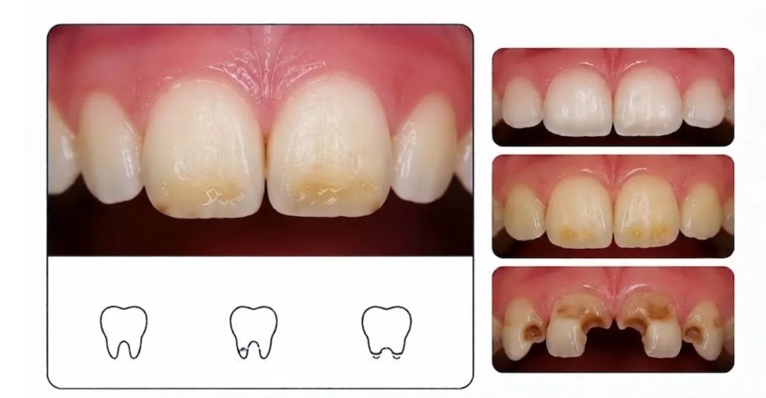
Note : All images used are for editorial and illustrative purposes only and may not originate from the original news provider or associated company.
Why Enamel Hypoplasia Is Sometimes Mistaken for Tooth Decay
Both conditions can produce visible depressions and discoloration. Developmental defects are present because enamel formation was interrupted. Caries develop after eruption through a disease process associated with bacterial acids. The surface characteristics can overlap when decay forms within a developmental pit. Timing and lesion activity become important clues. Dentists assess plaque accumulation, cavitation, borders, and changes over time. Early decay can look like a subtle white or brown area. A developmental defect may have a stable shape that was present after eruption. Radiographs can help when deeper decay is suspected. Careful differentiation prevents unnecessary removal of healthy tissue. It also helps determine whether preventive or restorative treatment is required.
Why Hypoplasia and Hypomineralization Should Not Be Treated as the Same Condition
The two defects involve different aspects of enamel formation. Hypoplasia primarily describes reduced enamel quantity or thickness. Hypomineralization primarily involves altered enamel quality and mineralization. Their clinical appearances can overlap, especially after enamel breaks down. Diagnosis therefore requires attention to surface thickness and translucency. Treatment may also differ because the structural problems are not identical. Qualitative defects can present with sensitivity and post-eruptive breakdown. Quantitative defects can present with pits, grooves, or missing enamel. Using the terms interchangeably can create confusion during treatment planning. Accurate terminology helps clinicians communicate the patient's condition more precisely.
Clinical Note
A detailed assessment should identify the pattern before treatment begins. The same visible discoloration can have several possible explanations. The dentist should therefore examine enamel thickness, surface structure, distribution, and associated symptoms. Medical and dental history can provide additional context. Imaging may be used when it answers a specific clinical question. Treatment should prioritize preservation of healthy tooth structure. Preventive measures remain important even when cosmetic treatment is planned. Patients should understand why a particular restoration has been recommended. Clinical decisions should also account for age and future dental development. A personalized approach helps match treatment intensity to the actual structural problem.
Dr. Rifat Alsaman
Dr. Rifat Alsaman, emphasizes individualized assessment when enamel defects are present. Each affected tooth should be examined according to its structure, symptoms, and functional demands. The visible appearance alone may not show the full extent of structural changes. Assessment can include teeth, gums, and surrounding oral tissues. Digital X-rays, CBCT, or 3D intraoral scanning may be considered when clinically appropriate. Digital planning can help compare restorative options before treatment begins. Conservative treatment may be suitable when adequate natural structure remains. More extensive restoration can be considered when structural integrity is compromised. The treatment plan should also consider aesthetic expectations and long-term maintenance. This individualized process supports safer and more predictable dental care.
Average Cost of Enamel Hypoplasia Treatment in the US, UK, and Turkey
Treatment costs vary because different patients need different procedures. A mild defect may need prevention or simple bonding. A severely weakened tooth may need a crown. Cosmetic concerns can also introduce additional restorative procedures. The following figures are planning benchmarks rather than fixed treatment quotes. They should not replace a personalized dental examination. U.S. figures use recent national cost data. UK figures separate NHS patient charges from private dental pricing. Turkey figures use published 2026 Istanbul pricing information. These markets use different healthcare systems and pricing structures. The final price can change after clinical assessment. Diagnostics, restorative materials, laboratory fees, and follow-up can also affect the total. Vitrin Clinic provides individualized treatment planning rather than applying one universal price.
Average Enamel Hypoplasia Treatment Cost in the United States
There is no single national price for treating a developmental enamel defect. Cost depends mainly on the procedure selected and the number of teeth treated. Dental bonding averaged $431 in CareCredit's published U.S. data. The reported bonding range was 288–915. Porcelain veneers averaged $1,455 per tooth in CareCredit's 2026 survey. The reported range was 1,020–2,506 per tooth. These figures can help patients build a preliminary budget. They do not represent a diagnosis-specific treatment fee. A dentist may recommend prevention instead of cosmetic restoration. Crown treatment may cost more when laboratory work and additional procedures are required. Insurance coverage also depends on whether treatment is considered restorative or cosmetic. Written estimates should identify every included service.
Treatment | Average Cost | Reported Range | Notes |
Dental bonding | $431 | 288–915 | Per procedure or tooth depending on provider |
Porcelain veneer | $1,455/tooth | 1,020–2,506 | Cosmetic treatment |
Dental crown | Varies | Varies | Depends on material, tooth, and complexity |
U.S. figures are planning benchmarks from published 2026 CareCredit cost data. Costs vary by location, provider, material, and insurance coverage.
Average Enamel Hypoplasia Treatment Cost in the United Kingdom
UK treatment costs depend on whether care is provided through the NHS or privately. In England, NHS Band 1 costs $27.90 from April 1, 2026. Band 2 costs $76.60, while Band 3 costs $332.10. Band 3 can include crowns and other complex restorative treatments. Private prices are considerably more variable. A 2026 survey covering more than 2,000 UK practices reported a $720 median private crown price. Its reported crown range was $350–$995. The same source reported a $614 median veneer price. These figures are market benchmarks rather than personalized quotes. Patients should confirm whether consultations, scans, temporary restorations, and follow-up are included.
Treatment | Average or Median Cost | Reported Range | Treatment Setting |
NHS Band 1 | $27.90 | — | NHS England |
NHS Band 2 | $76.60 | — | NHS England |
NHS Band 3 | $332.10 | — | NHS England |
Private dental crown | $720 median | $350–$995 | Private UK dental practices |
Private veneer | $614 median | Varies | Private UK dental practices |
UK figures use NHS England 2026 charges and published 2026 private-practice pricing research. Private costs vary by clinic, material, location, and treatment complexity.
Average Enamel Hypoplasia Treatment Cost in Turkey
Turkey uses private dental pricing for many international patients. Istanbul clinics publish prices that vary considerably between providers and materials. A 2026 Istanbul market review reported a median zirconia crown price of $205. The same review reported a median porcelain veneer price of $231. Another Istanbul provider lists zirconia crowns at $120 and E-Max crowns at $165. It lists composite bonding at $105 per tooth. Published Turkish tariffs can also differ from international-patient package prices. Some clinics combine treatment, accommodation, transfers, and coordination. Patients should compare the complete package rather than one treatment line. Final prices depend on examination findings, materials, laboratory choices, and treatment complexity. Vitrin Clinic prepares treatment plans according to individual clinical findings.
Treatment | United States | United Kingdom | Turkey |
Dental bonding | $431 average | Private prices vary | $105 published example |
Porcelain veneer | $1,455 average | $614 median | $231 median review |
Crown | Varies by material | $720 median private | $205 median zirconia review |
NHS crown pathway | Not applicable | $332.10 Band 3 | Not applicable |
What Determines the Final Treatment Cost?
Several factors can change the total price of dental treatment. The number of affected teeth is one important factor. One tooth usually requires less material and clinical time. Multiple teeth may require broader planning and additional appointments. Structural severity also influences the recommended procedure. Minor defects may need preventive care or composite bonding. Extensive structural damage may require crowns or other restorations. Diagnostic imaging can add costs when deeper problems require assessment. Existing cavities may require treatment before cosmetic procedures. Root canal treatment may also increase the overall cost when deeper disease is present. Material selection can affect laboratory and restoration fees. International patients may also have travel, accommodation, and transportation expenses. A written treatment plan should separate dental fees from optional travel services.
Cost Factor | How It Can Affect the Price |
Number of Teeth | More treated teeth usually increase materials, chair time, and appointments. |
Defect Severity | Minor defects may need prevention, while extensive damage may require restorations. |
Treatment Type | Bonding, crowns, microabrasion, and veneers have different costs. |
Diagnostic Imaging | X-rays or CBCT may add costs when clinically necessary. |
Existing Dental Problems | Cavities, infection, or gum problems may require additional treatment. |
Material Selection | Composite, ceramic, zirconia, and other materials have different laboratory costs. |
Additional Procedures | Root canal treatment or other procedures can increase the total fee. |
Follow-Up and Maintenance | Repairs, reviews, and long-term maintenance may create additional costs. |
International Travel | Flights, accommodation, transfers, and other travel expenses are separate costs. |
Costs should always be confirmed after an individual dental examination. Vitrin Clinic can provide a personalized treatment plan based on clinical findings and treatment needs.
Number of Teeth Requiring Treatment
Treatment costs generally increase when more teeth require restoration. Each additional tooth can require clinical preparation, material, and finishing. Multiple teeth may also require additional planning appointments. However, some practices provide package pricing for several procedures. Package discounts should not automatically determine treatment selection. Every tooth should first be assessed individually. Some teeth may need prevention rather than restoration. Others may require different materials because their functional demands differ. Front teeth often have greater aesthetic considerations. Posterior teeth commonly experience higher chewing forces. Treatment planning should therefore consider each tooth's role. Vitrin Clinic evaluates the affected teeth before determining the number of procedures required.
Severity of Enamel Loss or Structural Damage
Severity strongly influences the type of treatment required. Small surface irregularities may require no restoration. Deeper defects can expose dentin or weaken the crown. Structural loss may make composite bonding less suitable. A severely compromised tooth may require full coverage. Active decay can further increase treatment complexity. Sensitivity can also influence the sequence of care. The dentist first evaluates whether the tooth remains structurally stable. Preserving healthy tissue remains important during treatment selection. More extensive preparation can increase both procedure time and cost. Patients should ask why a specific restoration has been recommended. The answer should relate directly to the tooth's clinical condition.
Bonding, Crowns, Microabrasion, or Preventive Treatment
Different procedures have different costs and clinical purposes. Preventive care is generally less complex than restorative treatment. Bonding can repair small defects with relatively conservative preparation. Microabrasion targets selected superficial discoloration rather than missing structure. Crowns provide greater coverage for severely weakened teeth. Veneers may address selected aesthetic concerns in suitable permanent teeth. The treatment should match the problem rather than the patient's preferred material. Using a more expensive restoration does not automatically provide greater clinical benefit. The dentist should explain the expected advantages and limitations. Material selection should also consider longevity, function, and maintenance. Cost should be evaluated alongside tooth preservation and clinical suitability.
Diagnostic Imaging and Additional Dental Procedures
Some patients need imaging before restorative treatment. Basic radiographs can help identify hidden cavities or deeper structural problems. CBCT may be considered when three-dimensional information could alter treatment planning. It should not be used routinely without a clinical indication. Existing decay may require fillings before aesthetic procedures. Root canal treatment may be needed when pulpal disease is present. Gum treatment may also be necessary when periodontal inflammation exists. These additional procedures can increase the final cost. Patients should request an itemized estimate before starting complex treatment. Vitrin Clinic uses digital imaging when clinically appropriate. The purpose is to answer specific diagnostic questions.
Follow-Up and Maintenance
Restorations require monitoring after treatment. Composite restorations may need polishing, repair, or replacement over time. Crowns require checks for margins, wear, and surrounding tooth health. Patients with grinding habits may need additional protection. Follow-up can identify small problems before they become larger failures. International patients should also ask about remote support after returning home. Travel planning should include enough time for clinically necessary appointments. A low initial price may become less useful when aftercare is excluded. Patients should compare the full treatment pathway rather than one procedure price. Vitrin Clinic includes follow-up planning as part of individualized care.
Enamel Hypoplasia Treatment Cost Comparison: US vs UK vs Turkey
Cost comparisons should focus on equivalent treatments. A single bonding procedure is not directly comparable with a crown. Material quality, laboratory standards, diagnostics, and aftercare also vary. U.S. national data show relatively high average cosmetic treatment prices. CareCredit reported a $1,455 average for a single porcelain veneer. UK private crown pricing reached a $720 median in 2026 data. Istanbul published prices can be substantially lower for some restorative procedures. A 2026 Istanbul review reported a $205 median zirconia crown. These differences do not establish equal clinical value between providers. Patients should compare materials, clinician qualifications, diagnostics, guarantees, and aftercare. International travel costs also belong in the total budget. A personalized written quotation remains the most reliable comparison.
When Should You See a Dentist About Enamel Hypoplasia?
Professional evaluation is appropriate when enamel looks unusually thin, irregular, or damaged. Assessment becomes more important when symptoms are developing. Sensitivity can indicate exposed dentin or another dental condition. New chips may indicate increasing structural weakness. Food trapping around pits can increase plaque retention. Visible cavities require treatment rather than cosmetic masking. Children with multiple affected teeth should receive developmentally appropriate assessment. A dentist can distinguish developmental defects from fluorosis, hypomineralization, erosion, and decay. Treatment is not always required immediately. Some mild findings can be monitored with preventive care. Prompt evaluation can reduce the risk of avoidable structural complications.
Warning Signs That Need Professional Evaluation
Some symptoms should not be ignored. Persistent sensitivity can indicate underlying structural exposure. Increasing pain may indicate decay or pulpal involvement. New chips can reveal progressive structural weakness. Rapid wear may indicate excessive mechanical stress. Food trapping can make plaque control more difficult. Visible cavities require direct examination and treatment planning. Bleeding gums can also indicate a separate periodontal issue. A change in color should be assessed when its cause is uncertain. Children may need earlier evaluation because developing teeth remain vulnerable. Patients should also seek care when symptoms interfere with eating or brushing. Professional assessment provides a clearer explanation than appearance alone.
Persistent or Increasing Tooth Sensitivity
Short sensitivity can occur when dentin becomes more exposed. Persistent symptoms deserve professional assessment. Sensitivity that increases over time may reflect worsening structural protection. Decay can also produce temperature-related discomfort. Cracks can create similar symptoms during eating. The dentist will assess the duration and trigger of pain. Brief discomfort differs from lingering or spontaneous pain. Treatment depends on the underlying cause. Desensitizing toothpaste may help uncomplicated sensitivity. Fluoride treatment may also support prevention in selected cases. Severe symptoms may require restorative or endodontic treatment. Patients should report changes rather than continuously increasing home treatments.
New Chips, Cracks, or Rapid Wear
New chips can indicate that a vulnerable surface is under excessive stress. Cracks may involve enamel, dentin, or deeper structures. Rapid wear can result from grinding or other mechanical forces. Acid exposure can also contribute to progressive surface loss. The dentist should determine whether the damage is developmental or acquired. Bite assessment may be useful when wear is extensive. A night guard can sometimes reduce grinding-related damage. Restorative protection may be considered when structure is compromised. Early treatment can prevent a small defect from becoming more extensive. Patients should avoid chewing hard objects with affected teeth.
Visible Pits With Food or Plaque Trapping
Deep pits can retain food and plaque after eating. This can make affected surfaces harder to keep clean. Persistent trapping may increase caries risk in susceptible patients. A dentist can determine whether a sealant is appropriate. Some deeper defects require restorative material instead. The surface should be cleaned and examined before deciding. Regular fluoride exposure can provide additional preventive support. Children may need parental assistance with brushing. Interdental cleaning can help when defects involve contacting surfaces. Patients should mention recurring food trapping during dental appointments. Early protection can reduce the need for larger restorations.
Signs of Dental Cavities or Tooth Pain
Cavities may appear as white, brown, or darkened changes. Cavitation can create a hole or softened surface. Tooth pain may occur when decay reaches deeper tissues. Developmental enamel defects can sometimes make diagnosis more complex. A dentist assesses lesion activity and structural appearance. Radiographs may be appropriate when decay cannot be assessed visually. Pain that occurs without stimulation deserves prompt attention. Lingering temperature pain can indicate deeper pulpal irritation. Treatment may involve a filling, root canal treatment, or another procedure. Cosmetic treatment should wait until active disease is controlled.
When Should a Child With Suspected Enamel Defects Be Evaluated?
Children should be evaluated when unusual enamel appears after eruption. Early assessment is particularly important when several teeth show similar changes. Sensitivity, chipping, or food trapping also justify examination. A pediatric dentist can monitor developing permanent teeth. The clinician can compare affected teeth with normal neighboring teeth. Medical and dental history may reveal relevant developmental events. Parents should mention previous dental trauma or primary tooth infections. Significant childhood illness may also be worth documenting. Early preventive care can include fluoride, sealants, or monitoring. Treatment intensity depends on caries risk and structural severity. Parents should not delay assessment until pain develops.
When Is a Pediatric Dentist Referral Appropriate?
Referral can be appropriate when defects are extensive or generalized. Multiple affected teeth may require specialized developmental assessment. Young children can also benefit from pediatric behavior management. Complex restorative treatment may need coordination across dental specialties. Suspected inherited enamel disorders can warrant additional specialist evaluation. A pediatric dentist can monitor tooth eruption and growth. They can also coordinate preventive care during vulnerable developmental stages. Referral may be especially useful when routine dental treatment has been difficult. The decision depends on clinical findings and patient needs. Parents should receive a clear explanation for any recommended referral.
Tips for Patients
Good daily care can protect vulnerable tooth surfaces. Patients should follow their dentist's fluoride recommendations. Gentle brushing can reduce unnecessary mechanical abrasion. Frequent sugary and acidic exposures should be limited. Desensitizing products may help when sensitivity is present. Patients should monitor new chips, cracks, or changes in discomfort. Children may require parental assistance with brushing. A written treatment plan can clarify which teeth need protection. Patients should ask about restoration longevity and maintenance. International patients should also understand aftercare arrangements before traveling. Vitrin Clinic can coordinate individualized treatment planning for international patients.
What to Ask Your Dentist About Thin or Weak Enamel
Ask what type of enamel defect is present. Ask whether the defect affects thickness, mineral quality, or both. Ask whether decay is already present. Ask whether imaging is clinically necessary. Ask which treatment is preventive and which is restorative. Ask how much healthy tooth structure will be preserved. Ask how long the recommended restoration may last. Ask about sensitivity after treatment. Ask whether maintenance visits will be needed. Ask whether alternative conservative options are available. These questions help patients understand the treatment pathway. They also make cost comparisons more transparent.
What Information to Bring to Your Appointment
Bring previous dental records whenever they are available. Older radiographs can help establish whether a finding is longstanding. Parents should document previous trauma involving baby teeth. Medical history can include significant childhood illnesses and hospitalizations. Family dental history may be useful when several relatives have similar defects. Medication history should include relevant long-term treatments. Patients can also bring photographs showing when a visible change first appeared. A list of sensitivity triggers can help explain symptoms. International patients should bring details of previous restorations. Clear information helps clinicians build a more complete assessment.
How to Track Changes in Sensitive or Damaged Teeth
Photographs can help monitor visible changes over time. Take pictures under similar lighting conditions when possible. Record when sensitivity occurs and what triggers it. Note whether symptoms are brief or prolonged. Record new chips, rough areas, or visible surface changes. Keep track of restorations and their treatment dates. Parents can maintain a simple dental history for children. Avoid taking daily photographs unless a dentist recommends closer monitoring. The goal is to identify meaningful changes rather than create unnecessary records. Bring useful photographs to follow-up appointments. They can support clinical discussion when visual changes are subtle.
Why Regular Follow-Up Matters
Regular appointments help monitor vulnerable teeth before problems become extensive. Dentists can check restorations, plaque control, and new symptoms. Follow-up also identifies recurrent decay around restorations. Children require monitoring because permanent teeth continue developing and erupting. Changes in bite can alter stress on restored teeth. Grinding can also increase restoration wear. Preventive treatment may need renewal or adjustment. Sealants can require checking for partial loss. Composite restorations may need polishing or repair. Regular care can reduce the chance of unexpected treatment. The appropriate recall interval depends on individual risk.
Enamel Hypoplasia Assessment and Treatment at Vitrin Clinic
Vitrin Clinic approaches developmental enamel concerns through individualized clinical assessment. The evaluation considers tooth structure, symptoms, aesthetics, and functional requirements. Healthy tissue is preserved whenever clinically possible. Digital diagnostics may support assessment when additional information is needed. Treatment options are selected according to the actual condition of each tooth. Preventive procedures can be considered for mild abnormalities. Restorative treatment can protect teeth with greater structural compromise. Cosmetic options can be discussed when appearance is a significant concern. Existing decay or gum problems are addressed before elective cosmetic care. International patients receive coordinated planning throughout their treatment journey. The objective is a practical plan based on clinical findings.
Individual Assessment of Tooth Enamel and Existing Enamel Defects
Each affected tooth may present a different structural pattern. Vitrin Clinic examines thickness, surface integrity, color, and defect distribution. The surrounding gums and oral tissues are also assessed. Symptoms such as sensitivity and discomfort are documented. Existing restorations are evaluated for function and margins. Dental history can provide additional developmental information. The assessment does not rely on age alone. The number of affected teeth also helps guide diagnostic thinking. A localized defect may require a different approach from generalized involvement. The final recommendation reflects the specific clinical situation.
Digital Dental Imaging When Clinically Appropriate
Digital imaging can provide additional information that visual examination cannot show. Periapical or panoramic X-rays may help identify hidden dental problems. CBCT can provide three-dimensional information for selected clinical questions. It should only be used when its benefits justify the radiation exposure. Vitrin Clinic considers imaging according to clinical need. Digital images can also support treatment planning and communication. Imaging is interpreted alongside clinical examination and patient history. A scan should not replace direct dental assessment. This approach helps avoid unnecessary imaging while retaining useful diagnostic information.
Evaluating Tooth Structure Before Bonding, Restorations, or Crowns
Restorative treatment requires sufficient supporting tooth structure. Vitrin Clinic evaluates remaining enamel and dentin before choosing materials. Bite forces are also considered. Moisture control can influence adhesive restoration outcomes. Small defects may be suitable for conservative composite treatment. Larger structural defects may require greater coverage. Crowns can protect severely weakened teeth when indicated. Healthy tissue should not be removed unnecessarily. Existing decay must be treated before elective restorative care. The final restoration should support function and long-term maintainability. Patients receive treatment recommendations after clinical evaluation.
Personalized Treatment Planning for Aesthetic and Functional Concerns
Some patients primarily need structural protection. Others are concerned about visible discoloration or irregular contours. Treatment planning should address both goals when appropriate. Vitrin Clinic can evaluate cosmetic and functional priorities together. Digital photographs and intraoral scanning can support visual planning. DSD previews may help selected patients understand potential aesthetic changes. Treatment should remain biologically appropriate despite cosmetic goals. Conservative options are considered before more invasive procedures. Material selection depends on tooth condition and functional demands. The final plan should include expected maintenance. Patients can then understand what each procedure is designed to achieve.
When Enamel Defects Are Associated With Other Dental Treatments
Developmental defects can coexist with other dental problems. Affected teeth may also have cavities, sensitivity, or structural wear. Gum inflammation may need treatment before restorative procedures. Orthodontic treatment can sometimes change how vulnerable teeth are loaded. Missing teeth may require implant planning alongside restoration of existing teeth. Cosmetic treatment may need coordination with periodontal care. Vitrin Clinic evaluates the overall oral condition before combining treatments. Treatment sequencing can reduce complications and improve predictability. Not every procedure should be performed during the same appointment. The final sequence depends on clinical findings and patient priorities.
How Vitrin Clinic Supports Your Treatment Journey
Vitrin Clinic coordinates dental care through assessment, diagnostics, treatment planning, and follow-up. International patients can receive assistance with appointment coordination. Clinical findings remain the basis of treatment recommendations. Digital workflows can support communication between different stages of care. Treatment options are discussed according to tooth condition and patient goals. Conservative procedures may be used when they provide sufficient protection. Restorative or cosmetic treatment can be considered when clinically appropriate. International patients can also receive support with treatment logistics. Aftercare planning is included when restorations or complex procedures are performed. The process aims to provide continuity throughout the dental journey.
Consultation and Initial Clinical Assessment
The first appointment begins with a comprehensive dental evaluation. Teeth, gums, and surrounding oral tissues are examined. Existing restorations and visible defects are documented. Symptoms such as sensitivity or pain are reviewed. Dental history can help identify earlier developmental events. The dentist also evaluates oral hygiene and carries risk. Photographs may document visible structural changes. The initial assessment determines whether additional diagnostics are required. Patients receive information about possible treatment pathways. No restorative procedure should be selected solely from a photograph. Clinical examination remains the foundation of treatment planning.
Digital Diagnostics and Treatment Planning
Digital tools can improve visualization during treatment planning. Intraoral scanning can document tooth surfaces and occlusion. Digital radiography can reveal areas hidden from direct examination. CBCT may be used when three-dimensional information is clinically relevant. Vitrin Clinic uses digital diagnostics selectively. The objective is to collect information that changes treatment planning. Digital records can also support communication between clinical teams. Treatment simulations may help explain proposed aesthetic changes. Technology does not replace clinical judgment. It supplements examination, history, and professional assessment.
Personalized Selection of Conservative or Restorative Treatment
Treatment should correspond to structural severity. Preventive measures may be sufficient for mild defects. Bonding can restore selected localized surface losses. Composite restorations may provide additional structural coverage. Crowns can protect severely weakened teeth. Cosmetic procedures may address visible defects after health concerns are controlled. Vitrin Clinic considers preservation of healthy structure before selecting restorative materials. Bite forces and tooth location also influence material selection. Patients receive an individualized treatment pathway. The plan can be adjusted when clinical findings change.
Coordinating Treatment for International Patients
International dental care requires more than scheduling clinical appointments. Patients may need help coordinating treatment timing and travel logistics. Vitrin Clinic supports international patients throughout the planned dental journey. Appointment scheduling can be coordinated around treatment stages. Digital records can help prepare the clinical team before arrival. Patients can discuss diagnostic information before travelling when appropriate. Treatment plans should still be confirmed after direct clinical examination. Travel schedules should allow time for necessary procedures and reviews. Accommodation and transportation can be coordinated where included in the selected service. Patients should understand exactly what their package covers.
Follow-Up and Aftercare Planning
Aftercare helps protect restorations and monitor healing when procedures are performed. Patients receive guidance about brushing and dietary habits. Sensitivity should be reported when it persists or worsens. Restorations require monitoring for wear, chipping, or marginal changes. International patients should know how follow-up is handled after returning home. Vitrin Clinic can coordinate planned aftercare and treatment communication. Long-term dental maintenance remains important after cosmetic or restorative treatment. The patient should continue routine dental examinations after completing treatment. Follow-up requirements vary according to the selected procedure. Good maintenance supports the longevity of dental work.
Key Takeaways
Developmental enamel defects begin while teeth are forming. Their appearance can vary from shallow pits to extensive structural loss. Causes can include genetic, systemic, nutritional, prenatal, and local influences. Some cases remain without a clearly established cause. Diagnosis depends on clinical appearance, distribution, history, and selected investigations. Treatment ranges from prevention to restorative and cosmetic procedures. Early management can reduce secondary sensitivity, caries, and structural wear. Children may require closer monitoring during dental development. Treatment decisions should preserve healthy tissue wherever possible. Vitrin Clinic uses individualized assessment to determine suitable treatment options. Regular follow-up remains important after treatment.
Enamel Hypoplasia Is a Developmental Defect of Tooth Enamel
The condition develops while enamel is forming before tooth eruption. It primarily reflects reduced enamel quantity or thickness. The resulting surface may contain pits, grooves, or missing areas. Different developmental disturbances can produce overlapping appearances. Accurate diagnosis therefore depends on more than color alone. A dentist considers tooth distribution and developmental history. This helps separate quantitative defects from other enamel abnormalities. Treatment focuses on protecting the tooth after eruption. Missing enamel cannot naturally regenerate once mature tooth structure is lost. Restorative materials can replace selected areas when required.
Thin Enamel Can Increase the Risk of Sensitivity, Wear, and Dental Cavities
Thinner enamel provides less physical protection for underlying dentin. Irregular surfaces may also retain plaque more easily. These factors can increase susceptibility to sensitivity and caries. Not every affected tooth will develop complications. Risk depends on oral hygiene, diet, fluoride exposure, and defect severity. Chewing forces can also contribute to progressive surface damage. Preventive treatment can reduce avoidable risks. Fluoride and sealants may be appropriate for selected teeth. Regular examinations help identify changes before major damage develops.
The Cause and Severity Determine the Appropriate Treatment
Treatment should not be selected solely from appearance. Dentists consider structural severity, symptoms, tooth location, and age. The likely cause can also influence further evaluation. Mild defects may need monitoring and prevention. Larger defects can require bonding or full-coverage restoration. Generalized inherited conditions may require specialist evaluation. Active decay must be treated before elective cosmetic procedures. A conservative approach can preserve healthy natural tissue. The treatment plan should also consider future maintenance needs.
Early Dental Assessment Can Help Protect Affected Teeth
Early assessment provides an opportunity for preventive intervention. Sealants can protect selected pits and grooves. Fluoride can support resistance against caries-related mineral loss. Oral hygiene coaching can improve plaque control. Regular monitoring can identify chipping or decay early. Children benefit from assessment while permanent teeth continue developing. Early treatment does not always mean immediate restoration. Observation can be appropriate for stable and asymptomatic findings. Professional monitoring determines whether treatment becomes necessary.
Professional Evaluation Is Important When Pain, Sensitivity, Chipping, or Decay Develops
Symptoms can signal complications that require direct examination. Sensitivity may reflect exposed dentin or another dental problem. Pain can indicate decay, cracks, or pulpal inflammation. Chipping can reveal structural weakness. Visible cavities require professional treatment planning. A dentist can distinguish these problems from the underlying developmental defect. Delaying assessment may allow preventable problems to become more extensive. Prompt evaluation supports timely and conservative management.
At Vitrin Clinic
Vitrin Clinic provides individualized dental assessment for patients concerned about enamel appearance and structure. Treatment planning considers tooth health before cosmetic goals. Clinical examination can include teeth, gums, and surrounding oral tissues. Digital X-rays, CBCT, and 3D intraoral scanning are used when clinically appropriate. Digital planning can support restorative and aesthetic decision-making. Our Treatments options may include preventive care, bonding, restorations, or crowns. Cosmetic procedures are considered only when the tooth is suitable. International patients can receive coordinated treatment planning before arrival. The Vitrin team also supports aftercare planning. Each treatment pathway is developed according to the patient's dental findings.
Reference
FAQs
Dr. Rifat Alsaman has more than 5 years of clinical experience in dentistry and currently serves as the Head of the Medical Team at Vitrin Clinic. He is dedicated to providing exceptional patient care, overseeing treatment planning, and ensuring the highest clinical standards across the team. His expertise, attention to detail, and commitment to continuous professional development have helped countless patients achieve healthier, more confident smiles.


