Oral Surgery
October 11, 2026
Coronectomy: Is It Safer Than Wisdom Tooth Extraction When a Nerve Is Nearby?
 Dr. Rifat Alsaman
Coronectomy is a type of partial wisdom tooth removal. It may be recommended when the roots of an impacted wisdom tooth lie close to the inferior alveolar nerve. You may need a wisdom tooth removed but worry about numbness, tingling, or other nerve-related complications. This technique removes the crown and intentionally leaves the roots in place when appropriate. This guide explains who may benefit, how dentists assess nerve proximity, what recovery involves, and which risks matter.
Coronectomy: What Is It, and When Is It Recommended?
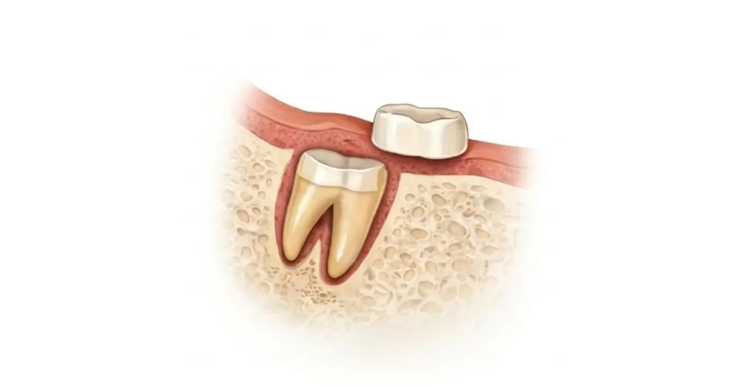
Coronectomy is a planned surgical alternative to taking out a whole lower wisdom tooth. The surgeon removes the crown and leaves healthy roots undisturbed in the jawbone. It is recommended only in selected cases. The usual reason is that imaging shows wisdom tooth roots near nerve structures in the lower jaw. In that situation, pulling the roots could bruise or tear the nerve. Leaving them avoids that contact. The sections below explain the technique, the problems impacted teeth cause, and why root position matters.
What Is a Coronectomy?
A wisdom tooth coronectomy is an operation that removes only the crown, the upper part of the tooth. The roots are deliberately left in the jawbone. Complete removal, by contrast, takes out the crown and every root. The technique is mainly used for selected impacted lower wisdom teeth. In these cases, removing the roots may pose a greater risk to the inferior alveolar nerve than leaving them. It is not suitable for every wisdom tooth or every patient. Each case needs its own assessment.
What Problems Can an Impacted Wisdom Tooth Cause?
An impacted wisdom tooth is trapped beneath the gum or bone and cannot erupt fully. A partly covered tooth can trap food and bacteria. This may cause pain, swelling, and gum inflammation called pericoronitis. Decay or bone loss can also affect the neighboring second molar. Repeated infection, persistent pain, or damage to the adjacent tooth needs assessment. However, many impacted teeth stay symptom-free for years. Impaction alone does not automatically mean surgery is necessary. Your dentist weighs the evidence of disease before recommending treatment.
Why Does the Position of Wisdom Tooth Roots Matter?
The inferior alveolar nerve runs through a canal inside the lower jaw. It supplies feeling to the lower teeth, lower lip, and chin on that side. When roots touch or wrap around this canal, complete removal can stretch or compress the nerve. This is the main cause of wisdom tooth extraction nerve damage affecting the lip and chin. The lingual nerve is different. It lies in the soft tissue on the tongue side of the jaw. Injury there can alter feeling or taste on one side of the tongue.
How Dentists Assess Nerve Risk Before a Coronectomy
Assessment happens in stages. First, your dentist confirms whether the wisdom tooth needs treatment at all. Next, a clinical examination records symptoms and the state of nearby tissues. Then imaging shows where the roots sit in relation to the nerve canal. A standard panoramic X-ray is usually the starting point. A three-dimensional scan may follow if the first image raises concern. Together, these findings allow the surgeon to estimate risk and explain your options. No test predicts the outcome with certainty, but careful planning lowers avoidable harm.
What Does a Dental Examination Look For?
Your dentist begins by asking about pain, swelling, bad taste, and difficulty opening your mouth. The examination then checks the gum over the tooth for redness, pus, or trapped food. The dentist notes how the tooth is angled and how much has erupted. Signs of spreading infection are recorded. The neighboring second molar is checked for decay and gum pocketing. This step answers a basic question. Does the wisdom tooth need treatment at all? If no disease is present, monitoring may be the safer path.
When Is a CBCT Scan Before Wisdom Tooth Removal Useful?
Cone-beam computed tomography, or CBCT, is a three-dimensional dental imaging technique. It shows the jaw in thin slices from several directions. A panoramic X-ray is flat, so overlapping structures can be hard to interpret. When that image suggests the roots touch the inferior alveolar canal, CBCT may clarify the true relationship. It can show whether the canal runs beside, below, or between the roots. However, it usually involves more radiation than a standard panoramic X-ray. It is therefore used only when the result may change treatment planning.
How Do Dentists Estimate the Risk of Nerve Injury?
Dentists look for recognized warning signs on the panoramic radiograph. These include darkening of the root, a break in the white outline of the canal, and a canal that changes course. When indicated, CBCT adds detail about contact between root and canal. These findings relate to the inferior alveolar nerve. The lingual nerve sits in soft tissue and does not show on X-rays. Its protection depends mainly on careful surgical technique. Imaging supports clinical judgment. It cannot guarantee a complication-free procedure.
Tips for Patients
A little preparation makes your consultation and recovery easier. Consider these practical steps:
Ask whether the tooth is causing disease or whether monitoring is a reasonable option.
Ask how close the roots are to the inferior alveolar nerve and what the images show.
Confirm whether CBCT would change the treatment decision in your case.
Discuss possible retained-root migration and future surgery before consenting.
Save your postoperative instructions and emergency contact details.
Record any change in lip, chin, or tongue sensation for your follow-up.
What Happens During the Coronectomy Procedure?
The operation is usually performed under local anesthesia in a dental or oral surgery setting. Planning is completed beforehand, so the appointment itself focuses on surgery. The surgeon exposes the tooth, removes the crown, and checks that the roots remain firm. The gum is then closed over the site. You stay awake but should not feel sharp pain. Pressure and vibration are normal. The exact sequence depends on how the tooth lies and what the surgeon finds. The steps below outline a typical appointment.
Coronectomy Procedure Steps
Assessment and planning. Your surgeon reviews symptoms, examination findings, and imaging.
Local anesthesia. The area is numbed. Sedation may be considered when appropriate.
Access to the tooth. A gum incision exposes the crown.
Crown removal. The crown is cut away while surrounding tissues are protected.
Root assessment. The surgeon confirms the roots are firm and trims them below the bone edge.
Wound closure. The site is rinsed, and sutures are placed when indicated.
The precise technique varies with tooth anatomy and the surgeon's judgment.
What Happens to the Roots After Coronectomy?
The retained roots stay beneath the gum. In uneventful healing, new bone gradually forms over them. Many roots also move slowly away from the inferior alveolar nerve. This is known as root migration after coronectomy. Studies report that most movement occurs in the first one to two years and then slows. The distance varies between patients and is often only a few millimeters. Retained roots do not always need further surgery. Follow-up X-rays remain important, because they reveal infection or roots moving toward the surface.
What Symptoms Can You Expect After Coronectomy?
Recovery usually feels similar to recovery after a surgical wisdom tooth removal. Expect some soreness, swelling, and stiffness in the first days. These effects are a normal response to surgery on gum and bone. They should ease gradually rather than worsen. Some patients develop complications, such as infection or delayed healing. Knowing what is expected helps you recognize what is not. The following answers cover pain, other common symptoms, and dry socket. Contact your treating clinician whenever a symptom worries you.
Is Pain After Coronectomy Normal?
Yes. Soreness is expected once the anesthetic wears off. Swelling and limited jaw opening are also common. Swelling often peaks around the second or third day. Discomfort is usually strongest early and then eases over the following days. The pace differs between people, so no fixed timeline applies. Pain relief recommended by your clinician normally keeps you comfortable. Some signs need assessment. These include pain that worsens instead of improving, fever, or swelling that keeps increasing. Such changes may indicate infection or another healing problem.
What Other Symptoms Might Occur During Recovery?
Slight oozing of blood during the first day is common. Bruising may appear on the cheek or jaw. The jaw can feel stiff for a short time. Chewing may be uncomfortable, so soft foods are easier at first. These effects are short-lived and settle as healing progresses. Numbness is different. The anesthetic normally wears off within hours. Numbness or tingling of the lower lip, chin, or tongue that persists warrants prompt contact with your treating clinician. Heavy bleeding or pus also needs review.
Can You Develop Dry Socket After Coronectomy?
Dry socket is a painful healing complication. It occurs when the blood clot in an extraction site is lost or breaks down too early. This procedure does not remove the entire tooth, and the roots still fill much of the socket. Even so, dry-socket-like healing problems have been reported. Other postoperative complications, including infection, may also occur. Typical warning signs are severe, throbbing pain and a bad taste a few days after surgery. Seek professional assessment if pain becomes severe or increases after initially improving.

Coronectomy Recovery Time: What Should You Expect?
Recovery happens in phases rather than on a single date. The first days involve swelling and tenderness. Over the next one to two weeks, eating and jaw movement usually become easier. Deeper healing of the bone continues for months without you noticing. Monitoring of the retained roots extends further still. Many people return to routine activities within a few days, although this varies. Your surgeon will advise you according to the difficulty of your operation. The answers below describe each stage and what influences it.
What Happens During the First 24–72 Hours?
Rest on the day of surgery, and keep your head slightly raised. A cold compress on the cheek may limit swelling when your clinician recommends it. Avoid vigorous rinsing on the first day so the clot stays in place. After that, gentle cleaning keeps the area healthy. Good coronectomy aftercare also means choosing soft foods and taking medication as directed. Swelling and discomfort often build during the first two to three days. They then begin to settle. Follow your written postoperative instructions closely throughout this period.
How Does Recovery Progress Over the Following Weeks?
Most people notice steady improvement after the first few days. Eating becomes more comfortable, and the jaw opens more freely. Everyday activities feel normal again for many patients within one to two weeks. Gum and bone heal on different timelines. The gum usually closes over the site within a few weeks. Bone remodels much more slowly, over several months. Sutures may dissolve or be removed at a review visit. Monitoring of the retained roots continues beyond this early recovery period, usually with periodic X-rays.
What Factors Can Affect Your Recovery?
Several factors shape healing. A deeply impacted tooth requires more surgery, which can mean more swelling. Existing gum inflammation may slow early progress. Smoking reduces blood supply and raises the risk of healing problems. General health matters too, including conditions such as poorly controlled diabetes. Clean teeth and careful adherence to postoperative instructions support recovery. Because of these variables, healing times differ. Your progress should be judged by the overall clinical course, not a fixed number of days. Steady improvement is the most reassuring sign.
.webp)
Coronectomy Risks and Complications You Should Understand
Every surgical procedure carries risk, and this one is no exception. It is designed to lower one specific risk, not all of them. General risks include pain, swelling, bleeding, and infection. Specific risks include nerve disturbance, delayed healing, and problems with the retained roots. Some patients later need a second operation to remove those roots. Your surgeon should explain how likely each outcome is in your case. Understanding these points is part of informed consent. The sections below describe the main complications in more detail.
Inferior Alveolar Nerve Damage
Injury to this nerve can cause numbness, tingling, or altered feeling in the lower lip and chin. Lip movement is not affected, because this nerve supplies feeling there, not muscle control. In selected high-risk cases, leaving the roots may lower this risk compared with complete extraction. However, the risk is not zero. The nerve can still be disturbed by swelling, instruments, or roots that loosen and must be removed. Most sensory changes after wisdom tooth surgery improve with time. Some are permanent, so report any change.
Lingual Nerve Damage
The lingual nerve supplies feeling to the front two-thirds of the tongue on one side. It also carries taste fibers from that area. The nerve lies in soft tissue close to the inner side of the lower wisdom tooth. It can be affected by tissue retraction, the incision, or instruments during surgery. This is true even when the roots are retained, because the crown must still be reached and removed. Injury may cause numbness, tingling, or altered taste. Most cases recover, but lasting changes are possible.
Infection, Root Exposure, and Root Migration
Infection can develop around the surgical site or the retained roots. Signs include increasing pain, swelling, pus, or a bad taste. Healing of the gum is sometimes delayed. Occasionally, a root works its way toward the surface and becomes exposed. Exposed or infected roots usually need removal. By then, the root has often moved away from the nerve, which can make that second operation less risky. Root migration by itself is expected and often harmless. Symptoms or visible root exposure always require clinical review.
What Is the Success Rate of Coronectomy?
Published outcomes vary, so one percentage would be misleading. Results depend on patient selection, surgical technique, follow-up duration, and how each study defines success. Clinical reviews generally report a low rate of inferior alveolar nerve injury in suitable cases. They also report that a minority of patients need later root removal. Avoiding nerve injury is one measure of success. Freedom from every complication is another. Any quoted coronectomy success rate should name its evidence source. Ask your surgeon which outcomes the figures describe.
How Can You Reduce Complications Before and After Coronectomy?
You cannot control your anatomy, but you can influence several other risk factors. Good preparation helps the surgeon plan safely. Careful aftercare protects the wound while it heals. Honest communication matters at both stages. Tell your clinician about your health before surgery, and report unexpected symptoms afterwards. Stopping smoking around the time of surgery is one of the most useful steps. Good oral hygiene is another. The guidance below is general. Your own instructions from the treating clinician always take priority.
Before Surgery
Share an accurate medical history with your clinician. Include every medication, supplement, allergy, and any previous reaction to anesthesia. Blood thinners and some bone medicines are especially relevant. Ask why this procedure is being considered for you. Ask whether the imaging findings support it. Discuss smoking, because stopping before surgery supports healing. Mention any existing infection or recent swelling around the tooth. Raise any concerns about nerve sensation, including previous numbness. Finally, arrange time to rest afterwards, and clarify whom to contact if problems arise.
After Surgery
Take prescribed or recommended medication exactly as directed. Follow the wound-care instructions you were given. Keep your mouth clean, but brush gently near the surgical site. Avoid vigorous rinsing, especially on the first day. Do not smoke, because smoking interferes with healing. Avoid strenuous exercise for the first few days, as your clinician advises. Choose soft, cool foods, and chew on the opposite side. Attend every scheduled review, including later X-ray checks of the roots. Report worsening pain, swelling, fever, or changes in sensation promptly.
Prevention: Can You Avoid Wisdom Tooth Surgery?
Sometimes, yes. Not every wisdom tooth needs to come out. A tooth that is healthy, symptom-free, and not harming its neighbor can often be monitored. However, you cannot prevent impaction itself. It results from jaw size, tooth position, and the direction of eruption. What you can do is reduce the problems that lead to surgery. Regular checkups and good cleaning lower the chance of decay and gum infection. If disease does develop, early assessment gives you more options. The answers below explain both approaches.
Can Regular Dental Checkups Help?
Yes. Routine examinations allow your dentist to watch how wisdom teeth develop and erupt. Appropriate X-rays, taken when clinically justified, can reveal decay, cysts, or damage to the second molar. Finding these problems early means treatment can be planned calmly. It need not be arranged during a painful infection. Early review also allows time to consider imaging and discuss surgical options. Checkups do not mean surgery is inevitable. Not every impacted wisdom tooth requires removal. Many are simply monitored for changes over time.
Can You Prevent Problems Around Impacted Wisdom Teeth?
You can lower the risk, although you cannot remove it completely. Clean carefully around the back molars twice a day. A small-headed or single-tuft brush reaches the area behind the last tooth. Clean between the teeth daily as well. Watch for pain, swelling, bad taste, or bleeding gums near impacted wisdom teeth. Seek assessment if inflammation keeps returning. Recurrent infection is a common reason for recommending treatment. Hygiene helps control local problems. It cannot change the anatomical position of an impacted tooth.
What We Notice Clinically
Symptom severity alone does not establish the safest surgical approach. A very painful tooth may be simple to remove. A quiet tooth may lie against the nerve. Dr. Rifat Alsaman, Head of the Medical Team and cosmetic dentist at Vitrin Clinic, emphasizes assessing three things before recommending a procedure. These are the tooth itself, the surrounding tissues, and the relationship between roots and nerve. The appropriate choice balances two concerns. One is the need to treat the tooth. The other is the risk of surgery and of retained roots.
Why Follow-Up Matters Even When the Procedure Goes Well
Feeling comfortable in the first weeks is encouraging. It does not confirm that every aspect of healing is complete. Bone healing and root position cannot be judged by symptoms alone. Dr. Rifat Alsaman, Head of the Medical Team and cosmetic dentist at Vitrin Clinic, describes two purposes for postoperative reviews. They help identify healing concerns early. They also show whether retained roots require further assessment. The timing and number of reviews should reflect individual clinical findings, not a fixed schedule for everyone.
Clinical Note
This procedure is a specialized option. It is primarily considered for selected mandibular wisdom teeth with a close relationship to the inferior alveolar nerve. It should not be presented as universally safer or as appropriate for every impacted tooth. Diagnosis, imaging, consent, surgical findings, and follow-up all influence the treatment pathway. Plans can also change during surgery if the roots loosen. Dr. Rifat Alsaman, Head of the Medical Team and cosmetic dentist at Vitrin Clinic, is the clinical authority for this article. It offers general education and cannot replace a personal examination.
Vitrin Clinic: An Individualized Approach to Dental Assessment
At Vitrin Clinic, assessment comes before any treatment recommendation. A wisdom tooth is evaluated in the context of your whole mouth and your general health. The aim is to understand what the tooth is doing now and what it may do later. Only then are options compared and discussed according to your individual needs. You should leave a consultation understanding why an option was suggested. You should also know its limits. The sections below explain how a plan is developed and what to ask.
How Is Your Treatment Plan Developed?
A treatment plan starts with information. Your symptoms, medical history, clinical findings, and appropriate imaging are reviewed before options are discussed. Vitrin Clinic approaches planning according to individual needs. Each option is explained with its potential benefits, limitations, risks, and follow-up requirements. Dr. Rifat Alsaman, Head of the Medical Team and cosmetic dentist at Vitrin Clinic, provides clinical oversight and supports patient education. Some cases need additional imaging or a specialist surgical opinion before a decision is made. You are encouraged to ask questions until the reasoning is clear.
What Should You Discuss During Your Consultation?
Arrive with questions. Ask how your wisdom tooth roots relate to the nerve and what the images show. Ask whether partial removal is appropriate or whether complete extraction is the better option. Discuss possible complications and how likely they are for you. Ask about recovery, time away from work, and follow-up visits. Request a clear explanation of costs, including imaging and reviews. Mention any medical conditions and medications. If you would like an assessment and an individualized treatment plan, you are welcome to contact Vitrin Clinic.
Is Coronectomy the Right Option for You?
It may be, if your lower wisdom tooth needs treatment and its roots lie against the nerve. The technique removes the crown of a selected tooth and leaves the roots in place. Its purpose is to help manage nerve-related risk. It is not a shortcut, and it brings its own follow-up needs. Depending on the clinical findings, complete extraction, partial removal, or monitoring may each be appropriate. Seek an individualized dental assessment. Make sure you understand the benefits, risks, and follow-up requirements before you decide.
Ready to transform your smile?
Talk to our team and get a FREE treatment plan.
References:
FAQs
Dr. Rifat Alsaman has more than 5 years of clinical experience in dentistry and currently serves as the Head of the Medical Team at Vitrin Clinic. He is dedicated to providing exceptional patient care, overseeing treatment planning, and ensuring the highest clinical standards across the team. His expertise, attention to detail, and commitment to continuous professional development have helped countless patients achieve healthier, more confident smiles.

.webp&w=3840&q=75)


