Dental Implant
October 12, 2026
What Causes Posterior Mandibular Bone Loss with Fixed Implant Prostheses, and How Can It Be Treated?
 Dr. Rifat Alsaman
Losing bone around lower back implants can feel alarming, especially when the restoration still looks completely normal. Posterior mandibular bone loss with fixed implant prostheses is a recognized clinical problem with several possible causes. Some bone change is expected after implant placement, while other patterns signal active disease. Systematic reviews suggest peri-implantitis affects roughly one in five patients with dental implants. This guide explains the causes, warning signs, diagnosis, treatment options, side effects, and realistic costs. It also explains how individualized planning helps protect both the jawbone and the restoration over many years.
What Is Posterior Mandibular Bone Loss with Fixed Implant Prostheses?
Put simply, bone loss around lower back implants means shrinking jawbone around posterior restorations. These implants usually support crowns or bridges that patients cannot remove themselves. The bone loss may be minor and stable, or it may be progressive and harmful. Understanding this difference guides every later treatment decision. The sections below explain the anatomy, the types of bone loss, and why bone support matters so much.
Understanding the posterior mandible and its role in implant-supported restorations
The posterior mandible is the lower jaw region behind the canine teeth. It supports the premolars and molars, which handle most chewing forces. Adult molar bite forces often range between 400 and 800 newtons in research measurements. This region also contains the inferior alveolar nerve, which limits how deep implants can safely go. Implants placed here must therefore balance length, width, strength, and safety. Dense cortical bone usually provides good initial stability, yet its blood supply can be relatively limited.
What is marginal bone loss around a dental implant?
Marginal bone loss means reduced bone height at the implant neck, where bone meets the implant collar. Dentists measure it on X-rays from a fixed reference point on the implant. Classic success criteria accept about 1 to 1.5 millimeters of loss during the first year. After that, annual loss should generally stay below 0.2 millimeters. Loss beyond these ranges deserves closer investigation, especially when inflammation is also present. Marginal bone loss is therefore a measurement, not automatically a diagnosis of disease.
How does posterior mandibular bone resorption differ from peri-implantitis?
Bone resorption is a broad term describing bone being broken down and removed by the body. In the posterior mandible, resorption often follows tooth loss, even before any implant is placed. Peri-implantitis is different because it is an infection-driven inflammatory disease around an existing implant. It involves inflamed soft tissue plus progressive bone loss beyond initial remodeling. A 2018 international consensus defined it using bleeding or pus on probing with increasing bone loss. Distinguishing the two matters, because treatment approaches differ significantly.
Normal bone remodeling versus progressive pathological bone loss
After implant placement, the bone adapts to surgical trauma and the new biological seal around the implant. This early remodeling usually stabilizes within the first year after the restoration is loaded. Progressive pathological loss continues beyond that point and may even accelerate. It is typically accompanied by bleeding, deepening pockets, or discharge. Comparing serial X-rays remains the most reliable way to tell these patterns apart. Bone loss around lower back implants is concerning mainly when it keeps progressing.
How fixed implant-supported crowns and bridges rely on adequate bone support
Fixed crowns and bridges transfer chewing forces through the implants into the surrounding bone. Adequate bone height and width distribute these forces safely. When supporting bone shrinks, the crown becomes longer relative to the implant, increasing leverage. Bridges spanning several implants share loads, but each implant still needs a stable foundation. Long-term studies often report ten-year implant survival above 90% when bone support remains healthy. Protecting that bone is therefore central to the long-term success of any fixed restoration.
What Causes Bone Loss Around Mandibular Dental Implants?
Bone loss around lower implants rarely has a single cause. Bacterial infection, mechanical overload, surgical factors, and general health often interact. Identifying the main drivers is essential, because treating the wrong cause rarely stops progression. The following sections explain the most common contributors seen in clinical practice and published research. Most patients have more than one contributing factor, so assessment must look at the whole picture.
Peri-implantitis and bacterial plaque accumulation
Bacterial plaque is the leading cause of peri-implant disease. Plaque first causes peri-implant mucositis, a reversible inflammation limited to the soft tissue. Systematic reviews estimate mucositis affects around 43% of implant patients. If untreated, it may progress to peri-implantitis, reported in roughly 22% of patients. Fixed bridges in the posterior mandible can be harder to clean, which encourages plaque buildup. Posterior mandibular bone loss with fixed implant prostheses frequently starts with this silent soft-tissue inflammation.
Previous periodontal disease and pre-existing jawbone loss
A history of periodontitis is one of the strongest predictors of peri-implantitis. Several studies suggest affected patients face roughly two to three times higher risk. The same bacteria and immune responses that destroyed tooth support can also attack implant support. Many patients lose their back teeth because of periodontitis, so bone is already reduced at placement. Stabilizing gum disease before implant treatment is therefore strongly recommended. Ongoing periodontal care remains important after the implants are restored.
Excessive occlusal forces and biomechanical overload
Chewing and grinding forces are highest in the molar region. Clenching or bruxism can multiply these forces, particularly during sleep. Overload may contribute to screw loosening, component fractures, and possibly marginal bone stress. Evidence linking overload alone to bone loss is mixed, but it appears to worsen existing inflammation. A carefully balanced bite and a protective night guard may reduce harmful loading. Dentists look for wear facets, muscle tenderness, and previous fractures as useful clues.
Poor implant positioning and unfavorable prosthesis design
Implants placed too close together, too close to teeth, or at awkward angles can compromise bone. Bone walls thinner than about 1.5 to 2 millimeters may resorb more readily. Prosthesis design also matters a great deal. Over-contoured crowns, wide emergence profiles, and long cantilevers may trap plaque or increase leverage. Studies associate restorations that block hygiene access with higher peri-implantitis rates. Peri-implant bone loss around fixed bridges is sometimes partly a design problem.
Smoking, poorly controlled diabetes, and other patient-related risk factors
Smoking reduces blood flow and impairs healing around implants. Meta-analyses suggest smokers face roughly double the risk of implant failure compared with non-smokers. Poorly controlled diabetes, particularly with HbA1c above about 8%, is linked to more inflammation and bone loss. Other factors include radiotherapy to the jaw, certain bone medications, and poor oral hygiene. Irregular attendance at maintenance visits may also contribute. Many of these factors can be improved with medical cooperation and patient commitment.
Bone resorption following tooth loss and changes in the posterior mandibular ridge
After tooth extraction, the ridge starts shrinking quickly. Research shows ridge width can reduce by about 50% within twelve months. Roughly two-thirds of this change happens during the first three months. Horizontal loss is usually greater than vertical loss in the early period. Over years, the posterior mandible may become narrow and low, bringing the nerve closer to the crest. Progressive bone height loss in the lower jaw may therefore partly reflect ridge changes before implant placement.
How implant surface, restoration fit, and maintenance may influence marginal bone stability
Moderately rough implant surfaces support osseointegration, yet exposed rough threads may harbor bacteria more easily. The connection between implant and abutment also matters, since micro-gaps may allow bacterial leakage. Platform-switched designs are associated with slightly less early bone loss in several studies. Poorly fitting restorations or residual cement can trigger inflammation. Clinical reports have found excess cement in many peri-implantitis cases involving cemented crowns. Regular maintenance helps detect these hidden problems early.
What Are the Symptoms and Warning Signs of Posterior Mandibular Bone Loss?
Recognizing early warning signs helps patients seek care before bone damage becomes severe. However, symptoms are often subtle or completely absent in early stages. The following sections describe what patients may notice at home. They also explain which signs should prompt urgent evaluation rather than waiting for a routine visit. Knowing these signs empowers patients to act sooner and protect their investment.
Why bone loss around dental implants may develop without noticeable symptoms
Implants lack the periodontal ligament that gives natural teeth sensation and early mobility. As a result, bone can be lost for months or years without pain. Osseointegrated implants often stay firm until very little bone remains. Many cases are therefore detected only on routine X-rays. Bone resorption in the lower back jaw can progress quietly beneath a healthy-looking bridge. Routine professional reviews remain the most reliable early detection method.
Bleeding, redness, swelling, and tenderness around an implant
Bleeding when brushing or flossing around an implant is an early inflammation sign. The gums may appear redder, shinier, or puffier than the surrounding tissue. Some patients notice tenderness when pressing on the gum or chewing on that side. Bleeding on gentle probing is a key diagnostic criterion for peri-implant disease. These signs alone may indicate reversible mucositis rather than bone loss. They still deserve prompt professional assessment, because early treatment is far more predictable.
Pus, persistent bad taste, or discharge around the implant-supported restoration
Pus or discharge around an implant suggests active infection. Patients may notice a salty or unpleasant taste, or persistent bad breath. Suppuration is strongly associated with peri-implantitis and progressive bone loss. Studies suggest pus on probing appears more often in advanced lesions. Discharge should never be ignored, even when pain is mild or absent. Early treatment usually gives better outcomes than delayed care.
Changes in bite, discomfort while chewing, or a loose implant bridge
A bridge that suddenly feels high, rocks, or clicks may have a loose screw or failing implant. Discomfort while chewing could reflect inflammation, overload, or a fractured component. Screw loosening affects a notable minority of fixed implant restorations over five years. A loose bridge does not always mean bone loss, but it always requires assessment. Continuing to chew on a loose bridge may overload the remaining implants.
When symptoms may indicate peri-implantitis rather than uncomplicated healing
Mild soreness and swelling are normal for a few days after implant surgery. Symptoms appearing months or years later are different. Bleeding, pus, deepening pockets, or new recession around a long-established implant suggest disease. Pain that returns after a long symptom-free period also warrants concern. Lower jaw implant bone loss linked to infection usually shows these inflammatory signs together. A professional examination is the only reliable way to confirm the cause.
When to seek urgent dental evaluation for implant-related symptoms
Most implant problems are not emergencies, but some require quick attention. Spreading infection, rapidly worsening pain, or new mobility should be assessed promptly. Delaying care may allow further bone destruction and limit future treatment options. The following warning signs help patients decide how quickly to act. When in doubt, contacting the clinic for advice is always reasonable.
Warning signs requiring prompt assessment, including pus, increasing swelling, and new implant mobility
Contact your dentist within a day or two if pus appears around an implant. Increasing swelling over several days also needs prompt review. New movement of an implant or bridge, or a broken component, should be checked quickly. Fever accompanied by gum swelling may indicate a spreading infection. Keep the area clean and avoid chewing on that side until you are seen.
Emergency symptoms such as rapidly spreading facial swelling, difficulty swallowing, or difficulty breathing
Rapidly spreading facial or neck swelling requires emergency medical care immediately. Difficulty swallowing, difficulty breathing, or high fever with swelling are serious warning signs. These may indicate a spreading infection that can become life-threatening. Go to an emergency department or call emergency services rather than waiting for a dental appointment. Dental follow-up can be arranged once you are medically safe.
What We Notice Clinically
In everyday practice, bone loss around lower back implants follows recognizable patterns. Understanding these patterns helps patients make sense of their own findings and X-ray reports. The following observations reflect common clinical experience combined with published evidence. They also explain why careful interpretation matters before any treatment decision is made.
Common clinical patterns in patients with posterior mandibular bone loss
Many patients present with a fixed bridge that looks intact but bleeds on probing. Bone loss often appears as a saucer-shaped defect around the implant neck. Lingual and buccal walls in the posterior mandible are frequently thin, so they tend to resorb first. Patients often have a history of periodontitis or irregular maintenance. Bone degradation under fixed restorations also appears commonly under bulky, hard-to-clean bridges.
Why radiographic bone loss does not always mean an implant has failed
An implant can lose some bone and still function well for many years. Failure usually means loss of integration, mobility, or inability to support a restoration. Early remodeling of up to about 1.5 millimeters is commonly considered acceptable. Even moderate loss may stabilize once inflammation is controlled. Implant survival and implant success are therefore different measures. Treatment focuses on stopping progression rather than reacting to every millimeter of change.
How dentists distinguish stable marginal remodeling from progressive bone destruction
Dentists compare current X-rays with baseline images taken when the prosthesis was delivered. Stable remodeling shows little change between consecutive images. Progressive destruction shows increasing loss together with bleeding or suppuration. Probing depths are also compared with earlier clinical records. Without baseline images, the 2018 consensus considers bone levels of 3 millimeters or more with inflammation as disease. Probing depths of 6 millimeters or more with bleeding further support that diagnosis.
Why posterior implant treatment requires attention to chewing forces and bone anatomy
Molars face heavy forces, while the bone above the nerve may be limited. This combination makes the posterior mandible biomechanically demanding. Reduced bone height can mean higher crown-to-implant ratios and greater leverage. The lingual concavity of the jaw may increase the risk of perforating the inner bone plate. Posterior mandibular bone loss with fixed implant prostheses therefore requires careful force and anatomy planning. Ignoring either factor can compromise even a well-placed implant.
Clinical Note: Dr. Rifat Alsaman's Perspective on Diagnosis-Led Implant Treatment
At Vitrin Clinic, treatment begins with understanding why bone loss happened in the first place. The clinical team considers inflammation, implant position, bite forces, and prosthesis design together. Treating only one factor may leave the underlying problem unresolved. This diagnosis-led approach helps avoid unnecessary procedures and supports more predictable outcomes. It also ensures patients understand the reasoning behind every recommendation.
Dr. Rifat Alsaman, on assessing bone support, inflammation, implant position, and prosthesis design before choosing treatment
Dr. Rifat Alsaman leads the medical team at Vitrin Clinic and works as a cosmetic dentist. His approach emphasizes assessing several factors before recommending any treatment. These include remaining bone support, active inflammation, implant position, and how the prosthesis loads the implants. A bridge that cannot be cleaned is redesigned rather than simply replaced with a copy. Treatment choices are based on documented clinical and radiographic findings rather than assumptions.
How Is Posterior Mandibular Bone Loss Diagnosed?
Accurate diagnosis combines history, examination, probing, and imaging. No single test tells the whole story. Each finding helps identify the cause, severity, and likely prognosis of the bone loss. The sections below explain each diagnostic step and why it matters for treatment planning. A thorough diagnosis also prevents unnecessary procedures when bone levels are actually stable.
Dental and medical history, including previous implant treatment and periodontal disease
Your dentist will ask when the implants were placed and by whom. Records of the implant system, previous X-rays, and any complications are valuable. A history of periodontitis, smoking, diabetes, or bone medications affects risk and treatment choices. Posterior mandibular bone loss with fixed implant prostheses is easier to assess when complete records exist. Mention any previous grafting procedures or nerve-related symptoms. Even small details can change the treatment plan.
Clinical examination of the gums and tissues surrounding the implant
The dentist inspects the soft tissue for color, swelling, recession, and texture. The width of firm keratinized tissue around the implant is also checked. Some studies associate less than 2 millimeters of keratinized mucosa with more plaque and brushing discomfort. The bridge is examined for plaque, cracks, and cleaning access. Gentle palpation can reveal tenderness or hidden discharge. These observations help separate soft-tissue problems from deeper bone involvement.
Measuring peri-implant probing depths and checking for bleeding or suppuration
A gentle probe measures pocket depth around the implant at several points. Light force is used to avoid damaging the delicate soft-tissue seal. Healthy sites usually show shallow depths without bleeding. Increasing depth compared with baseline suggests progressing disease. Bleeding or pus on probing indicates active inflammation. Fixed bridges sometimes need temporary removal to allow accurate probing around every implant.
Dental X-rays and comparison with previous baseline images
Periapical X-rays show bone levels along the implant in fine detail. Standardized, parallel images allow reliable comparisons over time. Baseline images taken at bridge delivery are particularly valuable. Changes are measured in millimeters from the implant shoulder or another fixed reference. Posterior mandibular bone loss with fixed implant prostheses is usually tracked using these serial X-rays. Bitewing or panoramic views may complement them when broader assessment is needed.
When is cone-beam computed tomography (CBCT) useful for posterior mandibular implant assessment?
Cone-beam computed tomography provides three-dimensional images of the jaw. It is useful before placing new implants, planning grafts, or assessing defects in all directions. CBCT can show buccal and lingual bone, which standard X-rays cannot display. Its radiation dose is generally lower than conventional medical CT. However, metal implants can create artifacts that limit image clarity. Dentists use it when the added information is likely to change treatment.
Assessing available bone height, ridge width, and the inferior alveolar nerve
Bone height is measured from the ridge crest down to the mandibular canal. Many clinicians keep a safety margin of about 2 millimeters above the nerve. Ridge width should typically allow at least 1.5 millimeters of bone around each implant. The position of the mental foramen is also identified carefully. These measurements decide whether standard implants, short implants, or grafting are appropriate. Accurate measurement is one of the most important steps in preventing nerve injury.
Evaluating implant stability, prosthesis fit, and occlusal loading
Implant stability may be checked by gentle tapping, mobility testing, or resonance frequency devices. Any mobility of an integrated implant usually indicates failure. The bridge is examined for passive fit, loose screws, and open margins. Bite contacts are checked during normal closing and side-to-side movements. Posterior mandibular bone loss with fixed implant prostheses may worsen when the bite loads implants unevenly. Correcting these mechanical issues often forms part of treatment.
How diagnostic findings help determine the severity and likely cause of bone loss
Findings are combined to classify the problem accurately. Minimal bone change without inflammation usually needs monitoring only. Inflammation without bone loss suggests mucositis, which is generally reversible. Bone loss with inflammation indicates peri-implantitis. Defects are often graded as early, moderate, or advanced based on the percentage of implant length affected. Severity then guides whether non-surgical, surgical, or prosthetic changes are needed.
What Are the Treatment Options for an Atrophic Posterior Mandible?
An atrophic posterior mandible has lost significant bone height, width, or both. Treatment aims to provide stable, cleanable, and functional restorations. Options range from short implants to bone grafting and prosthesis redesign. The right choice depends on anatomy, general health, and patient priorities. The following sections compare the main approaches, including their benefits and limitations.
How treatment decisions depend on bone volume, implant stability, and disease activity
Treatment begins with controlling any active disease. Placing new implants beside untreated peri-implantitis risks repeating the original problem. Next, the dentist assesses remaining bone volume and the stability of existing implants. Patient factors, such as smoking, diabetes, and preferences regarding surgery, are also considered. Posterior mandibular bone loss with fixed implant prostheses is managed best by addressing causes before rebuilding. This sequence protects both new and existing implants.
Can short dental implants treat limited posterior mandibular bone height?
Short implants, generally 6 millimeters or less in length, can avoid the nerve without grafting. Modern rough-surface designs achieve good stability in many patients. Systematic reviews report five-year survival rates often above 90% for short implants. Results are comparable to longer implants placed in grafted bone in several clinical trials. Careful case selection remains essential for predictable outcomes.
Potential advantages of short implants in selected patients
Short implants usually require less surgery, lower cost, and shorter treatment time. Patients may avoid a separate grafting procedure and its months of healing. Studies report fewer surgical complications than with vertical bone augmentation. Nerve injury risk may be lower because drilling stays farther from the canal. Many patients also find the simpler recovery more acceptable.
Limitations, biomechanical considerations, and potential complications
Short implants offer less surface area for bone contact. Survival may be slightly lower over longer follow-up, especially in very soft bones. Higher crown-to-implant ratios increase leverage, although studies suggest this is often tolerated. Splinting short implants together within a bridge may help share loads. Patients with heavy bruxism may need extra protection with a night guard.
Is bone grafting necessary before posterior mandibular implant placement?
Grafting is not always required. It becomes necessary when bone is too narrow or too low for a safe implant. The choice also depends on load expectations and long-term stability goals. Posterior mandibular bone loss with fixed implant prostheses sometimes requires grafting before replacement implants can be placed. The type of graft depends on whether width, height, or both are missing.
Horizontal ridge augmentation for insufficient bone width
Horizontal augmentation widens a narrow ridge. Techniques include guided bone regeneration with membranes, ridge splitting, or bone block grafts. Studies report average width gains of roughly 3 to 4 millimeters. Implants may be placed at the same time when initial stability is achievable. Otherwise, a staged approach places implants after the graft has healed.
Vertical bone augmentation for insufficient bone height
Vertical augmentation increases ridge height and is technically more demanding. Methods include guided bone regeneration, block grafts, and distraction osteogenesis. Reported vertical gains often range around 3 to 5 millimeters. Complications such as membrane exposure are reported in roughly 10 to 20% of cases in some reviews. Experienced surgeons and careful soft-tissue management improve outcomes significantly.
Bone graft materials, healing time, and staged implant placement
Graft materials include the patient's own bone, donor bone, animal-derived bone, and synthetic substitutes. Combinations are commonly used to balance strength and healing. Healing typically takes about four to nine months before implant placement. Implants then usually need around three months to integrate in the mandible. Staged treatment may therefore extend total treatment time beyond one year.
Short implants versus bone grafting followed by standard-length implants
Both approaches can succeed in properly selected patients. Randomized trials comparing short implants with grafting plus longer implants show similar survival over three to five years. Grafting is associated with more complications, higher cost, and longer treatment time. Longer implants may offer advantages for very long-term stability, though evidence remains limited. Posterior mandibular bone loss with fixed implant prostheses should be discussed with both options compared openly.
What alternatives may be considered when bone loss is severe?
When bone loss is extensive, standard approaches may not be suitable. Alternatives depend on anatomy, health, and patient expectations. Some options use different implant positions, while others change the restoration type entirely. Each option has trade-offs in cost, complexity, and long-term maintenance. A specialist assessment helps identify which alternatives suit your anatomy.
Individualized implant positioning and other specialist approaches when anatomically appropriate
Implants can sometimes be angled or positioned in areas with more available bone. Narrow-diameter implants may suit thin ridges in selected situations. Nerve lateralization exists but carries a significant risk of altered sensation, so it is rarely a first choice. Custom subperiosteal implants are used in some specialist centers, though long-term evidence remains limited. These approaches require specialist expertise and careful patient selection.
When a fixed implant bridge may not be suitable and other tooth-replacement options should be discussed
In some patients, a removable implant-retained denture may be more practical. It allows easier cleaning and can replace lost gum and bone contours. Removable partial dentures or tooth-supported bridges may suit other situations. The shortened dental arch concept accepts fewer back teeth when chewing function remains adequate. These options should be discussed honestly with each patient.
Can fixed implant bridges work for patients with severe mandibular bone loss?
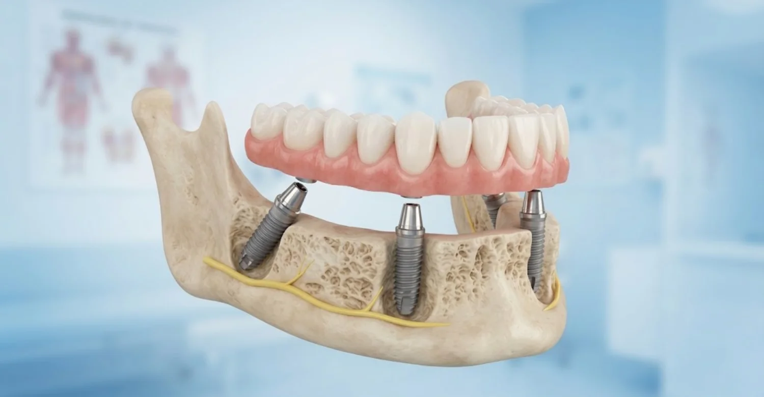
Fixed bridges can work in many cases with careful planning. Success depends on implant number, distribution, loading, and hygiene access. Posterior mandibular bone loss with fixed implant prostheses does not automatically rule out another fixed solution. However, some patients need augmentation or a different design first. The following factors determine whether a fixed bridge is realistic.
Evaluating the number and distribution of implants supporting the bridge
More implants spread chewing loads across a wider area. Implants placed at each end of a bridge reduce leverage on the structure. Two or three implants commonly support a posterior three- or four-unit bridge. Weak or compromised implants should not be relied upon as key supports. The planned layout should also leave space for effective cleaning.
Managing chewing forces, cantilevers, and prosthesis fit
Cantilevers extend beyond the last implant and increase leverage. Many clinicians limit posterior cantilevers to one small premolar-sized unit. Narrower chewing tables can reduce forces transmitted to the implants. Passive fit prevents constant stress on screws and surrounding bone. Occlusal guards may protect bridges in patients who clench or grind.
Preserving cleaning access beneath the fixed prosthesis
The bridge underside should allow floss, interdental brushes, or water flossers to pass easily. Convex, smooth undersurfaces are easier to clean than concave designs. Embrasures between implants should not be blocked by bulky contours. Studies link restorations that prevent hygiene access with more peri-implantitis. Cleaning access should be checked before the final bridge is fitted.
When additional augmentation or a different restoration design may be required
If bone cannot support adequate implants, grafting may be needed before a new bridge. If the bridge repeatedly fails, the design itself may need rethinking. Converting to a removable implant overdenture is sometimes the more sustainable choice. Decisions are reviewed with the patient's priorities, budget, and health in mind.
Note : All images used are for editorial and illustrative purposes only and may not originate from the original news provider or associated company.
How Is Peri-Implantitis Treated When Bone Loss Occurs?
Peri-implantitis treatment aims to stop infection and halt further bone loss. Treatment usually progresses step by step. Each stage is reassessed before moving to the next. Non-surgical care comes first, followed by surgery if inflammation persists. Long-term maintenance is essential for every approach. Posterior mandibular bone loss with fixed implant prostheses caused by infection follows this staged pathway.
Professional implant cleaning and non-surgical biofilm removal
The first step is thorough mechanical cleaning around the implant. Clinicians use specialized curettes, ultrasonic tips, or air-polishing devices designed for implants. Antiseptic rinses may be used as adjuncts. Non-surgical therapy reliably improves mucositis. For established peri-implantitis, studies show it often reduces bleeding but rarely resolves deep lesions completely. Reassessment usually follows after about six to eight weeks.
Improving oral hygiene and controlling periodontal inflammation
Daily plaque control is central to any successful treatment. Patients receive personalized instructions for cleaning around and beneath their bridge. Any periodontitis affecting natural teeth is treated at the same time. Untreated pockets elsewhere in the mouth can reinfect implant sites. Smoking cessation and diabetes control further strengthen results. Hygiene improvements are reviewed and reinforced at every visit.
When surgical treatment for peri-implantitis may be considered
Surgery is considered when bleeding, pus, or deep pockets persist after non-surgical care. It is also used for deeper bony defects that instruments cannot reach. Studies report surgical treatment can resolve disease in roughly half to two-thirds of cases, depending on definitions. Results depend on defect shape, implant surface, and patient risk factors. Surgery is usually followed by closer maintenance during the first year.
Access surgery and removal of inflammatory tissue and deposits
The gum is lifted to expose the implant surface and bone defect. Inflamed tissue is removed, and the implant surface is decontaminated thoroughly. In some cases, rough threads are smoothed to reduce future plaque retention. The gum may be repositioned lower to reduce pocket depth. Healing is monitored closely during the following weeks.
Selected regenerative procedures for suitable bone defects
Contained defects with surrounding bone walls may be filled with graft materials. Membranes may cover the graft to support regeneration. Some studies report radiographic bone fill in favorable defect shapes. However, complete re-osseointegration of the previously contaminated surface remains unpredictable. Regeneration is therefore reserved for carefully selected defects.
When is implant removal considered?
Removal is considered when an implant is mobile or has lost most of its supporting bone. It may also be advised when repeated treatment fails to control infection. Sometimes removal preserves remaining bone for future implants. Specialized kits allow removal with minimal additional bone damage. Posterior mandibular bone loss with fixed implant prostheses may then require grafting before replacement.
How prosthesis modification or replacement may support disease management
The restoration often contributes to disease and must be modified. Bulky contours may be reduced to improve cleaning access. Screw-retained bridges allow easier removal for treatment and maintenance. Cemented restorations with residual cement may need replacement. Correcting the bite also reduces loading on recently treated implants. These changes often improve the success of infection treatment.
Why treatment success depends on ongoing maintenance and risk-factor control
Peri-implantitis can recur even after successful treatment. Studies show patients attending regular supportive care have fewer recurrences. One five-year study found peri-implantitis in about 18% of maintained patients versus 44% without maintenance. Recall intervals of three to six months are common after treatment. Smoking and diabetes control remain lifelong priorities for implant patients.
Relief: How Can Patients Manage Implant-Related Discomfort Safely?
Discomfort around implants can come from surgery, inflammation, or mechanical problems. Simple measures may ease mild symptoms safely. However, relief should never replace professional diagnosis. The sections below explain safe self-care and its important limits. Persistent or worsening symptoms always need professional review.
What may help relieve mild discomfort after implant surgery?
Cold packs applied to the cheek during the first day may reduce swelling. Rest, soft foods, and avoiding chewing on the surgical site support healing. Avoid hot drinks, alcohol, and smoking during the first days. Gentle warm saltwater rinses are often advised from the second day. Keeping the head elevated may reduce swelling overnight. Always follow your surgeon's specific instructions, which may differ for grafting procedures.
Following prescribed pain-relief instructions and postoperative care guidance
Your dentist may recommend over-the-counter pain relievers or prescribe specific medications. Take them exactly as directed, respecting dose limits and timing. Inform your dentist about other medicines, allergies, and health conditions. Antiseptic mouthwash may be prescribed for a limited period. Follow cleaning guidance carefully around the surgical site. Never exceed the stated daily dose of any pain reliever.
Why pain relief does not treat the underlying cause of progressive bone loss
Painkillers reduce symptoms but do not remove plaque, infection, or overload. Bone loss may continue even when discomfort settles. Posterior mandibular bone loss with fixed implant prostheses needs treatment aimed at its cause. Masking symptoms for weeks can delay appropriate care and allow further damage. Professional assessment identifies whether infection, overload, or a loose component is responsible.
When persistent pain, swelling, or discharge requires professional assessment
Pain lasting more than a few days after surgery, or worsening over time, needs review. Swelling that increases after the third day may suggest infection. Any discharge, bad taste, or new mobility requires professional assessment. Long-standing implants that suddenly become painful should always be examined. Early assessment can limit bone damage and simplify treatment.
Why patients should not self-prescribe antibiotics or attempt to adjust a loose implant bridge
Antibiotics without diagnosis may mask symptoms and encourage resistant bacteria. They also cannot remove the biofilm attached to implant surfaces. Tightening a loose bridge at home can damage screws, components, or implants. Glue or similar home fixes may cause infection and harm tissues. Contact your dentist instead for a safe assessment. Your dentist can safely remove, clean, and refit the bridge when appropriate. Early professional care often prevents a minor issue from becoming a major repair.
.jpg)
.jpg)
.jpg)
.jpg)
What Side Effects and Complications Can Occur During Treatment?
All surgical and restorative treatments carry some risk. Most side effects are temporary and manageable. Understanding possible complications helps patients make informed decisions and recognize problems early. Posterior mandibular bone loss with fixed implant prostheses treatment involves specific anatomical risks discussed below.
Possible side effects after dental implant surgery
Most patients experience mild to moderate discomfort for a few days. Side effects are usually greatest within the first 48 to 72 hours. Serious complications are relatively uncommon in properly planned treatment. Following aftercare instructions reduces the chance of problems. Most people return to normal daily activities within one or two days. Recovery may take longer after grafting or when several implants are placed together.
Temporary pain, swelling, bruising, and minor bleeding
Swelling usually peaks around the second or third day after surgery. Bruising may appear on the cheek or jaw and fade within one to two weeks. Minor oozing of blood is common during the first day. Pain is usually controlled with recommended medication. Most discomfort settles within a week. Sleeping with the head raised can help limit overnight swelling. Contact your dentist if bleeding continues despite gentle pressure with gauze.
Risks associated with bone grafting and augmentation procedures
Grafting adds surgical complexity and healing time. Larger and vertical grafts carry higher complication risks than small horizontal grafts. Patient factors, such as smoking, strongly influence outcomes. Your surgeon should explain the specific risks of your planned procedure. Written consent should describe expected benefits, possible complications, and alternatives. Patients should feel free to ask for clarification before agreeing.
Infection, graft exposure, delayed healing, or insufficient bone regeneration
Graft infection may cause swelling, pain, or discharge. Membrane or graft exposure is reported in roughly 10 to 20% of some vertical procedures. Exposure can reduce bone gain or lead to partial graft loss. Sometimes less bone forms than planned, requiring short implants or additional grafting.
Potential nerve-related complications in the posterior mandible
The inferior alveolar nerve runs through the lower jaw beneath the molars and premolars. Injury may occur from drilling, implant placement, or swelling compressing the nerve. Careful CBCT planning and safety margins reduce this risk. Posterior mandibular bone loss with fixed implant prostheses brings the nerve closer to the surgical field.
Numbness, tingling, or altered sensation affecting the lower lip, chin, or tongue
Nerve injury may cause numbness, tingling, burning, or altered sensation in the lower lip, chin, or tongue. Reported incidence varies widely between studies and techniques. Most disturbances are temporary and improve over weeks or months. Persistent changes are less common but can be lasting. Report any numbness to your surgeon immediately after surgery.
Complications involving implant-supported crowns and bridges
Prosthetic complications are more common than implant loss. Reviews of fixed implant bridges report technical complications in a substantial share of patients over five years. Most are repairable, but they require timely attention. Regular checks allow small technical issues to be corrected before they damage implants. Screw-retained designs usually make these repairs simpler and less invasive.
Prosthesis loosening, material damage, unfavorable loading, and hygiene difficulties
Screw loosening, veneer chipping, and fractured components are the most frequent problems. Ceramic chipping has been reported in roughly 10 to 20% of fixed bridges over five years. Unfavorable loading may stress implants and components. Bridges that block cleaning increase inflammation risk over time. Most technical problems can be repaired chairside or in the laboratory. Prompt reporting of any change helps limit further damage.
Factors that may increase the risk of treatment complications
Smoking, uncontrolled diabetes, and poor oral hygiene raise complication rates. Bruxism increases mechanical failures of bridges and screws. Previous radiotherapy and certain bone-modifying drugs require special caution. Limited bone and complex grafting also increase risk. Posterior mandibular bone loss with fixed implant prostheses is more challenging to treat when these factors combine.
What Is the Average Cost of Treating Posterior Mandibular Bone Loss with Fixed Implant Prostheses?
The cost of treating Posterior Mandibular Bone Loss with Fixed Implant Prostheses varies depending on the severity of bone loss, the number of implants required, the type of restoration, and whether bone grafting or additional surgery is necessary. The following tables provide approximate costs for general guidance. Actual prices depend on the clinic, country, materials, and individualized treatment plan.
Average Cost of a Single Dental Implant with a Crown in the US, UK, and Turkey
The following table compares estimated costs for a single dental implant with a crown and additional bone grafting, where necessary.
Country | Single Implant with Crown | Bone Grafting per Site |
United States | 3,000–6,000 | 300–3,000 |
United Kingdom | 1,800–3,500 | 200–1,500 |
Turkey | 500–1,800 | 130–600 |
Note: All figures are approximate USD estimates as provided and may vary by clinic and case complexity. Verify the currency and local pricing before publication. Implant packages may exclude CBCT scans, extractions, grafting, or temporary restorations.
Average Cost of a Fixed Implant-Supported Bridge Compared with a Single Implant Crown
A fixed implant-supported bridge can replace several missing teeth using fewer implants than individual implant crowns. The total cost depends on the bridge length, implant number, materials, and whether additional procedures are required.
Country | Estimated Cost of a Three-Unit Bridge on Two Implants |
United States | 6,000–15,000 |
United Kingdom | 4,000–8,000 |
Turkey | 1,500–4,000 |
Zirconia and metal-ceramic restorations may have different prices. The quoted amounts should be checked to confirm whether they include both implants, abutments, the final bridge, and any necessary bone grafting.
How Much Can Severe Posterior Mandibular Bone Loss Increase Treatment Costs?
Severe posterior mandibular bone loss may require additional surgical procedures before or during implant placement. These procedures increase the total cost and can extend the treatment timeline.
Additional Treatment Requirement | Potential Effect on Total Cost |
Minor bone grafting | Adds the cost of graft materials and the procedure |
Extensive or vertical bone augmentation | May substantially increase surgical fees |
Peri-implantitis treatment | Adds non-surgical or surgical treatment costs |
Implant removal and replacement | Increases surgical and restorative expenses |
Temporary restorations | Adds laboratory and clinical fees |
Extended healing and follow-up | May require additional appointments and maintenance |
In complex cases, treatment costs can be two or three times higher than those of straightforward implant treatment. However, this is not a fixed multiplier; the final estimate depends on the clinical findings and the procedures required.
Additional Expenses for Scans, Graft Materials, Temporary Restorations, and Follow-Up Care
The implant and final restoration are not always the only expenses. Diagnostic imaging, surgical materials, temporary teeth, and maintenance appointments may be charged separately.
Additional Expense | Approximate Cost or Pricing Consideration |
CBCT scan in the United States | 250–600 |
Bone graft materials and membranes | Varies by material, quantity, and procedure |
Temporary restoration | Depends on the type of temporary teeth and laboratory work |
Follow-up appointments | May be included or charged separately |
Professional maintenance cleaning | Depends on the clinic and maintenance plan |
Night guard for teeth grinding | Depends on material and fabrication method |
Travel and accommodation | Depends on destination, trip length, and season |
Patients should request an itemized estimate that clearly separates surgical, restorative, diagnostic, and aftercare expenses. This is particularly important when comparing international dental treatment packages.
What Should Patients Compare When Reviewing Treatment Quotes in Different Countries?
A lower advertised price does not necessarily represent the final cost of treatment. Patients should compare the complete treatment plan, the materials used, the clinician's experience, and the arrangements for follow-up care.
What to Compare | Questions to Ask |
Implant system | Which implant brand and model will be used? |
Final restoration | Is the bridge made from zirconia, metal-ceramic, or another material? |
Bone grafting | Is grafting included, and could additional augmentation be needed? |
Diagnostic imaging | Are CBCT scans and other necessary assessments included? |
Temporary teeth | Are temporary restorations included in the package? |
Follow-up care | How many visits and maintenance appointments are covered? |
Complications and repairs | Who pays for repairs, remakes, or additional surgery? |
Warranty terms | What conditions, exclusions, and time limits apply? |
Confirming Whether Prices Include the Implant, Abutment, Final Bridge, Bone Graft, and Follow-Up
Before accepting a quote, confirm whether the price includes the implant fixture, abutment, final bridge or crown, bone grafting, CBCT scans, temporary restorations, and follow-up visits. Some advertised prices cover only the implant placement or exclude the final prosthesis.
Request a written, itemized estimate and clarify the payment schedule. For Posterior Mandibular Bone Loss with Fixed Implant Prostheses, the treatment plan should also explain whether bone augmentation, implant removal, or additional surgery may be necessary.
Accounting for Additional Appointments, Travel, Accommodation, and Aftercare
Dental treatment abroad may require multiple visits separated by a healing period. Flights, accommodation, transport, time off work, and follow-up care can therefore affect the total budget.
Travel-Related Expense | Budgeting Consideration |
Flights | May be required for more than one visit |
Accommodation | Depends on the length of each stay |
Airport transfers and local transport | Check whether these are included |
Time away from work | Consider lost income or paid leave |
Aftercare at home | Local assessments or maintenance may incur separate fees |
Unexpected complications | Allow for additional visits or treatment if needed |
Patients should confirm how the clinic manages complications after they return home and whether local dental care is included in any aftercare arrangement.
Important: These figures are planning estimates, not guaranteed treatment quotes. A clinical examination and appropriate imaging are needed to determine the extent of posterior mandibular bone loss, whether grafting is necessary, and which fixed implant-supported restoration is suitable.
How Can Patients Support Long-Term Bone Stability Around Posterior Mandibular Implants?
Long-term stability depends on daily care, professional maintenance, and health management. Patients play a central role in protecting their implants. The habits described below can significantly lower the risk of future bone loss. Small daily efforts often make the biggest long-term difference.
Daily cleaning around fixed implant-supported crowns and bridges
Brush twice daily with a soft brush, angling the bristles toward the gum line. Use interdental brushes or implant floss beneath the bridge every day. Water flossers can help reach difficult areas around back implants. Low-abrasive toothpaste protects restoration surfaces. Posterior mandibular bone loss with fixed implant prostheses is less likely with thorough daily cleaning.
Professional implant maintenance and periodontal reviews
Most patients benefit from professional maintenance every three to six months. Visits include probing, plaque removal, and checking the bite and components. Periodic X-rays help track bone levels over time. Higher-risk patients may need shorter intervals between visits. Missing maintenance is consistently linked with higher peri-implantitis rates. Professional cleaning also removes hardened deposits that home care cannot reach.
Managing smoking, diabetes, and other relevant health factors
Quitting smoking improves healing and lowers implant risk. Diabetes control, with HbA1c targets agreed with your physician, protects gum health. Tell your dentist about new medications, especially those affecting bone. Stress and poor sleep may increase clenching and grinding. Healthy routines support both oral and general health. Regular medical check-ups help identify conditions that may affect healing.
Monitoring changes in gum health and comparing radiographs over time
Watch for bleeding, swelling, or changes in how the bridge feels. Report concerns early rather than waiting for the next scheduled visit. Keep copies of implant X-rays and treatment records. Comparing images over years shows whether bone remains stable. Photographs of your gums can also help track visible changes. Posterior mandibular bone loss with fixed implant prostheses is detected earliest through consistent monitoring.
Why an individually designed bite and maintainable prosthesis matter
A bite designed for your jaw movements reduces harmful forces on implants. Bridges should allow easy cleaning beneath and around them. Screw-retained designs simplify future maintenance and repairs. Night guards may protect patients who grind their teeth. Periodic bite adjustments may be needed as natural teeth wear or shift. These details protect both the bone and the restoration.
Long-term bone stability: what patients can realistically expect
Most well-maintained implants keep bone stable for many years. Small changes after the first year are common and not necessarily harmful. Ten-year implant survival often exceeds 90% in published studies. However, peri-implant disease can develop at any stage. Posterior mandibular bone loss with fixed implant prostheses remains manageable when detected and treated early.
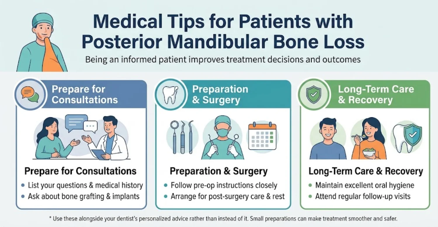
Questions to ask before agreeing to short implants or bone grafting
Ask why short implants or grafting is recommended in your specific case. Request success rates, risks, and healing times for each option. Ask how many similar cases the clinician has treated. Clarify what happens if a graft does not produce enough bone. Understanding alternatives helps you choose with confidence. Ask for the treatment plan and estimate in writing.
What dental records and previous implant X-rays should patients bring to a consultation?
Bring previous X-rays, CBCT scans, and implant records if available. Implant brand, size, and placement dates are especially helpful. Include your medical history, medication list, and recent blood tests if relevant. Posterior mandibular bone loss with fixed implant prostheses is assessed more accurately with baseline images.
How to prepare for implant surgery and follow postoperative instructions
Stop smoking as early as possible before surgery. Arrange soft foods and time to rest for the first few days. Take prescribed medications exactly as instructed. Avoid strenuous exercise for several days after surgery. Attend all follow-up visits, including suture removal and healing checks. Arrange transport home if sedation is planned. Prepare cold packs and prescribed medications before the surgery day.
What to avoid when an implant-supported bridge becomes uncomfortable or loose
Avoid chewing hard foods on the affected side. Do not attempt to tighten or glue the bridge yourself. Avoid pressing the bridge with your tongue or fingers. Keep the area gently clean, even if it feels sore. Contact your dentist promptly to prevent further damage. Keep any broken pieces and bring them to your appointment.
Why regular maintenance remains important even when the implant feels normal
Implants often feel completely normal despite active bone loss. Posterior mandibular bone loss with fixed implant prostheses frequently progresses without pain. Regular visits detect hidden problems before they become severe. Early treatment is usually simpler, less costly, and more predictable. Keep every scheduled visit, even when everything feels fine.
Personalized Treatment Planning for Posterior Mandibular Bone Loss at Vitrin Clinic
At Vitrin Clinic, every treatment plan starts with a detailed assessment. The team considers bone, gums, bite, and general health together. Plans aim for stable, cleanable restorations that suit each patient's priorities and budget. Every recommendation is explained before treatment begins.
Dental implant assessment for patients with limited posterior mandibular bone
Assessment includes examination, probing, X-rays, and review of previous treatment. Existing implants and bridges are evaluated for stability and fit. Posterior mandibular bone loss with fixed implant prostheses is assessed for both cause and severity. Findings are explained clearly so patients understand their situation.
How CBCT imaging can help assess bone volume and nearby anatomical structures
CBCT shows bone height, width, and density in three dimensions. It locates the mandibular canal and mental foramen precisely. Digital planning software can simulate implant positions before surgery. This helps reduce nerve risk and identifies any grafting needs in advance. Patients can also see their own anatomy, which helps explain the proposed plan.
Personalized planning for short implants, bone grafting, and fixed implant-supported restorations
The team compares short implants, grafting, and alternative restorations for each patient. Choices consider bone volume, bite forces, and health risks. Prosthesis design is planned alongside implant positions from the start. Posterior mandibular bone loss with fixed implant prostheses is addressed with restoration-driven planning.
Assessing periodontal health and implant maintenance needs before treatment
Active gum disease is treated before new implants are placed. Oral hygiene is reviewed and improved where needed. A maintenance schedule is planned from the beginning of treatment. This reduces the risk of recurrent inflammation and future bone loss. Patients leave with clear guidance on cleaning tools and recall intervals.
How Vitrin Clinic Supports Your Treatment Journey
Treatment for complex bone loss can involve several stages. Vitrin Clinic aims to make each step clear and well organized. Support continues from the first consultation through long-term follow-up. Each stage is explained in advance so patients know what comes next.
Initial consultation and review of existing dental records
The first visit reviews your concerns, medical history, and previous dental records. Existing X-rays and scans are compared with new findings. Posterior mandibular bone loss with fixed implant prostheses is evaluated before any treatment is proposed. Patients are encouraged to ask questions freely. Treatment goals and concerns are recorded to guide later planning.
Digital diagnostics and individualized assessment before treatment planning
Digital X-rays, CBCT scans, and intraoral scanning support accurate assessment. These tools help plan implant positions and restoration design. Individual risk factors are documented carefully. The resulting plan reflects your specific anatomy and needs. Digital records also make future comparisons more accurate.
Discussing treatment alternatives, expected limitations, and estimated costs
Each suitable option is explained with its benefits, risks, and limitations. Estimated healing times and the number of visits are discussed. Costs are provided in itemized form wherever possible. Patients choose with a full understanding of what to expect. No treatment begins until the patient feels informed and ready.
Coordinating appointments and treatment stages for international patients
International patients often need staged visits planned around healing periods. Appointments can be arranged to reduce the number of trips. Guidance on travel timing and recovery is provided. Records are shared to support continued care at home. Remote communication can help answer questions between visits.
Providing postoperative guidance and planning appropriate follow-up
Patients receive written aftercare instructions following each procedure. Follow-up visits check healing and implant integration. Maintenance plans are agreed before treatment ends. Patients know whom to contact if symptoms appear after treatment. Posterior mandibular bone loss with fixed implant prostheses requires lifelong follow-up for lasting results.
Ready to transform your smile?
Talk to our team and get a personal FREE treatment plan.
Key Takeaways
Bone loss around lower back implants is common, but progressive loss is not inevitable. Understanding causes, signs, and options helps patients act early. The key points below summarize the most important messages of this guide. Use them as a quick reference when discussing treatment.
The main causes of posterior mandibular bone loss around fixed implant prostheses
Peri-implantitis driven by plaque is the leading cause. Previous periodontitis, smoking, and poorly controlled diabetes increase risk. Overload, poor positioning, and uncleanable designs also contribute. Ridge resorption after tooth loss limits available bone. Posterior mandibular bone loss with fixed implant prostheses usually reflects several interacting factors.
Why diagnosis must distinguish normal remodeling from progressive disease
Early remodeling of about 1 to 1.5 millimeters is usually normal. Progressive loss with bleeding or pus suggests disease. Comparing baseline and current X-rays is essential. Correct diagnosis prevents both overtreatment and undertreatment. Clinical signs and X-rays should always be interpreted together.
How short implants, bone grafting, and prosthesis redesign may address different clinical needs
Short implants can avoid grafting when bone height is limited. Grafting rebuilds width or height when standard implants are preferred. Prosthesis redesign improves cleaning and reduces harmful loads. Posterior mandibular bone loss with fixed implant prostheses often needs a combination of these approaches.
Why warning signs and regular professional maintenance should not be overlooked
Bleeding, pus, bad taste, and loosening deserve prompt review. Facial swelling with breathing or swallowing difficulty is an emergency. Regular maintenance substantially lowers peri-implantitis risk over time. Implants that feel normal can still have hidden bone loss.
The importance of individualized treatment planning and a complete cost estimate
Every jaw and every patient is different. Plans should address causes, anatomy, and personal priorities together. Itemized quotes help patients compare clinics fairly and avoid unexpected costs. Clear plans support confident decisions and better long-term outcomes.
At Vitrin Clinic
If you are concerned about bone loss around your lower implants, a detailed assessment is the best next step. Vitrin Clinic offers diagnosis-led planning, digital imaging, and clear treatment options. Posterior mandibular bone loss with fixed implant prostheses can often be managed successfully with timely, individualized care. Contact the team to arrange your consultation and review your existing records.
Reference
FAQs
Dr. Rifat Alsaman has more than 5 years of clinical experience in dentistry and currently serves as the Head of the Medical Team at Vitrin Clinic. He is dedicated to providing exceptional patient care, overseeing treatment planning, and ensuring the highest clinical standards across the team. His expertise, attention to detail, and commitment to continuous professional development have helped countless patients achieve healthier, more confident smiles.


.webp&w=3840&q=75)


