Dental Implant
September 15, 2026
How Much Do Digital Dental Implants Cost? A Guide to 3D Planning and Guided Implant Surgery
 Dr. Rifat Alsaman
The cost of treatment depends on the number of implants, materials, diagnostics, surgical complexity, and restoration selected. Digital Dental Implants may involve CBCT imaging, intraoral scanning, computer-based planning, and guided surgery. These technologies can improve visualization before treatment, but they do not create one universal treatment price. A single implant differs greatly from full-arch rehabilitation with All-on-4 or All-on-6.
Additional procedures can also affect the final quotation. Bone grafting, sinus augmentation, tooth extraction, temporary teeth, and advanced restorations may increase the overall cost. In the United States, a complete single implant treatment commonly falls within several thousand dollars.
Forbes cites an ADA Health Policy Institute survey range of approximately $3,100 to $5,800 for an implant, abutment, crown, and related procedures. In the UK, Bupa lists single implant treatment from $2,400 at selected practices. Bookimed's September 2026 data reports Turkey averages around $593 for a dental implant, although actual treatment packages vary considerably.
What Are Digital Dental Implants?
Digital Dental Implants describe an implant treatment approach that uses digital diagnostic and planning technologies before and during implant placement. The implant itself is still a physical fixture placed into the jawbone. The term mainly describes the digital workflow surrounding diagnosis, planning, surgical guidance, and restoration.
CBCT imaging can provide three-dimensional information about bone anatomy and nearby structures. An intraoral scanner can create a digital model of the teeth and surrounding tissues. These datasets can then be combined in specialized planning software.
The clinician can evaluate potential implant positions before surgery rather than relying only on conventional two-dimensional images. Digital workflows can also support communication between implant dentists, surgeons, and restorative teams.
At Vitrin Clinic, digital assessment can include CBCT imaging, digital X-rays, three-dimensional intraoral scanning, and treatment planning. However, technology should support clinical decision-making rather than replace professional examination. The appropriate implant design and surgical approach remain dependent on individual anatomy, oral health, systemic factors, and restorative objectives.
How Digital Technology Changes Implant Treatment
Digital technology changes implant treatment mainly by improving how information is collected, combined, visualized, and transferred between treatment stages. Traditional workflows may rely on physical impressions, conventional radiographs, and manual surgical planning.
Modern workflows can incorporate CBCT scans, intraoral scans, digital photographs, and computer-based treatment simulations. These records can create a more comprehensive representation of the patient's anatomy and proposed restoration.
Digital planning can also help clinicians examine implant angulation, depth, spacing, and proximity to anatomical structures. A surgical guide may then transfer the planned position to the clinical environment. The degree of guidance depends on the system and the patient's circumstances.
Digital technology can also make patient communication easier because proposed implant positions can be displayed visually. This can help patients understand why particular implant locations are recommended.
However, digital planning does not eliminate surgical uncertainty. Bone quality, soft tissues, access, healing, and intraoperative findings can still influence treatment. This is a defining characteristic of Digital Dental Implants.
Digital vs. Traditional Dental Implant Placement
The main difference between digital and traditional implant workflows is how diagnostic information and surgical positioning are planned. Traditional placement may use conventional radiographs and clinical measurements, with the dentist determining implant position during surgery.
Digital workflows can use three-dimensional imaging and virtual planning before the procedure. A surgical guide may then help reproduce the planned trajectory. This can improve consistency when the planned position is clinically appropriate.
Traditional methods are not inherently inferior, however. Experienced clinicians have successfully placed implants using conventional techniques for many years.
Digital planning becomes particularly useful when anatomy is complex or when the restorative outcome requires precise positioning. The final choice depends on the clinician, available technology, patient anatomy, and treatment goals.
Digital workflows can also require additional equipment, software, planning time, and technical expertise. Therefore, patients should evaluate the complete treatment strategy rather than assuming that digital treatment automatically produces a better outcome.
Who Can Benefit From Digital Implant Treatment?
Many patients requiring implant-supported restorations may benefit from digital planning, particularly when accurate spatial information is important. Patients replacing one tooth can use digital planning to evaluate available bone and the relationship between the implant and neighboring teeth.
Multiple missing teeth may require more detailed planning because implant positions must work together. Full-arch cases can require even greater coordination between implant placement and the final prosthesis.
Patients with limited bone volume may also benefit from detailed three-dimensional assessment. Digital planning can help identify anatomical limitations before surgery and support discussions about grafting or alternative approaches.
Patients seeking cosmetic outcomes may benefit from connecting the proposed restoration with implant positioning. Nevertheless, not every patient needs the same level of digital guidance. Some straightforward cases can be successfully managed with conventional techniques. A qualified implant dentist should determine whether digital planning adds meaningful clinical value for each patient.
Learn why your teeth may feel tight
How Does Digital Implant Planning Work?
Digital implant planning begins with collecting diagnostic information about the teeth, jawbone, soft tissues, and intended restoration. The clinician typically reviews the patient's dental history and performs an oral examination before selecting appropriate imaging.
CBCT can provide three-dimensional information about bone anatomy and important structures. An intraoral scanner can create a digital representation of the dental arches and occlusal relationships. These datasets may then be imported into specialized planning software.
The clinician can virtually position implants according to bone availability and restorative requirements. The plan may also show distances from nerves, sinuses, adjacent roots, and other anatomical structures.
For selected cases, the final plan can be used to design a surgical guide. The guide may help transfer the planned implant trajectory into the mouth during surgery.
The process is therefore more than simply obtaining a digital scan. It is a coordinated workflow that connects diagnosis, implant positioning, surgery, and restoration. This principle underlies most approaches to Digital Dental Implants.
What Is Digital Implant Planning?
Digital Implant Planning is the process of using computer-based diagnostic information to determine the proposed location, orientation, and dimensions of dental implants. The process usually begins with collecting appropriate clinical and imaging records.
CBCT provides three-dimensional anatomical information, while intraoral scanning can provide detailed information about teeth and soft-tissue relationships. These datasets can be combined within specialized planning software. The clinician can then assess possible implant locations virtually.
Important factors include bone height, bone width, anatomical structures, prosthetic space, implant dimensions, and the planned restoration. The software can display measurements that may be difficult to appreciate using conventional radiographs alone.
In complex cases, digital planning may also support the creation of a surgical guide. The purpose is not simply to make treatment look technologically advanced. Its clinical value comes from using accurate information to support appropriate decisions. The final plan should always be reviewed by a qualified dental professional before treatment proceeds.
How CBCT Scans Are Used for Implant Planning
CBCT imaging provides three-dimensional information that can help clinicians evaluate jawbone anatomy before implant surgery. Conventional two-dimensional radiographs can show important structures, but they may not provide the same spatial information.
CBCT can help assess bone dimensions and identify structures such as the mandibular canal, mental foramen, and maxillary sinus. It can also assist with evaluating the relationship between proposed implant locations and neighboring anatomical structures.
The scan should be prescribed when clinically justified because imaging involves radiation exposure. The American Dental Association emphasizes that dental imaging should be selected according to individual diagnostic needs rather than used automatically.
CBCT data can then be imported into implant planning software. The clinician can measure available bone and simulate potential implant positions. This information may influence whether grafting, sinus augmentation, a different implant dimension, or an alternative treatment is appropriate. CBCT therefore provides valuable anatomical information, but it remains one component of a complete clinical assessment.
How 3D Intraoral Scanning Supports Implant Planning
A 3D intraoral scanner captures digital information about the patient's teeth and oral surfaces without requiring a conventional physical impression. The resulting digital model can show tooth contours, occlusion, spacing, and restorative surfaces.
For implant treatment, this information can complement CBCT imaging. The CBCT primarily provides anatomical and bone information, while the intraoral scan provides detailed information about the visible oral structures. Combining both datasets can help clinicians evaluate the relationship between the proposed implant and the intended restoration.
This concept is especially important when implant placement needs to support a crown or bridge with appropriate emergence and aesthetics. Digital scanning can also make records easier to transfer between members of a treatment team.
At Vitrin Clinic, three-dimensional intraoral scanning can form part of a digital assessment workflow. The goal is to create an integrated digital record that supports diagnosis and treatment planning. Scanner accuracy can depend on technique, equipment, scanning conditions, and the specific clinical situation. Digital Dental Implants rely heavily on this type of planning.
Combining CBCT and Digital Scans
Combining CBCT imaging with an intraoral scan can create a more comprehensive digital representation of the treatment area. CBCT contributes three-dimensional information about the jawbone and anatomical structures. The intraoral scan contributes detailed surface information about teeth, gums, and occlusion.
When the datasets are accurately aligned, the clinician can evaluate the proposed implant in relation to both bone and the intended restoration. This supports the principle of prosthetically driven implant planning.
Instead of selecting an implant position based only on available bone, the clinician considers where the final tooth should ideally emerge. The plan must then determine whether adequate bone exists for that restorative position. If not, additional procedures may be considered.
Digital integration can also help identify potential conflicts before surgery. Accurate registration remains essential because errors in the digital datasets can affect the resulting plan. Therefore, technology improves information management only when the underlying records are reliable and clinically interpreted.
Virtual Dental Implant Planning
Virtual dental implant planning allows clinicians to simulate implant placement before entering the surgical environment. The clinician can examine the jaw from different angles and evaluate proposed implant dimensions.
The software can display the planned implant relative to bone boundaries and anatomical structures. The proposed restoration can also be considered when restorative data are available. This approach can be especially useful for cases involving multiple implants.
Implant positions can be evaluated collectively rather than individually. The clinician may also assess spacing, parallelism, emergence profiles, and prosthetic access.
In guided cases, the virtual plan can be used to create a surgical guide. The guide is designed to help transfer the planned position into the patient's mouth. Digital planning can therefore connect diagnostic imaging with the practical surgical workflow.
Nevertheless, the virtual plan represents a proposed treatment strategy rather than a guarantee. Clinical conditions during surgery can require modifications. The dentist must remain prepared to adapt treatment when patient safety or anatomical findings require it.
Evaluating Bone Volume
Evaluating bone volume is one of the most important functions of three-dimensional implant planning. An implant requires adequate bone for stability and long-term support. The available bone must be considered in three dimensions rather than only from a single radiographic view.
CBCT can help clinicians evaluate bone height, width, and anatomical limitations. The quality and density of bone also influence treatment decisions, although imaging alone cannot provide every clinical answer.
If bone volume is inadequate, the dentist may consider augmentation procedures. These can include guided bone regeneration, ridge augmentation, or sinus augmentation depending on the location and clinical need. The final decision should be based on the patient's complete examination.
Digital planning can help identify limitations before surgery and can make discussions about additional procedures clearer. Patients should ask whether the quoted treatment includes grafting when indicated. Understanding these details can prevent unexpected costs and treatment delays. Bone evaluation is therefore both a clinical and financial planning consideration. Digital Dental Implants depend on getting this step right.
Planning Implant Position, Angle, and Depth
Implant position, angle, and depth influence both surgical safety and the final restoration. The implant should ideally be positioned within a suitable bone while supporting the intended prosthetic design.
Digital planning software allows the clinician to adjust the proposed implant virtually. The dentist can examine the implant from multiple views and assess its relationship with nearby anatomical structures. Angulation can be evaluated relative to neighboring teeth and the planned restoration.
Depth can also be assessed to support appropriate implant emergence and prosthetic connection. In guided surgery, these parameters may be incorporated into the surgical guide. The guide can help control the drilling and implant trajectory according to the selected system.
However, guidance does not eliminate the need for surgical judgment. Tissue conditions, visibility, access, and intraoperative findings may require modifications. The objective is not maximum technological control. The objective is an implant position that is biologically appropriate, surgically safe, and compatible with the final restoration.
Identifying Nerves and Other Anatomical Structures
Identifying important anatomical structures is a major reason three-dimensional imaging can be valuable for implant planning. In the lower jaw, the inferior alveolar nerve and mental nerve require careful consideration. In the upper jaw, the maxillary sinus can influence implant placement.
Other structures may also affect surgical planning depending on the patient's anatomy. CBCT can help clinicians visualize these structures in three dimensions. The dentist can then assess the proposed implant location in relation to them.
This information can influence implant length, position, angulation, or the need for an alternative approach. Digital planning software can provide measurement tools that support this evaluation. However, software measurements should not replace clinical interpretation.
Imaging quality, patient movement, anatomical variation, and registration accuracy can influence the reliability of digital measurements. The clinician must therefore combine imaging findings with examination and professional judgment. Patient safety remains the primary consideration when selecting an implant position.
Clinical Note: Why Digital Planning Matters
Digital planning matters because implant treatment involves several interconnected decisions rather than simply placing a titanium fixture into bone. The final restoration, available bone, anatomical structures, surgical access, and long-term maintenance should all be considered.
A digital workflow can bring these factors together before surgery. It can also improve communication between the surgical and restorative teams. This is particularly valuable for multiple implants and full-arch rehabilitation.
Digital planning may reveal limitations that would otherwise become apparent later in treatment. It can also help patients understand why additional procedures may be recommended. However, digital planning should never be presented as a guarantee of perfect implant placement.
Clinical experience remains essential. Dr. Rifat Alsaman, Head of the Medical Team at Vitrin Clinic and cosmetic dentist, emphasizes the importance of connecting digital information with clinical examination and restorative objectives. In practice, the best workflow is one where technology supports professional judgment rather than attempting to replace it. This step is central to how Digital Dental Implants are performed.
Find out why teeth can hurt when you have the flu
What Is 3D Dental Implant Planning?
3D dental implant planning uses three-dimensional diagnostic information to simulate proposed implant placement before surgery. The process can incorporate CBCT imaging, intraoral scanning, digital photographs, and restorative information.
The clinician can examine bone anatomy and virtually position implants according to the planned restoration. This approach provides more spatial information than conventional two-dimensional planning alone. It can be particularly helpful when several implants must work together.
Each implant can be positioned according to the overall prosthetic design rather than treated as an isolated fixture. Digital planning can also support surgical guide fabrication. The guide transfers selected elements of the virtual plan to the clinical setting.
The level of guidance depends on the technology and surgical system used. Three-dimensional planning does not automatically mean that treatment is safer or more successful in every case. Its value depends on accurate data, appropriate planning, experienced clinical interpretation, and proper execution. These principles apply whether treatment involves one missing tooth or an entire arch.
Creating a 3D Digital Model
Creating a three-dimensional digital model involves combining diagnostic information into a virtual representation of the patient's oral structures. CBCT contributes information about the jawbone and deeper anatomical structures. An intraoral scanner captures detailed surface information from the teeth and surrounding tissues.
Digital photographs may add information about facial aesthetics and smile characteristics. When appropriate, these datasets can be aligned using specialized software. The resulting model can provide a more complete basis for treatment planning.
The clinician can rotate the model, examine cross-sectional views, and measure relevant anatomical distances. The digital model may also allow the planned restoration to be visualized. This helps connect the surgical plan with the prosthetic result.
The quality of the model depends on the quality of the source records. Errors during scanning or data alignment can affect the planning process. Therefore, digital models should always be checked by qualified clinicians before they are used for surgical decisions.
Planning the Ideal Implant Position
Planning the ideal implant position requires balancing bone anatomy with the intended restorative result. The implant should be placed where it can achieve appropriate stability while supporting the final crown, bridge, or prosthesis.
Digital software allows clinicians to test different implant positions virtually. The dentist can assess angulation, depth, spacing, and restorative access. The proposed position can also be evaluated relative to anatomical structures.
This process supports prosthetically driven planning because the final tooth position influences where the implant should ideally be located. In some cases, the available bone does not match the desired restorative position. The clinician may then discuss bone augmentation or an alternative implant position.
For multiple implants, the positions must also be coordinated with each other. The final goal is not simply geometric accuracy. The goal is a clinically appropriate position that supports function, biological health, aesthetics, and future maintenance. Individual anatomy always determines the final treatment strategy. Digital Dental Implants are built around this kind of coordination.
Using 3D Planning for Complex Implant Cases
Complex implant cases often involve multiple anatomical, surgical, and restorative considerations. Examples include severe bone loss, multiple missing teeth, full-arch rehabilitation, and implant placement near important anatomical structures.
Three-dimensional planning can help clinicians visualize these challenges before surgery. It allows proposed implant positions to be evaluated against bone anatomy and restorative requirements.
For multiple implants, the software can display the complete arrangement simultaneously. This can help identify spacing conflicts or unfavorable trajectories before treatment begins. Digital planning may also assist communication among surgeons, restorative dentists, dental technicians, and patients.
However, complex cases require more than sophisticated software. They require appropriate diagnosis, treatment sequencing, surgical skill, restorative planning, and contingency strategies. The technology should therefore be considered one component of multidisciplinary treatment.
At Vitrin Clinic, digital workflows can support assessment and planning for patients traveling internationally for implant treatment. Each case still requires individual evaluation before a treatment plan and quotation are provided.
Single-Tooth Implant Planning
Single-tooth implant planning focuses on replacing one missing tooth while protecting neighboring structures and achieving an appropriate restoration. The clinician evaluates bone availability, adjacent tooth roots, gum tissue, and the intended crown.
Digital imaging can help determine whether sufficient bone exists for the proposed implant. An intraoral scan can provide information about the surrounding teeth and occlusion. These records can be combined to evaluate the relationship between the implant and the future crown.
This is especially important in visible areas where implant position can influence the emergence profile and soft-tissue appearance. The clinician can also assess available restorative space.
If the bone is insufficient, augmentation may be recommended before or during implant treatment. The surgical approach may be guided or conventional depending on the case. Digital planning can provide useful visualization even when guided surgery is not necessary. The final plan should always consider long-term hygiene, function, aesthetics, and maintenance.
Multiple Implant Planning
Multiple implant planning requires coordination between several implant positions and the final restoration. Each implant must have an appropriate relationship with surrounding anatomy and the other implants.
Digital planning allows the clinician to visualize all proposed implants simultaneously. This can help assess spacing, parallelism, restorative access, and prosthetic design.
The number and distribution of implants depend on the number of missing teeth, available bone, occlusal requirements, and planned restoration. Bone deficiencies can complicate the treatment plan and may require staged procedures. Digital planning can help identify these issues before surgery.
It can also support surgical guide fabrication when guided placement is selected. For patients traveling internationally, detailed planning can be especially useful because treatment time may need to be coordinated carefully.
Nevertheless, planning cannot guarantee that every planned implant will be placed exactly as simulated. Clinical findings can require modifications. The dentist should explain possible alternatives before surgery. This reflects the broader philosophy behind Digital Dental Implants.
Full-Mouth Implant Planning
Full-mouth implant planning involves coordinating treatment across both arches or across extensive areas of the mouth. The clinician must consider bone anatomy, occlusion, facial aesthetics, implant distribution, and the final prosthetic design.
Digital planning can provide a comprehensive view of these relationships. CBCT can show bone availability throughout the arches. Intraoral scanning can capture the dental surfaces and occlusal relationships.
The planned restorations can then be incorporated into the digital workflow. This approach can help clinicians determine appropriate implant positions before surgery.
Full-mouth treatment may involve staged extractions, bone grafting, temporary teeth, implant placement, healing, and final prosthetic delivery. The sequence must therefore be carefully coordinated.
Digital technology can improve communication between the surgical and laboratory teams. It may also help patients understand the proposed treatment. However, full-mouth rehabilitation remains a highly individualized procedure. The number of implants and treatment stages cannot be determined accurately without clinical assessment and diagnostic imaging.
All-on-4 and All-on-6 Planning
All-on-4 and All-on-6 treatment requires coordinated planning because multiple implants support an entire fixed arch. The implant positions must be selected according to available bone and the design of the prosthesis.
Digital planning can help visualize the relationship between proposed implants and the final teeth. It can also assist evaluation of anatomical limitations. Angulation and implant distribution are particularly important because the implants work together as a system.
CBCT data can help assess bone anatomy, while intraoral scanning can contribute restorative information. A digital surgical guide may be designed when appropriate. The final approach depends on bone availability, systemic health, occlusion, restorative requirements, and clinician experience.
Current market data illustrate the wide price variation for full-arch treatment. Bookimed's September 2026 data reports an average All-on-4 price in Turkey of approximately $4,150, with reported prices ranging from $2,300 to $6,000. Its reported All-on-6 average is approximately $9,000, with a range of $4,200 to $13,800. These figures are not individualized quotations.
Explore the process of chewing step by step
What Are Computer Guided Dental Implants?
Computer guided dental implants refer to implant treatment where computer-based planning is used to determine the proposed implant position and guide surgical placement. The workflow generally starts with diagnostic imaging and digital records. The clinician then plans the implant position within specialized software.
When indicated, the digital plan can be used to design a surgical guide. The guide fits onto the patient's teeth, mucosa, or bone depending on the system. During surgery, it can help control the planned drilling trajectory and implant positioning.
The objective is to transfer the virtual treatment plan into the clinical environment with greater control. Guided treatment can be particularly useful when anatomical limitations or restorative requirements make positioning especially important.
However, guided surgery is not appropriate for every patient. The guide must fit accurately, the digital records must be reliable, and the clinical plan must be sound. Experienced clinicians must also monitor the surgical situation and adapt when necessary.
How Computer Guided Implant Placement Works
Computer guided implant placement begins with diagnostic information that is used to create a virtual treatment plan. The clinician identifies the proposed implant locations and evaluates their relationship with the bone and anatomical structures.
The implant dimensions and trajectories can then be selected within planning software. When guided surgery is appropriate, a surgical guide is designed from the digital plan. The guide incorporates sleeves or other components that help control the surgical instruments.
During the procedure, the guide is positioned and stabilized according to the selected system. The clinician then follows the guided sequence while maintaining direct clinical control. Some systems provide more extensive guidance than others.
The final implant position can still be influenced by guide fit, tissue conditions, surgical technique, and intraoperative circumstances. Guided placement therefore provides a method of transferring a planned position. It does not eliminate the need for a qualified implant dentist or surgeon.
Careful planning remains the foundation of successful guided treatment. Such planning is a hallmark of well-executed Digital Dental Implants.
What Is a Surgical Guide?
A surgical guide is a customized device designed to assist implant placement according to a predetermined treatment plan. It is commonly produced using digital planning and manufacturing technologies.
Depending on the system, the guide may be supported by teeth, mucosa, or bone. Its design is based on diagnostic records and the proposed implant positions. During surgery, the guide can help control the direction and location of drills.
Some systems can also help control implant insertion according to specific clinical parameters. The purpose is to improve the transfer of the digital plan to the patient's mouth. Surgical guides are not universal devices.
Their design depends on the patient's anatomy, implant system, surgical technique, and planned restoration. Accurate digital records and appropriate guide fabrication are essential. The clinician must also verify the guide's fit before proceeding.
If the guide does not fit as intended, surgery should be reassessed rather than performed blindly. This emphasizes why digital planning and clinical judgment must work together.
How Digital Planning Guides Implant Surgery
Digital planning guides implant surgery by establishing a proposed implant position before the patient enters the surgical stage. The clinician can determine the implant trajectory using three-dimensional imaging and restorative information.
If a surgical guide is fabricated, this plan can then be transferred to the mouth. The guide can help control drilling direction and positioning. This may reduce reliance on intraoperative estimation in selected cases.
It can also support restorative objectives when the implant needs to emerge from a specific location. The degree of guidance varies between systems. Some approaches provide partial guidance, while others control several surgical parameters.
Regardless of the system, the clinician remains responsible for assessing tissue conditions and surgical safety. If unexpected anatomy or inadequate access is encountered, the planned approach may need modification.
Digital planning therefore creates a structured surgical framework. It should not be interpreted as an automatic guarantee of accuracy or treatment success.
Computer Guided Implant Placement vs. Freehand Placement
Computer guided implant placement and freehand placement are different surgical approaches rather than universally better or worse methods. Freehand placement relies heavily on clinical examination, radiographic assessment, surgical landmarks, and the clinician's experience.
Guided placement adds a physical or digital method for transferring a predetermined plan into the surgical field. The choice depends on case complexity, clinician preference, available technology, and patient-specific factors.
Guided surgery may be especially useful when implant position must be closely coordinated with the final prosthesis. Freehand surgery can remain appropriate for many straightforward cases. Digital guidance may also involve additional planning and manufacturing costs.
The important question is whether the technology provides meaningful clinical value for the individual patient. Patients should therefore ask what problem guided surgery is intended to solve. They should also ask whether the surgeon has experience with the selected system. A sophisticated workflow is valuable only when it supports sound clinical decision-making.
Learn how thumb sucking can affect teeth
What Happens During Guided Dental Implant Surgery?
Guided dental implant surgery typically follows several coordinated stages beginning with diagnosis and ending with restorative planning. The process may vary depending on the patient's condition and the selected surgical system.
First, the clinician reviews the patient's medical and dental history and performs an oral examination. Diagnostic records such as CBCT and intraoral scans may then be analyzed. The implant positions are planned virtually according to bone anatomy and restorative requirements.
A surgical guide can be designed and manufactured when indicated. On surgery day, the guide is checked for proper fit and stability. The clinician then performs the surgical procedure according to the planned protocol.
Implant stability may be assessed using clinical methods or specialized measurements. The restorative team can then use the implant position to plan temporary or final teeth.
Not every patient follows the same sequence. Bone grafting, extraction, healing periods, and staged treatment can alter the timeline. This is one of the reasons Digital Dental Implants have become popular.
Digital Examination and Diagnosis
Digital examination begins by collecting information that helps the clinician understand the patient's oral condition. A clinical examination remains essential because digital records cannot show every biological factor.
The dentist evaluates teeth, gums, occlusion, oral hygiene, bone conditions, and other relevant findings. CBCT may be prescribed when three-dimensional imaging is clinically justified. Intraoral scanning can provide detailed digital records of the dental arches.
Digital photographs may also help assess aesthetics and facial relationships. These records can then be reviewed together during treatment planning. The goal is to identify the cause of tooth loss and determine whether implant treatment is appropriate.
Existing gum disease, infection, inadequate bone, or other issues may need management first. The digital workflow should therefore begin with diagnosis rather than technology selection.
At Vitrin Clinic, digital examination can be integrated into the treatment planning process. Each international patient's plan should still be individualized according to clinical findings and treatment goals.
3D Treatment Planning
3D treatment planning involves analyzing diagnostic data and creating a virtual representation of the proposed implant treatment. The clinician can position virtual implants within the available bone and assess their relationship with anatomical structures.
The final restoration can also be considered when restorative data are available. This allows the implant position to be evaluated from multiple perspectives before surgery. Measurements can be taken to assess spacing, angulation, depth, and anatomical clearance.
For multiple implants, the entire arrangement can be reviewed simultaneously. This can help identify potential conflicts before the procedure. If a guided approach is selected, the plan can be used to design a surgical guide.
The clinician must verify that the plan is clinically appropriate before manufacturing the guide. The virtual environment provides valuable information, but it does not reproduce every intraoral condition.
Soft tissue movement, surgical access, and intraoperative findings remain important. Therefore, 3D planning should be treated as a decision-support tool.
Surgical Guide Design
Surgical guide design translates the approved digital treatment plan into a physical device for surgery. The guide is designed according to the patient's anatomy and the planned implant positions. Its support can be tooth-based, mucosa-based, or bone-based depending on the clinical approach.
The guide contains features that help control the surgical instruments. Manufacturing can involve digital milling or three-dimensional printing. Before surgery, the clinician should verify that the guide matches the patient's anatomy and remains stable.
A poorly fitting guide can compromise the intended transfer of the digital plan. The guide design should also consider access, mouth opening, implant system requirements, and surgical visibility.
The technology therefore involves collaboration between the clinician and dental laboratory or digital manufacturing team. The guide is an aid rather than an independent surgical device. The dentist must continue to monitor the patient throughout the procedure.
Any discrepancy between the guide and clinical reality should trigger reassessment. Digital Dental Implants often depend on this kind of preparation.
Guided Implant Placement
Guided implant placement uses the surgical guide to help reproduce the planned implant trajectory during surgery. After appropriate anesthesia and surgical preparation, the clinician positions and stabilizes the guide.
The drilling sequence follows the selected implant system and guided protocol. The guide may control the direction and depth of instruments depending on the technology used. The dentist remains responsible for monitoring the surgical field and patient response.
Bone density can influence drilling and insertion decisions. If clinical findings differ from the virtual plan, the clinician may need to modify the approach. Guided surgery therefore improves control without removing professional responsibility.
After preparation of the implant site, the implant is placed according to the treatment plan and clinical conditions. Implant stability can then be evaluated.
The procedure may be minimally invasive in selected cases, but the actual surgical approach varies considerably. Patients should receive individualized instructions regarding anesthesia, medication, oral hygiene, and recovery.
Checking Implant Position and Stability
After implant placement, the clinician evaluates whether the implant has been positioned appropriately and whether it has adequate primary stability. The assessment can include clinical evaluation and imaging when indicated.
Stability is important because excessive movement during healing can compromise osseointegration. The clinician may use specialized devices to measure implant stability in appropriate cases. The planned position can also be compared with postoperative information when clinically necessary.
Digital planning may make this comparison easier because a predefined reference exists. However, the implant's long-term outcome depends on biological healing as well as initial positioning.
Bone quality, infection control, systemic health, loading forces, and oral hygiene can all influence results. The clinician therefore considers the complete clinical picture before deciding when the implant can support a restoration.
Patients should understand that immediate loading is not suitable for every case. The timing of temporary and final teeth depends on individual stability and healing factors.
Planning the Final Restoration
Planning the final restoration is an essential part of implant treatment because the implant ultimately exists to support a functional and aesthetic prosthesis. The crown, bridge, or full-arch prosthesis should be considered before implant placement whenever possible.
Digital workflows can connect the proposed restoration with the planned implant position. The clinician and dental technician can evaluate emergence, occlusion, tooth shape, and restorative access. This is particularly important in the aesthetic zone.
An implant placed only according to available bone may not produce the ideal restorative result. Prosthetically driven planning instead considers where the final tooth should be located first. The implant position is then planned to support that restoration within biological and anatomical limits.
Digital scanning can help capture the patient's existing dental relationships. The final prosthesis may be fabricated using CAD/CAM technology.
At Vitrin Clinic, digital planning can connect implant treatment with the restorative workflow. The exact restoration depends on the patient's clinical needs and selected materials. This consideration is especially relevant for Digital Dental Implants.
Discover what can cause a tight jaw
What Are the Benefits of Digital Implant Surgery?
Digital implant surgery can offer several potential advantages when appropriately selected and executed. One major benefit is improved visualization before surgery. Three-dimensional imaging allows clinicians to examine anatomy from multiple perspectives.
Digital planning can also support more deliberate implant positioning. Surgical guides may help transfer the planned trajectory into the mouth. Digital workflows can improve communication between clinicians and dental laboratories. They may also help patients understand their proposed treatment.
These advantages can be particularly useful for complex cases involving multiple implants or full-arch rehabilitation. However, benefits depend on accurate imaging, proper planning, guide fit, and clinician experience. Digital technology does not guarantee a successful outcome.
Biological healing remains essential after implant placement. Cost can also increase when advanced scanning, planning, guide fabrication, and laboratory services are included. Patients should therefore consider the complete treatment plan rather than selecting a clinic solely because it offers digital technology.
More Predictable Implant Positioning
Digital planning can improve control over the proposed implant position by allowing the clinician to define the trajectory before surgery. The implant can be evaluated relative to the available bone and planned restoration. A surgical guide may then assist with transferring the selected position.
This can be useful when multiple implants must maintain specific relationships. It can also be valuable when anatomical structures limit available space. However, predictable planning is not the same as guaranteed accuracy.
Guide movement, manufacturing tolerances, imaging errors, and clinical conditions can affect the final position. The dentist must therefore verify the guide and monitor the procedure. Digital planning is most valuable when it improves the quality of the clinical decision.
The implant position should always be biologically appropriate and prosthetically useful. Patients should ask their dentist how the planned position will be verified. Understanding this process can help patients evaluate the quality of the proposed treatment rather than focusing solely on technology labels.
Better Visualization Before Surgery
Three-dimensional visualization allows clinicians to examine anatomy in ways that conventional two-dimensional imaging cannot always provide. The dentist can rotate the virtual model and evaluate the jaw from different angles.
Cross-sectional views can show bone dimensions around a proposed implant. Important anatomical structures can also be assessed in relation to the planned trajectory. This information can help identify challenges before surgery.
It can also improve communication with patients because proposed implant locations can be shown visually. Better visualization may be especially useful in areas with limited bone or complex anatomy.
However, digital images are still representations of biological structures. The clinician must interpret them alongside physical examination findings. Imaging artifacts or registration errors can also affect the planning process.
Therefore, visualization is a support tool rather than a substitute for clinical assessment. The quality of the decision depends on the quality of both the data and the professional interpretation. Patients considering Digital Dental Implants should keep this in mind.
Improved Treatment Communication
Digital workflows can improve communication because treatment plans can be represented visually rather than described only verbally. Patients can see proposed implant positions and understand how they relate to the final restoration.
Dentists can also communicate more efficiently with laboratories and other members of the treatment team. Digital files can be transferred electronically, reducing reliance on physical models in suitable workflows.
This can be particularly useful for international patients coordinating treatment across different appointments. Clear communication is also important when discussing additional procedures such as bone grafting. Patients can understand why a particular sequence is recommended.
At Vitrin Clinic, digital records can support communication throughout the international treatment journey. However, visual simulations should be presented as planning tools rather than guaranteed final results.
Patients should ask whether the proposed digital outcome depends on additional procedures or assumptions. Transparent communication remains more important than the technology itself.
Supporting Complex Implant Cases
Digital planning can be particularly useful when implant treatment involves multiple anatomical and restorative considerations. Complex cases may include multiple missing teeth, extensive bone loss, full-arch rehabilitation, or implants near important anatomical structures.
A three-dimensional plan allows these factors to be assessed together. The clinician can evaluate implant distribution and restorative requirements before surgery. Digital guides can then help transfer the plan when guided surgery is selected.
This approach can also support communication between surgeons and restorative clinicians. Complex treatment often requires contingency planning because the clinical situation can change during surgery. Digital planning helps prepare for these possibilities by identifying anatomical constraints in advance.
Nevertheless, technology does not eliminate biological risk. Patients with severe bone loss may still require grafting or alternative treatment. The appropriate plan depends on clinical assessment rather than the availability of digital equipment alone. Experienced treatment teams remain essential for managing complex implant rehabilitation.
Creating an Efficient Digital Workflow
A digital workflow can connect diagnosis, planning, surgery, and restoration within one coordinated process. CBCT data can contribute anatomical information. Intraoral scanning can provide detailed surface information.
Planning software can combine these records and simulate implant placement. Surgical guides can then be produced when appropriate. Digital records can also support restorative design and laboratory communication. This can reduce repetitive impressions and improve information transfer in suitable cases.
Efficiency does not necessarily mean that every treatment becomes faster. Complex cases may still require healing periods, grafting, multiple appointments, and laboratory fabrication. The main advantage is often better coordination between treatment stages.
For international patients, efficient coordination can be especially valuable because travel schedules can affect treatment planning. Vitrin Clinic can use digital workflows to organize diagnostic and restorative information for patients seeking treatment in Istanbul.
Each patient's schedule remains dependent on clinical requirements and healing. This is a key part of the Digital Dental Implants workflow.
Clinical Note: Digital Technology Does Not Replace Clinical Judgment
Digital technology can improve information quality, but it cannot independently diagnose disease or determine the correct treatment. A computer model may show where an implant could be positioned, but the clinician must decide whether that position is biologically and prosthetically appropriate.
The dentist must also consider medical history, periodontal health, occlusion, bone quality, patient expectations, and long-term maintenance. Unexpected findings can arise during surgery and require changes to the plan. This is why experience remains essential even in highly digital workflows.
Dr. Rifat Alsaman, Head of the Medical Team at Vitrin Clinic and cosmetic dentist, views digital planning as a clinical support system rather than a replacement for professional expertise. Patients should therefore ask about the dentist's experience, treatment planning process, and follow-up protocol.
The presence of scanners and planning software alone does not demonstrate clinical quality. Technology should be evaluated according to how effectively it supports safe and individualized care.
Read more about tingling of the mouth
What Is Digital Dentistry for Dental Implants?
Digital dentistry for implant treatment describes the use of digital technologies throughout different stages of implant care. These technologies can include CBCT imaging, intraoral scanning, digital photographs, CAD/CAM restoration, virtual planning, and surgical guide manufacturing.
The objective is to connect diagnostic information with surgical and restorative decisions. Instead of treating each stage as an isolated process, a digital workflow can create a continuous information pathway.
CBCT can provide information about bone and anatomy. Intraoral scanning can capture dental surfaces and occlusion. Planning software can combine the records. CAD/CAM systems can then support restoration design. Surgical guides can help transfer planned implant positions to the mouth.
This approach can be particularly useful for complex cases and international treatment workflows. However, digital dentistry does not eliminate conventional clinical procedures.
Physical examination, anesthesia, surgery, healing, and professional follow-up remain essential. The technology should therefore be viewed as an integrated support system.
CBCT and Digital Diagnostics
CBCT is one of the most important digital diagnostic tools used in modern implant planning. It provides three-dimensional imaging that can help clinicians evaluate bone anatomy and nearby structures.
This information is particularly useful when implant positioning depends on limited anatomical space. Digital diagnostics can also include intraoral photographs, digital X-rays, and scanning. Each diagnostic method provides different information.
CBCT primarily supports three-dimensional anatomical evaluation. Intraoral scanning provides detailed surface information. Digital photographs can help evaluate aesthetic and facial considerations. Combining these records can create a more comprehensive treatment picture.
However, not every patient requires every diagnostic method. Imaging should be selected according to clinical need. The clinician should also explain why each scan is recommended.
At Vitrin Clinic, digital imaging can form part of an individualized implant assessment. The objective is to obtain enough information to make appropriate treatment decisions without relying on technology for its own sake. Digital Dental Implants generally follow this same principle.
3D Intraoral Scanning
3D intraoral scanning creates a digital representation of the patient's teeth and oral surfaces. The scanner captures a sequence of images and combines them into a virtual model. This model can be viewed on a computer and used for treatment planning.
For implant cases, the scan can show tooth positions, occlusion, restorative space, and visible soft-tissue contours. It can also replace some conventional impression procedures. The digital model may be combined with CBCT data when appropriate.
This allows clinicians to relate the visible dental structures to the underlying bone. Digital scanning can also help laboratories design crowns and prostheses using CAD/CAM systems. The technology can improve communication because digital files are easy to transfer.
However, scanning accuracy depends on technique and clinical conditions. Saliva, blood, limited access, and scanning errors can affect the digital model. Clinicians must therefore verify the quality of the scan before using it for important treatment decisions.
Digital Smile and Prosthetic Planning
Digital smile and prosthetic planning focuses on the final appearance and function of the restoration before implant placement. The dentist can evaluate tooth proportions, position, shape, and relationship with the patient's smile.
Digital photographs and intraoral scans can contribute to this process. The planned restoration can then be considered alongside the proposed implant position. This approach supports prosthetically driven implant placement.
The objective is to avoid placing implants solely according to bone availability when the restorative position requires something different. If the ideal restorative position lacks sufficient bone, the clinician may discuss augmentation or alternative strategies.
Digital planning can also help patients understand proposed changes before treatment. However, simulations are not guarantees of the final result.
The actual outcome depends on healing, tissue response, laboratory fabrication, occlusion, and clinical execution. At Vitrin Clinic, digital planning can be incorporated into smile and implant treatment workflows when clinically appropriate. The final restoration remains customized to each patient's anatomy and goals.
Computer-Aided Surgical Guide Design
Computer-aided surgical guide design uses digital planning information to create a guide for implant placement. The clinician first determines the desired implant positions within planning software. The software then generates the guide according to the patient's anatomy and selected implant system.
The guide can be manufactured using digital fabrication technologies. Before surgery, the dentist verifies its fit and stability. During surgery, the guide can help control the planned drilling trajectory.
This may be particularly useful when several implants need to be placed according to a coordinated restorative design. The guide can also assist when anatomical limitations make implant positioning more demanding.
However, the guide is only as accurate as the data and planning behind it. Registration errors or poor fit can compromise the intended transfer.
The dentist must therefore check every stage. Guided surgery should be performed by clinicians familiar with the system and its limitations. This is why Digital Dental Implants require careful planning.
Digital Implant Placement and Restoration
Digital workflows can connect implant placement with restoration design. The implant position is planned according to the intended crown, bridge, or full-arch prosthesis. The digital plan can then guide surgery when appropriate.
After healing or immediate loading when clinically indicated, digital scans can support restoration fabrication. CAD/CAM technology can be used to design prosthetic components with high levels of customization.
The digital record can also make communication between the dental clinic and laboratory more efficient. This is especially helpful for patients receiving complex restorations. The restoration still requires careful clinical evaluation before delivery.
Occlusion, contacts, aesthetics, fit, and hygiene access must all be assessed. Digital design does not eliminate the need for clinical adjustments.
At Vitrin Clinic, digital technologies can support the workflow from initial assessment through implant restoration. The exact sequence depends on whether the patient receives a single crown, multiple-unit bridge, or full-arch prosthesis.
From Digital Scan to Final Crown
The journey from digital scan to final crown can involve several coordinated stages. The patient's dental structures are captured using an intraoral scanner. CBCT information may be added when implant planning requires three-dimensional anatomical data.
The clinician then plans the implant position and restorative design. After implant placement and appropriate healing, another digital scan may be obtained. The dental laboratory uses digital records to design the crown.
CAD/CAM manufacturing can then produce the restoration from the selected material. The dentist evaluates the crown clinically before final placement. Contacts, bite, aesthetics, marginal adaptation, and hygiene access should all be assessed.
Digital workflows can reduce the need for physical impressions in suitable cases. However, each clinical stage remains important. The digital process does not mean the crown is automatically perfect after manufacturing. Clinical verification is essential before final cementation or screw retention.
Connecting Surgical and Restorative Planning
Connecting surgical and restorative planning means determining implant positions with the final prosthesis in mind. This is often called prosthetically driven implant planning. The final crown or prosthesis determines where the implant ideally needs to emerge.
Bone anatomy then determines whether that position is achievable. If the available bone does not support the desired position, augmentation or an alternative treatment may be required. Digital tools make it easier to visualize this relationship.
The clinician can place a virtual implant beneath the planned crown and evaluate the surrounding anatomy. This helps identify potential restorative or surgical conflicts before treatment. For multiple implants, the same concept applies across the entire restoration.
Implant positions must work together as a coordinated system. This approach can improve treatment planning because surgery is connected to the final clinical objective. Technology remains supportive, while the clinician determines whether the proposed plan is appropriate. Such attention to detail supports successful Digital Dental Implants.
Explore the connection between tinnitus and teeth
How Is Dental Implant Placement Using Digital Technology Different?
Dental implant placement using digital technology differs mainly in how treatment information is collected and used before surgery. Traditional workflows may depend heavily on clinical examination, conventional radiographs, physical impressions, and freehand surgical positioning.
Digital workflows can combine CBCT, intraoral scans, virtual planning, and surgical guides. This creates an opportunity to evaluate the proposed treatment before surgery. Implant position can be planned relative to both bone anatomy and the final restoration.
The approach can also improve information transfer between dentists and laboratories. However, digital treatment does not remove the biological stages of implant therapy. The implant still needs appropriate bone, surgical placement, osseointegration, and maintenance.
Some straightforward cases may not require extensive digital guidance. More complex cases can benefit more from three-dimensional planning. The best approach depends on clinical needs rather than technology alone. Patients should ask how digital tools specifically improve their treatment plan.
Traditional Implant Treatment Workflow
A traditional implant workflow generally begins with clinical examination and diagnostic imaging. The dentist assesses the missing tooth area and evaluates the patient's oral health.
Conventional radiographs may provide information about bone and nearby structures. Physical impressions may be taken to create dental models. Implant positioning can then be determined using clinical measurements and radiographic information.
During surgery, the clinician uses these records together with surgical experience to position the implant. The approach can be highly effective when performed appropriately.
However, conventional workflows may provide less three-dimensional visualization than modern digital workflows. The dentist may have to mentally combine different sources of information. Laboratory communication may also involve physical models and impressions.
Traditional treatment remains widely used because it can be practical and reliable. Digital technology should therefore be viewed as an evolution of the workflow rather than a replacement for proven clinical principles.
Digital Implant Treatment Workflow
A digital implant workflow begins with collecting digital diagnostic records. CBCT can provide three-dimensional anatomical information when clinically indicated. An intraoral scanner can capture the visible dental structures and occlusion. These records can be combined within planning software.
The clinician then determines proposed implant positions according to anatomy and restorative requirements. A surgical guide can be created when guided placement is selected. Digital files can also be sent to a laboratory for restorative planning.
After implant placement, digital scanning can support temporary or final restoration fabrication. This workflow can reduce repetitive information collection and improve coordination between treatment stages. It can also help patients visualize the proposed treatment before surgery.
The digital process still requires physical clinical examination and surgical care. The technology organizes information but does not make independent clinical decisions.
The dentist remains responsible for diagnosis, treatment selection, surgery, and follow-up. Such attention to detail is fundamental to Digital Dental Implants.
Comparing Digital and Traditional Implant Placement
Digital and traditional implant placement share the same fundamental biological objective. Both aim to place an implant in a suitable bone so it can support a functional restoration. The main difference lies in planning and information management.
Digital workflows provide three-dimensional visualization and can incorporate restorative information before surgery. Guided surgery can then help transfer the planned position. Traditional freehand surgery relies more heavily on clinical landmarks and operator experience.
Neither method guarantees success independently of clinician skill. Digital workflows may require higher initial costs because they involve scanners, imaging, software, laboratory services, and guide fabrication.
Traditional approaches may be more straightforward for simple cases. Complex cases can benefit from additional digital visualization. Patients should therefore focus on whether the selected workflow is appropriate for their anatomy. The quality of diagnosis and clinical execution remains more important than whether the treatment is marketed as digital.
When Digital Technology May Be Useful
Digital technology may be particularly useful when implant positioning requires detailed three-dimensional planning. Examples include multiple missing teeth, full-arch rehabilitation, limited bone volume, and implants near important anatomical structures.
It can also be useful when the restorative result requires precise implant positioning. Digital workflows may help coordinate surgical and laboratory teams. Patients receiving complex treatment can benefit from seeing a proposed treatment plan before surgery.
International patients may also benefit from efficient digital communication before traveling. However, digital technology may provide limited additional value in simple cases. The clinician should determine whether the technology meaningfully improves diagnosis or treatment.
Patients should ask what digital tools are being used and why. They should also ask whether guided surgery is planned and whether the guide is included in the treatment price. Clear answers can help patients distinguish useful technology from marketing language.
What Is the Average Cost of a Single Dental Implants in the USA, UK, and Turkey?
The average cost of a single dental implant varies considerably between the USA, UK, and Turkey. In general, Turkey offers lower treatment costs, while prices in the USA and UK are higher. The final price depends on whether the quoted amount includes the implant, abutment, crown, scans, surgery, and follow-up care.
Country | Average Cost of a Single Dental Implant |
USA | $3,000–$6,000 |
UK | $1,800–$3,500 |
Turkey | $500–$1,800 |
These figures are indicative 2026 market ranges and should not be considered fixed quotations. Published sources report US private prices around $3,000–$6,000, UK prices around $1,800–$3,500, and Turkish prices around $500–$1,800 for a single implant with a crown.
Additional treatments can increase the total cost. These may include bone grafting, sinus lifting, tooth extraction, temporary teeth, or premium implant systems. Patients should therefore compare what each dental implant package includes rather than choosing a clinic based only on the lowest advertised price.
At Vitrin Clinic, the final cost is determined after reviewing the patient's dental condition, diagnostic scans, treatment goals, and required procedures.
How Much Do Multiple Dental Implants Cost?
The cost of multiple dental implants depends on the number of implants, jaw condition, implant system, and type of final restoration. Replacing several missing teeth usually costs more than a single implant, but the cost per implant may be lower when multiple implants are placed during the same treatment.
Treatment | USA | UK | Turkey |
2 Dental Implants | $6,000–$12,000 | $3,600–$7,000 | $1,000–$3,600 |
4 Dental Implants | $12,000–$24,000 | $7,200–$14,000 | $2,000–$7,200 |
6 Dental Implants | $18,000–$36,000 | $10,800–$21,000 | $3,000–$10,800 |
Full-Mouth Implants | $20,000–$50,000+ | $15,000–$40,000+ | $6,000–$20,000+ |
These are approximate treatment ranges rather than fixed prices. Full-mouth treatment may involve temporary teeth, bone grafting, extractions, sedation, and a final fixed restoration. All-on-4 and All-on-6 treatments can also have different pricing because fewer implants support an entire dental arch.
When comparing costs, patients should check whether the quotation includes CBCT scans, implant placement, abutments, temporary teeth, final crowns or bridges, and aftercare. At Vitrin Clinic, treatment costs are determined through an individualized digital assessment and treatment plan.
Learn about tongue function and oral health
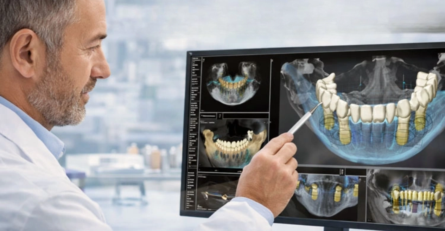
Note : All images used are for editorial and illustrative purposes only and may not originate from the original news provider or associated company.
Can Digital Dental Implants Be Used for Full-Mouth Rehabilitation?
Yes, digital planning can be used extensively for full-mouth rehabilitation involving multiple implants or complete-arch restorations. Full-mouth treatment requires careful coordination because implant positions must support the final prosthesis across a large area.
CBCT can help evaluate bone anatomy throughout the jaws. Intraoral scanning can provide detailed information about existing teeth, occlusion, and soft-tissue surfaces. These records can be combined with the planned prosthetic design. The clinician can then simulate implant positions before surgery.
Guided surgery may be selected when transferring those positions accurately is clinically beneficial. Full-mouth cases can involve All-on-4, All-on-6, individual implants, or other implant-supported designs.
The treatment sequence depends on bone condition, remaining teeth, periodontal health, and restorative goals. Digital planning can make the process more coordinated, but it does not eliminate healing periods or surgical risks. Each full-mouth case requires a personalized clinical assessment before treatment begins.
Digital Planning for Multiple Implants
Multiple implant planning requires careful coordination because implant positions influence one another. The clinician must consider bone volume, implant dimensions, restorative space, and occlusion.
Digital software allows several implants to be visualized simultaneously. This can reveal spacing or angulation issues before surgery. The proposed positions can also be evaluated relative to the final prosthesis.
This is important because implants should support the restoration rather than simply occupy available bone. CBCT can identify anatomical limitations. Intraoral scanning can show tooth positions and occlusal relationships. Combining the records can create a more complete treatment model.
A surgical guide can then be fabricated if appropriate. Multiple implant cases may also require temporary restorations while the implants heal. The clinician must therefore plan both surgical and restorative stages.
Digital planning can help organize this sequence and improve communication with the laboratory. Such coordination is fundamental to Digital Dental Implants.
Full-Arch Implant Planning
Full-arch implant planning involves designing an implant-supported prosthesis for an entire upper or lower arch. The clinician must consider implant number, distribution, bone availability, occlusion, aesthetics, and prosthetic design.
CBCT can help identify available bone and anatomical structures. Digital scans can capture the dental arch and restorative relationships. These datasets can then be used to simulate the proposed treatment.
Full-arch planning is often prosthetically driven because implant positions must support the final prosthesis. The clinician may evaluate whether a fixed bridge, overdenture, All-on-4, All-on-6, or another design is appropriate.
Bone grafting may be required in selected patients. Treatment can also involve extractions and temporary teeth. Digital planning can help coordinate these stages.
The final decision depends on clinical examination, bone anatomy, systemic factors, oral hygiene, and patient expectations. No single full-arch technique is appropriate for every patient.
Digital Planning for All-on-4 and All-on-6
Digital planning can support All-on-4 and All-on-6 treatment by coordinating implant positions with the planned full-arch prosthesis. The clinician can assess bone anatomy using CBCT and evaluate restorative information through digital scanning.
The proposed implants can be positioned virtually according to the available anatomy. Angulation and distribution can then be assessed. This can help identify anatomical constraints before surgery. Surgical guides may be used when appropriate.
The prosthetic team can also use digital records to design temporary and final restorations. Cost varies substantially depending on the number of implants, prosthesis material, diagnostic work, grafting, and included services.
Bookimed's September 2026 data reports All-on-4 prices in Turkey from approximately $2,300, compared with reported starting prices around $20,000 in the UK and $25,000 in the USA. These figures are comparison data rather than fixed clinic prices. A personalized quotation requires clinical assessment and treatment planning.
Coordinating Implant Placement With the Final Restoration
Coordinating implant placement with the final restoration is central to successful full-arch treatment. The final teeth determine where the implants should ideally support the prosthesis. Digital planning allows the clinician to visualize this relationship before surgery.
The implant positions can be adjusted according to bone anatomy and restorative requirements. This can help reduce situations where an implant is technically stable but poorly positioned for the final prosthesis. The laboratory can also participate earlier in treatment planning.
Digital files can be shared between the clinic and laboratory. The temporary restoration can be designed before surgery when appropriate. However, immediate loading is only suitable for selected cases.
Primary stability, bone quality, implant distribution, and occlusal factors must be considered. Patients should therefore avoid assuming that a digital workflow automatically means same-day permanent teeth. The treatment timeline depends on biological and clinical requirements. This is a critical checkpoint within the Digital Dental Implants process.
Dr. Rifat Alsaman's Clinical Opinion
Dr. Rifat Alsaman, Head of the Medical Team at Vitrin Clinic and cosmetic dentist, emphasizes that full-mouth implant treatment should begin with the final restorative objective. In his clinical perspective, implant positions should be evaluated according to bone anatomy, prosthetic requirements, aesthetics, and long-term maintenance.
Digital tools can make these relationships easier to visualize before surgery. CBCT can show the available bone, while digital scanning can provide detailed information about the dental surfaces. When these datasets are combined, the team can create a more comprehensive treatment plan.
However, the technology should support clinical judgment rather than determine treatment independently. Full-mouth rehabilitation requires careful diagnosis and sequencing.
Patients should understand why each implant is recommended and how it contributes to the final restoration. At Vitrin Clinic, the digital workflow can help connect diagnosis, implant planning, guided surgery, and restorative treatment within one coordinated pathway.
Discover how tongue thrust can affect oral development
What We Notice Clinically
Clinical outcomes depend on more than the presence of advanced digital equipment. The most important factor is whether the technology improves the actual treatment decision. A scanner can create detailed images, but those images must be interpreted correctly.
A surgical guide can reproduce a plan, but the plan must first be appropriate. Implant treatment should therefore remain centered on anatomy, biology, function, aesthetics, and long-term maintenance. Digital workflows can improve communication and visualization.
They can also make complex cases easier to coordinate. However, clinicians must recognize the limitations of every digital tool. Patient movement, scanning errors, guide instability, bone quality, and intraoperative findings can affect treatment.
This is why experienced clinical teams remain essential. Digital planning is most valuable when it reduces uncertainty without creating false confidence. Patients should evaluate the entire treatment pathway rather than focusing on a single technology or marketing term.
Why Implant Position Should Be Planned Around the Final Restoration
Implants replace tooth roots, but the ultimate goal is usually to support functional and aesthetic teeth. Therefore, implant position should be planned according to the final restoration whenever possible. A technically stable implant can still create restorative challenges if it emerges in an unfavorable location.
The implant may affect crown contours, hygiene access, aesthetics, occlusion, or prosthetic maintenance. Prosthetically driven planning reverses the traditional question. Instead of asking where bone is available first, the clinician considers where the final tooth should ideally be positioned.
The available bone is then evaluated around that restorative position. If bone is inadequate, augmentation may be discussed. Digital planning can make this relationship easier to visualize.
The approach is especially important in the aesthetic zone and full-arch rehabilitation. Patients should ask their dentist whether the final restoration was considered before implant placement. This simple question can reveal how comprehensively the case has been planned. Digital Dental Implants are only as reliable as this stage of planning.
Why Bone Anatomy Remains Important
Bone anatomy remains fundamental because an implant requires adequate biological support. Digital planning can visualize bone dimensions, but it cannot change the underlying anatomy. The clinician must assess bone height, width, quality, and relationship with nearby structures.
Bone loss can result from previous tooth loss, periodontal disease, trauma, infection, or other conditions. If insufficient bone is present, augmentation may be required. The type of augmentation depends on the location and severity of the deficiency.
In some cases, an alternative implant strategy may be possible. CBCT can help identify these conditions before surgery. This is valuable because patients can understand additional treatment requirements before committing to a procedure.
Bone anatomy also affects treatment timing. Grafting may require a healing period before implant placement. Digital planning therefore supports diagnosis but does not eliminate biological limitations. A personalized assessment remains essential for determining the appropriate treatment sequence.
Why Digital Data Must Be Combined With Clinical Examination
Digital data provide valuable information, but they cannot capture every aspect of a patient's health. Clinical examination can identify inflammation, periodontal disease, soft-tissue conditions, occlusal problems, and other factors. Medical history can also reveal conditions that affect healing or surgical risk.
These findings must be considered alongside imaging. A CBCT scan may show adequate bone while the patient has untreated gum disease. In that situation, implant placement may need to be delayed.
Similarly, a digital scan may show the teeth clearly but cannot determine every biological characteristic of the tissues. Combining different information sources produces a more complete assessment. This is one reason experienced implant dentists remain essential.
Digital workflows should enhance clinical examination rather than replace it. Patients should be cautious when clinics present scans as if they alone determine treatment suitability. A comprehensive diagnosis requires both technology and professional evaluation.
The Importance of Prosthetically Driven Implant Planning
Prosthetically driven planning connects implant surgery with the final restorative objective. The clinician determines where the final tooth or prosthesis should ideally be positioned. Implant placement is then planned to support that result within biological and anatomical limitations.
This approach can be particularly important for visible teeth. Implant angulation can influence the emergence profile and crown aesthetics. In full-arch cases, implant distribution can influence prosthetic stability and maintenance.
Digital planning can help visualize these relationships before surgery. It can also allow the laboratory to participate earlier in the process. However, prosthetic planning must remain compatible with bone anatomy.
If the ideal implant location lacks sufficient bone, augmentation or an alternative design may be necessary. The principle therefore involves balancing restoration, biology, and surgery. Digital technology makes that balance easier to visualize, but clinical judgment remains decisive. This is an essential consideration for anyone pursuing Digital Dental Implants.
Digital Dental Implants at Vitrin Clinic
Vitrin Clinic can integrate digital technologies into implant treatment planning for international patients visiting Istanbul. The workflow can include CBCT imaging, digital X-rays, three-dimensional intraoral scanning, and computer-based treatment planning.
These technologies can help the clinical team assess anatomy and visualize proposed implant positions. Digital records can also support communication between the implant team and dental laboratory. For selected cases, guided surgery may be incorporated into the treatment plan.
The clinic's international patient workflow can also coordinate treatment planning with accommodation, transportation, appointments, and follow-up. Vitrin Clinic offers implant treatments ranging from individual tooth replacement to complex full-arch rehabilitation.
The exact treatment depends on the patient's anatomy and clinical requirements. Patients should receive an individualized assessment before receiving a final quotation. Digital technology is used to support treatment decisions, not to replace professional diagnosis. Patients considering treatment in Istanbul can request a FREE Treatment Plan from Vitrin Clinic.
Vitrin Clinic's Digital Implant Planning Workflow
Vitrin Clinic's digital implant workflow can begin with an initial consultation and diagnostic assessment. The clinical team reviews the patient's dental concerns, medical history, previous treatment, and treatment goals.
CBCT imaging can then provide three-dimensional information when required. Digital scanning can capture detailed information about the teeth and occlusion. These records can be combined during treatment planning.
The clinician can evaluate potential implant positions according to bone anatomy and restorative requirements. For selected cases, computer-guided planning and surgical guide fabrication may be appropriate.
The restorative stage can then be coordinated with the implant plan. This workflow can be especially useful for international patients because treatment information can be organized before and during their visit.
Each case remains individualized. Some patients require additional procedures before implant placement. Others may be suitable for a more straightforward implant workflow. The treatment sequence is determined after clinical assessment.
CBCT and Digital Imaging
CBCT and digital imaging provide important information for evaluating implant candidates. CBCT can show three-dimensional bone anatomy and the relationship between proposed implant locations and anatomical structures. Digital X-rays can provide additional diagnostic information when appropriate.
The combination of imaging methods depends on the patient's needs. Vitrin Clinic can use digital imaging as part of the implant assessment workflow. The clinical team can review bone volume, tooth roots, sinus anatomy, and other relevant structures.
This information supports treatment planning and helps identify potential challenges before surgery. Imaging is selected according to clinical requirements rather than simply for technological purposes.
Patients should be informed about why a particular scan is needed. The resulting information can also contribute to a digital treatment plan. For international patients, digital records may help the clinical team prepare before the patient arrives. A final decision still requires appropriate clinical examination. Digital Dental Implants benefit directly from this type of assessment.
3D Intraoral Scanning
Vitrin Clinic can use three-dimensional intraoral scanning to capture detailed digital records of the patient's teeth and oral structures. The scanner creates a digital model that can be viewed and analyzed during treatment planning.
This can support implant positioning and restorative design. When combined with CBCT, the scan can provide information about both visible dental surfaces and underlying anatomy. Digital scanning can also support CAD/CAM laboratory workflows.
This may reduce the need for conventional physical impressions in appropriate cases. The digital model can be shared with the restorative team to coordinate crown or prosthesis design. Scanning can also help patients understand their current dental condition because the model can be displayed visually.
The accuracy of the final record depends on scanning technique and clinical conditions. Therefore, the dentist must verify the digital model before using it for treatment decisions. Digital scanning is a useful component of the broader treatment workflow.
Smart Digital Treatment Planning
Smart digital treatment planning combines diagnostic information into a structured treatment workflow. CBCT provides three-dimensional anatomical information. Intraoral scanning provides digital surface information.
Planning software allows the clinician to examine these datasets together. The proposed implants can then be positioned virtually. The clinician can evaluate spacing, angulation, depth, bone support, and restorative relationships.
For multiple implants, the entire arrangement can be reviewed before surgery. If guided surgery is appropriate, the digital plan can support surgical guide design. The same digital records may also contribute to restorative planning.
This creates continuity between diagnosis, surgery, and prosthetic treatment. Vitrin Clinic's digital workflow is designed to support international patients who want coordinated implant treatment in Istanbul.
However, the term smart does not mean automatic diagnosis. Every treatment plan requires professional review. The final decision depends on the patient's clinical condition and treatment objectives.
Digital Technology From Planning to Implant Restoration
Digital technology can support several stages from initial diagnosis through implant restoration. CBCT and digital scanning provide diagnostic records. Planning software can combine these records and support implant positioning.
Guided surgery can transfer selected elements of the digital plan to the clinical environment. After appropriate healing, digital scans can assist restorative fabrication. CAD/CAM technology can then support production of crowns, bridges, or full-arch prostheses.
This creates a connected workflow that can improve information transfer between the clinic and laboratory. The approach can also support treatment planning for international patients.
Vitrin Clinic can coordinate implant surgery and restorative stages according to the patient's individual plan. The exact timeline depends on healing and clinical requirements.
Same-day or immediate temporary teeth may be appropriate for selected patients but are not guaranteed. Final restorations require clinical verification before delivery. This underscores why Digital Dental Implants demand individualized care.
Dr. Rifat Alsaman's Clinical Opinion
Dr. Rifat Alsaman, Head of the Medical Team at Vitrin Clinic and cosmetic dentist, considers digital planning an important support tool when it provides clinically useful information. His clinical perspective emphasizes the relationship between implant position, bone anatomy, and the final restoration.
Digital scans can make this relationship easier to visualize before treatment. CBCT can help assess anatomical limitations, while intraoral scanning can provide restorative information. When combined, these records can support a more comprehensive plan.
However, the dentist must still evaluate the patient clinically. Digital planning cannot predict every aspect of healing or surgical conditions. The treatment plan should therefore remain adaptable.
Patients should also understand the difference between a planned outcome and a guaranteed outcome. At Vitrin Clinic, digital technologies are incorporated to support personalized implant treatment rather than replace clinical expertise.
The Vitrin Clinic Digital Implant Journey
The Vitrin Clinic digital implant journey is designed to connect diagnosis, planning, surgery, restoration, and aftercare. International patients can begin by providing information about their dental concerns and previous treatment.
The clinical team can review available records and determine which additional diagnostics may be required. During the Istanbul visit, CBCT and digital scanning can provide detailed information for treatment planning. The clinician can then create a personalized treatment strategy.
Depending on the case, guided surgery may be considered. Temporary and final restorations are planned according to the selected implant approach. The aftercare process remains important because implant success depends on long-term maintenance.
International patients may also receive support with treatment coordination, transportation, accommodation, and appointments. The exact journey differs between patients because implant treatment is not a single standardized procedure. Vitrin Clinic can provide a FREE digital treatment plan after reviewing the patient's clinical information.
Initial Consultation and Digital Assessment
The initial consultation establishes the patient's goals and clinical needs. The dentist reviews medical and dental history and performs an oral examination. Existing radiographs and dental records can also be reviewed.
When additional imaging is necessary, CBCT may be used to evaluate three-dimensional anatomy. An intraoral scan can provide a detailed digital model of the mouth. These records help the clinician understand the patient's current condition. They also provide the foundation for treatment planning.
Patients should use this stage to discuss previous dental procedures, medications, allergies, concerns, and expectations. International patients should also discuss their available travel dates.
Treatment timing may depend on whether grafting or staged implant placement is required. The consultation should result in a clear explanation of possible treatment options. Patients should understand both the benefits and limitations of each option before proceeding. Such factors directly influence the outcome of Digital Dental Implants.
Personalized Digital Treatment Plan
The personalized treatment plan brings together the patient's diagnostic information and treatment objectives. The clinician determines the appropriate number and type of implants based on the clinical findings.
CBCT information can help assess bone anatomy. Digital scans can help evaluate the restorative situation. The proposed implant positions can then be planned virtually.
If guided surgery is suitable, the plan can be used to create a surgical guide. The treatment plan may also include temporary teeth and final restorations. Additional procedures such as extraction or bone grafting can be incorporated when necessary.
Patients should receive an explanation of the expected treatment sequence and estimated costs. At Vitrin Clinic, the digital treatment plan can help international patients understand the proposed pathway before committing to treatment.
A personalized plan is more reliable than using a generic online price. Final costs depend on the actual clinical requirements.
Implant and Surgical Planning
Implant and surgical planning involves determining the appropriate implant positions, dimensions, and treatment sequence. The clinician evaluates bone availability and anatomical structures using diagnostic information.
The proposed implants are positioned according to the intended restoration. If several implants are planned, their positions are coordinated as a group. The surgical approach is then selected.
Some patients may be suitable for conventional placement. Others may benefit from guided surgery. If a guide is required, it is designed and manufactured before the procedure. The clinician checks the guide and confirms that it corresponds with the planned treatment.
Any additional surgical procedures are also considered. Patients should understand whether bone grafting or other procedures are included in their treatment plan.
Planning is particularly important for international patients because appointments may need to be coordinated with travel schedules. Biological healing remains the main factor determining the overall treatment timeline.
Guided Implant Surgery
Guided implant surgery uses the approved digital plan and surgical guide to assist implant placement. Before surgery, the clinician confirms the guide's fit and stability. Local anesthesia or another appropriate anesthetic approach is provided according to the procedure.
The surgical site is prepared following the selected implant protocol. The guide can then help control the drilling trajectory. Implant placement is performed according to the planned position and clinical findings.
The clinician continues to monitor bone quality and surgical conditions throughout the procedure. If unexpected findings occur, the treatment approach may need modification. Guided surgery therefore supports the planned workflow but does not prevent clinical adaptation.
After implant placement, stability is evaluated. The patient receives postoperative instructions and information about medication and oral hygiene.
Follow-up is arranged according to the treatment plan. The next stage may involve healing, temporary restoration, or immediate provisionalization when clinically appropriate. This remains an important part of planning Digital Dental Implants.
Temporary and Final Restoration
Temporary and final restorations serve different purposes during implant treatment. A temporary restoration may be used while implants heal or while the final prosthesis is being fabricated. In selected cases, an immediate temporary restoration may be possible.
However, immediate loading requires adequate stability and favorable clinical conditions. The final restoration is normally produced after the implant and surrounding tissues have reached the appropriate stage of healing. Digital scanning can support the final restorative process.
CAD/CAM systems can be used to design the crown, bridge, or full-arch prosthesis. The dentist checks the restoration for fit, aesthetics, occlusion, and function. Adjustments may be required before final delivery.
Material selection can include options such as zirconia or porcelain depending on the clinical case. Vitrin Clinic can coordinate restorative planning with the implant workflow. The final choice depends on the patient's anatomy, functional requirements, and treatment plan.
Aftercare and Follow-Up
Aftercare is essential because implant treatment does not end when the prosthesis is delivered. Patients must maintain excellent oral hygiene around the implant-supported restoration. Regular professional examinations can help identify inflammation or mechanical complications early.
The dentist may recommend periodic radiographic monitoring depending on the clinical situation. Patients should also avoid habits that place excessive forces on the restoration. Smoking can negatively affect oral and periodontal health and should be discussed with the dental team.
International patients should understand how follow-up will be managed after returning home. Vitrin Clinic's aftercare team can help coordinate follow-up communication for international patients.
Any pain, swelling, bleeding, mobility, or changes around an implant should be reported promptly. Long-term maintenance is a shared responsibility between the patient and dental team. Proper care can help protect both the implant and surrounding tissues.
Tips for Patients Considering Digital Dental Implants
Patients considering implant treatment should evaluate the entire clinical workflow rather than focusing only on digital technology. Ask which imaging systems are used and why they are required. Ask how implant positions are determined and whether the final restoration is considered during planning.
Ask whether guided surgery is recommended for your particular case. Patients should also ask about bone quality, gum health, and possible grafting. The quotation should clearly explain what is included.
This may include imaging, surgery, abutments, temporary teeth, final crowns, medications, and follow-up. International patients should also clarify accommodation, transportation, and appointment scheduling when applicable.
Most importantly, choose a qualified clinician with appropriate implant experience. A digital scanner cannot compensate for poor diagnosis or inadequate surgical skill.
Technology should make a sound treatment plan clearer and more coordinated. It should not be used as a substitute for professional evaluation.
Ask What Digital Technology Will Be Used
Patients should ask clinics to explain exactly which digital technologies are included in treatment. Terms such as digital, smart, advanced, and computer-guided can have different meanings.
Ask whether the clinic uses CBCT, intraoral scanning, virtual implant planning, surgical guides, or CAD/CAM restoration. Ask what each technology contributes to your specific case. This can help distinguish clinically useful technology from general marketing terminology.
Patients should also ask whether the digital planning is performed by the treating dentist or another professional. Understanding who interprets the scans is important. The clinic should be able to explain how digital information influences implant placement.
At Vitrin Clinic, patients can receive an explanation of the digital workflow as part of treatment planning. The goal should always be to connect technology with a clear clinical purpose. Patients should not assume that more technology automatically means better treatment. This is a defining feature of transparent Digital Dental Implants.
Ask How Your Implant Position Will Be Planned
Implant position is one of the most important factors in implant treatment. Patients should ask how the dentist determines the planned location. A good explanation should include bone anatomy and the final restoration.
The dentist may use CBCT, intraoral scanning, or other diagnostic records. Patients can ask whether their implant positions are planned virtually before surgery. They can also ask whether a surgical guide will be used.
If guided surgery is proposed, ask how the guide is designed and verified. Understanding the planning process can provide insight into the quality of the treatment strategy. The final implant should not simply be placed wherever bone happens to be available.
Its position should support the intended crown or prosthesis within biological limits. This is particularly important for aesthetic areas and full-arch rehabilitation. Clear communication before surgery can help patients understand the treatment.
Understand Your Bone and Gum Condition
Bone and gum health directly affect implant treatment. Patients should ask whether they have adequate bone volume for the proposed implant. CBCT imaging can help evaluate bone dimensions when clinically indicated.
Gum health is equally important because active periodontal disease can compromise the surrounding tissues. Any infection or inflammation should be addressed before implant placement. If bone loss exists, the dentist may recommend grafting.
This can affect both treatment time and cost. Patients should ask whether grafting is included in the quotation. They should also understand whether additional healing time is required.
Good oral hygiene is essential before and after implant surgery. Patients should discuss smoking and other habits that may affect oral health. A thorough assessment helps prevent unexpected changes during treatment.
Digital planning can identify anatomical limitations, but biological health still determines whether treatment can proceed safely.
Ask Whether Guided Surgery Is Suitable for You
Guided surgery is not automatically required for every implant patient. Its value depends on anatomy, restorative requirements, implant number, and clinician preference. Patients should ask why guided surgery is recommended in their particular case.
The dentist should explain what the guide will control and what it cannot control. Patients can also ask whether the surgical guide is included in the treatment price. Some clinics may charge separately for planning or guide fabrication.
The guide should be checked for accurate fit before surgery. Patients should understand that guided surgery does not eliminate surgical risk. The clinician still needs to monitor the surgical field and respond to unexpected findings.
Guided treatment can be particularly helpful in complex implant cases. For straightforward cases, freehand placement may remain clinically appropriate. The decision should be based on individual needs rather than marketing claims.
Discuss the Final Restoration Before Surgery
The final restoration should be discussed before implant placement whenever possible. The crown or prosthesis determines how the implant needs to support the tooth. Patients should ask what material will be used and how the restoration will be retained.
They can also ask whether temporary teeth are included. For full-arch treatment, ask whether the initial prosthesis differs from the final restoration. Digital planning can help connect the final tooth design with implant placement.
This can be particularly important for visible teeth. The dentist should explain how aesthetics, function, hygiene, and maintenance will be considered. Patients should also ask about replacement or maintenance costs in the future.
A low initial price may not include every restorative stage. Understanding the complete pathway makes price comparisons more meaningful. At Vitrin Clinic, restorative planning can be integrated into the digital implant workflow. This is a critical checkpoint within the Digital Dental Implants process.
Choose an Experienced Implant Dentist
The dentist's experience remains one of the most important factors in implant treatment. Patients should ask about the clinician's implant training and experience with cases similar to theirs.
A dentist who routinely manages full-arch rehabilitation may have different expertise from a clinician who primarily performs simple single implants. Patients can also ask how complications are managed. The clinic should explain its approach to follow-up and maintenance.
Digital equipment is valuable, but clinical expertise determines how that equipment is used. A sophisticated scanner does not independently create a treatment plan. The dentist must interpret the data and make appropriate decisions.
International patients should also consider how communication and aftercare are handled after returning home. At Vitrin Clinic, patients can discuss their treatment with the clinical team before traveling. A personalized assessment is preferable to choosing a provider based solely on advertised prices.
Are Digital Dental Implants Suitable for Everyone?
Not every patient requires the same digital implant workflow, and not every patient is immediately suitable for implant treatment. Suitability depends on oral health, bone anatomy, systemic health, treatment goals, and the ability to maintain good hygiene.
Patients with sufficient bone and healthy tissues may be suitable for straightforward implant placement. Patients with bone loss may require additional procedures before implant placement. Multiple missing teeth can benefit from coordinated digital planning.
Full-arch patients may require extensive diagnostic and prosthetic planning. Digital technology can assist many of these cases, but it does not remove biological limitations. Some patients may need periodontal treatment, extraction, grafting, or other preparation first.
A complete clinical assessment is therefore essential. Vitrin Clinic evaluates each international patient individually before recommending an implant treatment plan. The final decision should always be made by the treating dental professional after appropriate examination and diagnostic assessment.
Patients With Sufficient Bone Support
Patients with sufficient bone support may be candidates for relatively straightforward implant placement. The dentist still needs to assess bone dimensions, gum health, occlusion, and the position of neighboring structures.
CBCT can provide three-dimensional information when required. Digital scanning can help evaluate the restorative situation. The implant can then be selected according to the clinical requirements.
In some cases, guided surgery may provide useful additional control. In other cases, conventional placement may be entirely appropriate. The presence of adequate bone does not automatically determine the treatment method.
The final restoration and aesthetic requirements also matter. Patients should ask whether their treatment requires a surgical guide and why. They should also ask how implant stability will be evaluated. Even straightforward cases require careful planning. The goal is to create a stable biological foundation for a durable restoration.
Patients With Bone Loss
Patients with bone loss may still be candidates for implant treatment, but additional planning may be necessary. Bone loss can reduce the available height or width for implant placement. It can also alter the relationship between the proposed implant and anatomical structures.
CBCT can help clinicians assess the extent of the deficiency. Depending on the location and severity, the dentist may recommend bone grafting or another augmentation procedure. In selected cases, alternative implant designs or positions may be considered.
Treatment timing can become longer when grafting requires a healing period. Digital planning can help visualize the bone deficiency before surgery. It can also show the relationship between the desired restoration and available bone.
Patients should ask which additional procedures may be required. They should also ask whether those procedures are included in the quoted price. A detailed plan reduces the risk of unexpected treatment changes. Digital Dental Implants cannot succeed without this step.
Patients Requiring Bone Grafting
Patients requiring bone grafting need careful assessment before implant placement. Grafting may be recommended when bone volume is insufficient for the intended implant position. The specific procedure depends on the location and type of bone deficiency.
Some patients may require ridge augmentation, while others may require sinus augmentation. Healing time varies according to the procedure and patient. Digital imaging can help clinicians determine the extent of the deficiency.
It can also assist with planning the eventual implant position. However, the graft itself must heal biologically before the implant can provide long-term support. Patients should understand that digital planning cannot shorten every biological healing period.
The treatment may therefore require multiple visits. International patients should discuss whether grafting and implant placement can be performed during the same trip. The answer depends on the clinical situation. Vitrin Clinic can develop an individualized sequence after evaluating the patient's imaging and oral condition.
Patients With Multiple Missing Teeth
Patients with multiple missing teeth may benefit significantly from coordinated digital planning. Several implants must be positioned in relation to one another and the final restoration. The clinician must consider available bone, implant distribution, occlusion, and hygiene access.
Digital planning can display the proposed implants together. This can help identify spacing or trajectory problems before surgery. Intraoral scanning can also provide detailed information about the remaining teeth.
The final restoration can be incorporated into the planning process. A surgical guide may be appropriate when precise positioning is important. Treatment may involve individual crowns, bridges, or an implant-supported prosthesis.
The choice depends on the number and location of missing teeth. Patients should ask how many implants are recommended and why. They should also ask whether the final prosthesis is fixed or removable. Clear planning helps patients understand both treatment and cost.
Patients Requiring Full-Arch Rehabilitation
Patients requiring full-arch rehabilitation often need comprehensive planning because many teeth are missing or failing. The clinician must evaluate remaining teeth, bone anatomy, gum health, occlusion, and the desired restoration.
Digital planning can integrate these factors into a three-dimensional workflow. CBCT can help assess bone availability. Intraoral scanning can capture the existing dental situation. The proposed implants can then be planned around the intended full-arch prosthesis.
All-on-4 and All-on-6 are examples of full-arch approaches, but they are not appropriate for every patient. The number of implants depends on anatomy and the selected restorative strategy. Bone grafting may sometimes be required.
Temporary teeth may be used while the implants heal. Final prostheses are generally planned separately from immediate provisional restorations. Patients should receive a complete treatment explanation before beginning full-arch rehabilitation.
Why Individual Assessment Is Essential
Individual assessment is essential because implant treatment varies substantially between patients. Two people with the same number of missing teeth can require completely different treatment plans. Bone anatomy may differ significantly.
Gum health, occlusion, medical history, and previous dental treatment also influence the decision. Digital planning can reveal anatomical differences, but the clinician must interpret them. The final treatment may involve one implant, multiple implants, grafting, or full-arch rehabilitation.
Cost also depends on these variables. Online price ranges can therefore provide only general guidance. A personalized quotation should follow clinical examination and appropriate diagnostics.
At Vitrin Clinic, international patients can request a FREE Treatment Plan to begin discussing their potential treatment. The final plan should explain the recommended procedures and their sequence. Patients should never select an implant treatment solely from an advertised starting price. This is a defining characteristic of well-planned Digital Dental Implants.
How Long Does Digital Dental Implant Treatment Take?
Treatment time depends on the patient's anatomy, number of implants, surgical approach, healing response, and restoration. Digital planning itself can often be completed before surgery once the required diagnostic records are available.
The surgical appointment may be relatively short for a straightforward single implant. Complex full-mouth treatment can require substantially more planning and multiple procedures. Healing is often the longest biological stage because the implant must integrate with the surrounding bone.
Some patients may receive temporary teeth during healing. Immediate provisionalization is possible in selected cases when clinical conditions are favorable. The final restoration is usually placed after the dentist determines that the implant is ready.
Bone grafting can extend the timeline because grafted tissue may require healing before implant placement. International patients should therefore avoid assuming that every implant can be completed in a few days. A personalized schedule is necessary.
Digital Planning Time
Digital planning time depends on the complexity of the case and the amount of information required. A straightforward single implant may require less planning than a full-arch rehabilitation.
The clinical team must review CBCT images, intraoral scans, photographs, and other relevant records. Multiple implants require additional coordination because the proposed positions must work together.
If a surgical guide is required, additional laboratory or manufacturing time may be necessary. Restorative planning can also increase the planning stage. International patients may complete some preliminary assessment remotely before traveling.
However, final planning may require an in-person examination and updated diagnostic records. Vitrin Clinic can organize digital records as part of the patient's treatment journey. The planning process should never be rushed simply to meet a travel schedule. Accuracy and clinical safety are more important than minimizing preparation time. Patients should ask when their treatment plan will be finalized.
Implant Surgery and Healing
Implant surgery may take a relatively short time for a simple case, but the healing period is much longer. After placement, the implant needs to integrate with the surrounding bone.
The duration depends on bone quality, implant stability, surgical approach, and patient-specific healing. Some cases can support immediate or early loading. Other cases require a longer period before the final restoration is attached.
Bone grafting can add additional healing time. The clinician determines the appropriate timeline based on clinical evidence and the patient's condition. Patients should not pressure a dentist to accelerate biological healing for travel convenience.
International treatment planning should allow enough flexibility for follow-up and unexpected changes. The dentist may also recommend temporary restorations during the healing period. Regular communication is important after returning home. Patients should report unusual pain, swelling, mobility, or other concerning symptoms promptly.
Temporary and Final Teeth
Temporary teeth can help patients maintain function and appearance while implants heal. The type of temporary restoration depends on the implant stability and overall treatment plan.
Some patients may receive immediate temporary teeth on the day of surgery. Others may use removable or temporary restorations during healing. The final teeth are usually placed after the dentist confirms that the implants are ready.
Digital scanning can support the fabrication of the final restoration. The laboratory can use digital records to design crowns or full-arch prostheses. The final restoration must be checked for fit, occlusion, aesthetics, and hygiene access.
Adjustments may be required before final delivery. Patients should ask whether temporary and final restorations are included in the treatment quotation. They should also ask what materials will be used. Understanding these details can make treatment planning more transparent.
Factors That Affect Treatment Time
Several factors can affect implant treatment time. The number of implants is one important consideration. Bone quality and quantity can also influence the surgical approach and healing period.
Patients requiring bone grafting may need additional appointments and healing time. Active gum disease or infection should generally be managed before implant placement. Full-arch treatment requires more extensive planning than a single-tooth case.
The selected restoration can also influence laboratory time. International travel can add scheduling constraints. Some patients may complete treatment within a short visit, while others require multiple trips.
Digital technology can improve coordination, but it cannot eliminate biological healing requirements. Patients should therefore request a timeline based on their specific diagnosis. At Vitrin Clinic, treatment schedules are planned according to the patient's clinical requirements and selected treatment approach. Flexibility is important when complex implant treatment is involved.
How Should Patients Prepare for Digital Implant Surgery?
Preparation begins with a complete medical and dental assessment. Patients should provide accurate information about medications, allergies, previous surgeries, medical conditions, and previous dental treatment.
The dentist may request CBCT imaging, digital scans, photographs, and other records. Oral hygiene should be optimized before surgery. Existing gum disease or infection may need treatment first.
Patients should follow all preoperative instructions provided by their dentist. They should also discuss smoking, alcohol consumption, and medications that may influence healing or bleeding. International patients should plan travel around the recommended appointment schedule.
They should understand how long they may need to stay in Istanbul. Patients should also ask what happens if additional treatment becomes necessary. Having a clear plan reduces uncertainty during the treatment journey. Digital planning can organize the clinical information, but successful preparation still depends on patient cooperation.
Dental and Medical Assessment
Dental and medical assessment helps determine whether implant treatment is appropriate and safe. The dentist evaluates teeth, gums, bone, occlusion, and existing restorations. Medical history can reveal factors that may affect surgery or healing.
Patients should disclose all medications and relevant medical information. The dentist may request additional medical clearance in selected cases. Gum disease should be controlled because inflammation around implants can create long-term problems.
Existing infections may also require treatment before implant placement. CBCT can help assess bone anatomy when clinically indicated. Digital scans can provide detailed information about the dental structures. Together, these findings help determine the appropriate implant strategy.
Patients should ask questions if they do not understand why a particular test is required. Good communication before surgery improves preparation and informed decision-making. No digital planning system can replace a complete clinical assessment.
Records and Scans You May Need
The diagnostic records required for implant treatment vary by patient. Common records can include dental photographs, digital X-rays, CBCT scans, and intraoral scans.
CBCT is particularly useful when three-dimensional information about bone and anatomical structures is needed. Intraoral scanning can provide a detailed digital model of the teeth and occlusion. Existing dental records may also be reviewed.
The clinician determines which records are clinically necessary. Patients should avoid unnecessary imaging simply because a technology is available. Radiation exposure should always be considered when prescribing radiographic examinations.
Digital records can be transferred electronically when patients are planning treatment from another country. This may allow the clinic to perform preliminary treatment planning before arrival. However, updated in-person records may still be required. Accurate diagnostic information is the foundation of reliable implant planning.
Questions to Ask Your Implant Dentist
Patients should ask several questions before agreeing to implant treatment. Ask how many implants are recommended and why. Ask how the implant positions will be planned. Ask whether CBCT and intraoral scanning are required. Ask whether guided surgery is appropriate for your case. Ask whether bone grafting may be necessary. Ask what type of restoration will be used. Ask whether temporary teeth are included. Ask how long healing is expected to take. Ask what follow-up will be provided after treatment. Patients should also request a complete written quotation. International patients should clarify whether accommodation and transportation are included in a treatment package. These questions help patients understand both clinical and financial aspects of treatment. A trustworthy clinic should be willing to explain the plan clearly. Patients should feel comfortable asking for clarification before surgery.
What to Expect on Surgery Day
On surgery day, the clinical team confirms the treatment plan and reviews any relevant medical information. The dentist checks the surgical area and confirms the planned procedure. Appropriate anesthesia is then provided. If guided surgery is planned, the surgical guide is checked for proper fit. The procedure follows the selected implant protocol. The clinician monitors the surgical site throughout treatment. If unexpected findings occur, the dentist may adjust the plan. After implant placement, stability can be assessed. Temporary teeth may be placed when appropriate. The patient receives postoperative instructions regarding medication, eating, oral hygiene, and activity. Follow-up arrangements are also discussed. Patients should have a way to contact the clinic if concerns develop after surgery. International patients should understand the emergency and follow-up arrangements before leaving the clinic. Recovery instructions should always be individualized.
Digital Dental Implants: Key Takeaways
Digital implant treatment combines modern diagnostic and planning technologies with established surgical and restorative principles. CBCT can provide three-dimensional anatomical information. Intraoral scanning can provide detailed digital models of the teeth.
Virtual planning can help determine proposed implant positions. Surgical guides can assist with transferring the plan to the mouth. CAD/CAM workflows can connect implant surgery with restoration fabrication.
These technologies can be particularly useful for multiple implants and full-arch rehabilitation. However, digital treatment does not guarantee a successful outcome. Biological healing, bone quality, gum health, oral hygiene, and clinical expertise remain essential.
Patients should choose treatment based on a comprehensive clinical assessment. Cost should also be evaluated as a complete package rather than a single implant price. At Vitrin Clinic, digital workflows can support individualized implant planning for international patients in Istanbul. Patients can request a FREE digital treatment plan to discuss their potential treatment needs.
The Role of Digital Planning
Digital planning helps clinicians visualize implant treatment before surgery. It can combine CBCT imaging, intraoral scanning, and restorative information. The clinician can then evaluate proposed implant positions in relation to bone anatomy and the final prosthesis. This can be particularly useful for complex cases. Digital planning can also support surgical guide fabrication. The main benefit is improved information management and visualization. However, digital planning remains a support tool. The dentist must interpret the information and determine whether the proposed treatment is appropriate. Clinical examination remains essential. The patient's biological condition also influences the final outcome. Digital planning should therefore be integrated into a broader clinical workflow. Patients should ask how digital technology changes their treatment rather than simply whether a clinic uses it. This creates a more meaningful discussion about the value of technology.
What Guided Implant Surgery Can and Cannot Do
Guided implant surgery can help control planned implant positioning in selected cases. A surgical guide can assist with drilling direction and other surgical parameters. This may be useful when precise positioning is important. However, guided surgery cannot eliminate biological healing requirements. It also cannot guarantee perfect implant placement. Guide fit, manufacturing accuracy, digital registration, and clinical conditions can influence the result. Unexpected findings may require changes during surgery. The clinician must therefore remain in control throughout the procedure. Guided surgery is best viewed as a method for transferring a carefully designed plan. It does not replace experience or clinical judgment. Patients should ask what level of guidance their proposed system provides. They should also ask how the clinician verifies the guide before surgery. Understanding these limitations creates more realistic expectations.
Why Professional Clinical Assessment Matters
Professional clinical assessment remains essential because implant treatment involves biological and mechanical factors. Digital imaging cannot independently determine whether an implant is appropriate. The dentist must evaluate bone, gums, occlusion, systemic health, oral hygiene, and patient expectations. The final restoration must also be considered. A technically possible implant position may not be the best restorative or biological choice. Experienced clinicians can identify these issues during treatment planning. They can also modify the plan when clinical conditions change. Digital technology makes information easier to visualize, but interpretation remains a professional responsibility. Patients should therefore evaluate the qualifications and experience of the treating team. They should also consider follow-up and maintenance support. At Vitrin Clinic, the digital workflow is designed to support clinical assessment and individualized treatment planning. The technology is a tool within the treatment process rather than the treatment itself.
At Vitrin Clinic
Vitrin Clinic provides digital implant treatment planning in Istanbul with a focus on coordinated care for international patients. The clinic can integrate CBCT imaging, digital X-rays, three-dimensional intraoral scanning, and computer-based treatment planning.
These tools can support single implants, multiple implants, and full-arch rehabilitation. The digital workflow can connect surgical planning with restorative design. Selected patients may also benefit from guided implant surgery.
Vitrin Clinic's international patient experience can include treatment coordination, airport transportation, accommodation support, and aftercare communication. The clinic also offers digital treatment planning designed around individual clinical requirements.
Patients can discuss their goals and receive a FREE Treatment Plan before committing to treatment. The final treatment, number of implants, restoration type, and cost depend on clinical assessment. Patients should receive a personalized quotation rather than relying on generic online prices. Dr. Rifat Alsaman and the clinical team can evaluate the case and determine the most appropriate treatment pathway.
Reference
FAQs
Dr. Rifat Alsaman has more than 5 years of clinical experience in dentistry and currently serves as the Head of the Medical Team at Vitrin Clinic. He is dedicated to providing exceptional patient care, overseeing treatment planning, and ensuring the highest clinical standards across the team. His expertise, attention to detail, and commitment to continuous professional development have helped countless patients achieve healthier, more confident smiles.


