Dental Crowns
October 7, 2026
Digital vs Conventional Impressions for Implant Crowns: Which Is Better?
 Dr. Rifat Alsaman
Choosing the right impression method can influence implant restoration accuracy, comfort, and laboratory communication. Digital vs Conventional Impressions for Implant Crowns is not simply a comparison of old and new technology. The better method depends on implant number, implant position, access, scanner performance, and clinical conditions. Digital scanning can offer a comfortable workflow and direct electronic communication with the laboratory. Conventional impressions remain valuable when specific clinical situations favor physical impression materials. Research also shows mixed accuracy results across different implant configurations and study designs. Patients should therefore avoid assuming that newer technology always provides better results. A careful clinical assessment should guide the final choice of impression technique. At Vitrin Clinic, the selected workflow should match the planned restoration and individual oral conditions. The final objective is accurate implant position transfer and a crown that fits predictably.
What Are Digital and Conventional Impressions for Implant Crowns?
Digital and conventional impressions both record the three-dimensional position of a dental implant. That information allows the dental laboratory to design a restoration around the implant connection. Digital methods use an intraoral scanner together with a calibrated scan body. Conventional methods use impression copings and elastomeric impression materials inside a tray. When patients research Digital vs Conventional Impressions for Implant Crowns, they should remember both methods need careful execution. The impression itself is only one step within the complete restoration process. Implant position, tissue condition, occlusion, and laboratory design also influence the final result. Research suggests both techniques can produce clinically acceptable outcomes in suitable cases. A 2025 review found that scan-body design, implant location, scanner type, and protocol affect digital accuracy.
What Is a Digital Implant Impression?
A digital implant impression captures implant information through intraoral optical scanning. The dentist attaches a manufacturer-compatible scan body to the implant or abutment. The scanner then records the scan body's shape and its spatial relationship with nearby structures. Software converts the captured images into a detailed three-dimensional digital model. The laboratory can import this file directly into a computer-aided design workflow. Digital data can also include neighboring teeth, soft-tissue contours, and occlusal relationships. This process reduces the need for physical trays and setting impression materials. Digital records can be stored securely and transferred electronically within minutes. However, scanning quality depends on visibility, scan-body seating, scanner performance, and operator technique. Research identifies scan-body characteristics and scanning strategy as important accuracy variables.
What Is a Conventional Implant Impression?
A conventional implant impression records implant position using physical impression materials. The dentist first places an impression coping onto the implant connection. The coping becomes mechanically captured within the impression material as it sets. After setting, the impression is carefully removed from the patient's mouth. The laboratory then uses the impression to create an accurate working cast. An implant analog is positioned according to the recorded coping location. The resulting cast becomes the foundation for restoration design and fabrication. Conventional impressions remain widely recognized and trusted within implant prosthodontics. Their accuracy depends on tray stability, coping design, material properties, and handling. The selected technique may follow an open-tray or closed-tray protocol. Clinical evidence suggests splinted techniques can improve accuracy in multiple-implant situations.
How Implant Impression Techniques Transfer the Implant Position
The main purpose of any implant impression is accurate spatial transfer. The laboratory must know the implant's exact three-dimensional position and orientation. Small transfer errors can influence restoration fit and long-term prosthetic relationships. Digital workflows transfer position through scan bodies and digital reference geometry. Conventional workflows transfer position through impression copings and laboratory implant analogs. Within Digital vs Conventional Impressions for Implant Crowns, both approaches depend on secure component seating. Implant angulation can make position transfer more demanding for either method. Implant number and inter-implant distance can also affect the impression process. A 2024 systematic review found multiple variables influencing digital implant scanning accuracy. Those variables included implant position, inter-implant distance, scan-body design, scanner type, and technique.
Scan Bodies in Digital Implant Impressions
Scan bodies are calibrated components used during digital implant impressions. Their geometry gives the scanner recognizable reference surfaces to capture. The software uses this information to identify implant position and orientation. Correct seating is therefore critical for reliable digital data transfer. A loose, rotated, or incorrectly selected scan body can create inaccurate positioning. Manufacturer compatibility should always be confirmed before scanning begins. Scan-body shape can also influence how easily surfaces are captured optically. A 2025 systematic review found geometry affected both linear and angular trueness. Most included studies were laboratory-based, so clinical interpretation requires some caution.
Impression Copings in Conventional Techniques
Impression copings mechanically transfer implant location into the physical impression. They may be designed specifically for open-tray or closed-tray procedures. Open-tray copings remain connected within the impression during removal. Closed-tray copings stay on the implants and are repositioned after impression removal. Splinting can connect multiple copings into a more rigid and stable arrangement. Clinical studies generally support splinting for many multi-implant situations. However, technique selection should still reflect implant arrangement and restoration design. A systematic review found greater accuracy with splinted methods across several implant configurations. Careful coping selection therefore supports reliable conventional implant position transfer.
Compare full-arch restoration options in fixed prosthesis vs 3D printed overdenture
What Is the Difference?
Digital
Traditional Dental Impressions
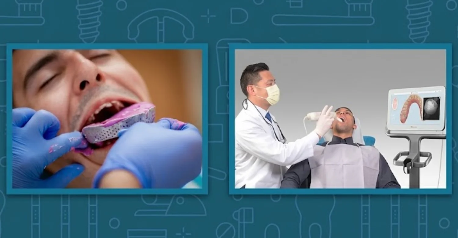
Digital scanning replaces physical impression material with optical data capture. Conventional impressions create a physical negative representation of oral structures. The difference between Digital vs Conventional Impressions for Implant Crowns extends beyond materials alone. Workflow speed, patient experience, laboratory communication, storage, and error sources can also differ. Digital systems allow immediate visualization of the areas already captured. Errors can therefore be corrected before the patient leaves the clinic. Conventional materials require mixing, loading, setting, removal, disinfection, and laboratory processing. Neither method eliminates clinical errors completely. Technology selection should therefore consider the restoration rather than marketing claims. A 2025 umbrella review found digital systems generally improved comfort and reduced procedure time.
Digital Scanning Workflow
Digital scanning begins with careful preparation of the treatment area. The dentist exposes the implant connection and attaches the correct scan body. The scanner then captures the implant region and surrounding anatomy. The operator follows a planned scanning pathway across the arch. Software stitches many individual images into one continuous digital model. The dentist inspects the scan for missing or distorted data. Additional scanning can be performed whenever defects are detected. The completed file can then be transferred electronically to the laboratory. This workflow reduces physical handling between the clinic and laboratory. Scan-body placement and scan strategy remain essential to final accuracy.
Conventional Impression Workflow
The conventional workflow begins with selecting an appropriate and rigid impression tray. The dentist connects the impression copings to the implants. Impression material is mixed and placed into the tray and around the copings. The tray is seated carefully and held steady until the material sets. After removal, the dentist checks the impression for voids and defects. The laboratory receives the impression and pours or scans the resulting model. Implant analogs reproduce implant locations within the working cast. Restoration design then proceeds using conventional or digital laboratory methods. Material handling and setting can introduce additional practical variables. Proper technique remains essential throughout every stage of this workflow.
Digital vs Conventional Impressions at a Glance
Both approaches aim to reproduce implant position as accurately as possible. Digital impressions remove the need for impression paste and physical stone casts. Conventional impressions provide a physical record that laboratories can inspect directly. Digital workflows can simplify communication and long-term file storage. Conventional workflows can remain predictable for selected complex implant cases. Looking at Digital vs Conventional Impressions for Implant Crowns side by side shows no universal winner. Accuracy depends on the complete workflow rather than one isolated component. The best option may therefore vary between individual patients. Current evidence does not support one universal method for every restoration. Recent reviews continue to report contradictory findings across full-arch studies.
Accuracy
Accuracy refers to how closely the recorded implant position matches the actual position. Digital systems measure deviations against digital reference models. Conventional systems are assessed through cast measurements and prosthetic fit. Studies report different results depending on scanner, technique, implant configuration, and test method. Some reviews favor digital methods, especially for selected short-span cases. Other findings support conventional approaches for specific extensive restorations. Accuracy should therefore be interpreted within the clinical context of each case. A laboratory-perfect result may still fail when component seating is incorrect. Proper verification remains more important than relying on technology labels.
Patient Comfort
Patient comfort often differs between digital and conventional impression procedures. Digital scanning generally avoids large amounts of impression material inside the mouth. This can reduce sensations of pressure, unpleasant taste, nausea, and breathing difficulty. A systematic review found patients preferred intraoral scanning over conventional impressions. The pooled difference in patient preference was statistically significant. Patients with strong gag reflexes may particularly appreciate digital scanning. However, holding the mouth open can still feel tiring during longer scans. Comfort should be considered alongside clinical accuracy and treatment requirements. In Digital vs Conventional Impressions for Implant Crowns, comfort is important but never the only factor.
Time and Workflow
Digital scanning can shorten several stages of impression handling. There is no need to wait for physical material to set. The dentist can also inspect the scan immediately at the chair. Laboratory transmission can happen electronically within minutes. Conventional impressions require more physical processing before laboratory fabrication begins. A meta-analysis reported shorter mean clinical times for digital scanning. The same review found comparable marginal fit between the two techniques. Time savings may become more important when multiple appointments are involved. However, rescanning can increase chair time when optical capture becomes difficult.
Materials and Laboratory Handling
Digital workflows rely on scanners, software, scan bodies, and compatible laboratory systems. Conventional workflows rely on trays, impression materials, copings, adhesives, and analogs. Digital files avoid shipping physical impressions between clinics and laboratories. Conventional impressions require transportation and careful storage before laboratory processing. Physical materials can also experience handling-related distortions during transport. Digital records require secure software storage and compatible file formats. Laboratories may prefer one method depending on equipment and established workflows. The most suitable process should support both clinical and laboratory communication. Clear handling protocols reduce avoidable errors in either workflow.
Error and Distortion Risk
Every impression technique has potential sources of error. Digital errors can result from scanning gaps, stitching problems, scan-body movement, or limited visibility. Conventional errors can result from material distortion, coping movement, tray instability, or improper setting. Digital scanning provides immediate opportunities to identify missing areas before submission. Conventional impressions allow direct visual inspection of the physical impression. Understanding Digital vs Conventional Impressions for Implant Crowns means accepting that neither workflow guarantees perfect transfer. Research continues to identify case-specific variables affecting both techniques. Careful execution and checking remain the best protection against distortion.
Learn about the effects of water fluoridation and oral health
Which Is More Accurate for Implant Crowns?
Digital vs Conventional Impressions for Implant Crowns cannot be judged through accuracy figures alone. Clinical evidence shows that performance varies by restoration type and implant configuration. Digital methods can perform very well for single and short-span implant restorations. Full-arch cases introduce greater scanning distances and more opportunities for stitching errors. Conventional impressions can remain highly reliable with appropriate open-tray and splinted techniques. A 2024 review of full-arch implants found broadly comparable outcomes between both methods. Another review reported mixed findings and substantial methodological variation between studies. The most accurate method is therefore the one properly matched to the clinical case.
What Does Digital Implant Impression Accuracy Mean?
Accuracy includes two related concepts called trueness and precision. Trueness describes closeness to the true reference position. Precision describes consistency among repeated measurements or repeated scans. Digital implant impressions can be evaluated using linear, angular, and three-dimensional deviations. These measurements do not always translate directly into clinical problems. A small laboratory deviation may remain clinically acceptable for a single crown. Larger errors can become more significant across multiple connected implants. Accuracy also depends on how the scanner captures and stitches separate images. A 2024 full-arch meta-analysis identified differences across scanner systems and implant arrangements. Patients should therefore view accuracy as a multifactorial concept.
What Affects Implant Crown Fit and Impression Accuracy?
Implant crown fit depends on accurate implant position and correct prosthetic design. Impression accuracy represents only one part of the overall workflow. Component compatibility and seating also matter substantially. Tissue contours can influence restoration emergence and access. Occlusion must be evaluated carefully before final delivery. Laboratory design and manufacturing tolerance can affect the completed crown. Operator technique influences both digital and conventional results. Digital scanning introduces additional variables involving hardware and software. Any discussion of Digital vs Conventional Impressions for Implant Crowns should include these wider factors. Clinical verification remains necessary even after an apparently excellent impression.
Number and Position of Implants
More implants can increase the complexity of implant position transfer. Long distances create more opportunities for cumulative scanning deviations. Implant location also affects visibility and instrument access. Posterior implants can be more difficult to scan consistently. Multiple nonparallel implants can increase registration challenges for the software. Conventional techniques may use splinted copings to stabilize several transfer components. Digital techniques may use specialized scanning strategies or splinted scan bodies. Research suggests implant number and inter-implant distance can affect digital accuracy. The dentist should therefore review implant distribution before choosing a method.
Implant Angulation and Depth
Implant angulation changes the orientation of the prosthetic connection. Greater angulation can make component seating and optical recognition more demanding. Implant depth can also limit visibility during digital scanning. Deep implants may leave little scan-body height above the gum line. Conventional coping access may also become more complicated in deep or angled implants. Evidence suggests angulation can influence conventional impression accuracy. Digital evidence remains variable across scanners and scanning techniques. Clinical planning should account for implant location before selecting the impression method.
Scan-Body Design and Position
Scan-body design directly affects digital implant position recognition. The geometry must provide enough reference surfaces for reliable optical detection. Incorrect positioning can create a significant transfer error. Different implant systems use different scan-body interfaces. The correct component must precisely match the implant connection. Recent systematic evidence found geometry influenced digital trueness and precision. Some designs may perform better under specific scanning conditions. Clinicians should follow validated manufacturer protocols whenever they are available. Careful scan-body handling is a simple step that protects final accuracy.
Scanner Type and Scanning Strategy
Intraoral scanners differ in optical systems, algorithms, and software processing. Accuracy can therefore vary between different scanner models. Scanning strategy also determines how the software combines individual images. Excessive scanning distance can increase cumulative alignment error. A planned path may improve data continuity across the arch. Recent evidence identifies scanner type and scanning strategy among key accuracy factors. In Digital vs Conventional Impressions for Implant Crowns, scanner choice shapes digital performance significantly. Vitrin Clinic can select scanning workflows according to restoration requirements and clinical access.
Operator Experience
Scanner performance does not replace clinical expertise. Operators must control moisture, retract tissues, position scan bodies, and recognize incomplete data. Conventional impression procedures also require considerable technical experience. Mixing, tray loading, seating, removal, and inspection all affect results. Research has identified operator experience as a factor in digital accuracy. Experienced clinicians may therefore reduce avoidable technical errors. Training remains important regardless of impression technology. A skilled operator often matters more than the newest equipment available.
Saliva, Tongue Movement, Soft Tissue, and Limited Access
Optical scanners perform best when important surfaces remain clearly visible. Saliva, blood, moving tissues, and tongue interference can interrupt data capture. Posterior implant regions may provide especially limited access. Conventional impression materials are less dependent on optical visibility. However, conventional impressions still require good moisture control. Tissue displacement can also affect physical impression quality. The clinical environment should therefore guide technology selection. A digital workflow should not be used simply because it appears more modern. Practical conditions inside the mouth often decide which technique is safer.
Are Digital Impressions as Accurate as Conventional Impressions for a Single Implant?
For single implant restorations, digital methods can provide highly reliable results. Short-span scanning limits the cumulative stitching challenges seen across longer arches. A systematic review found comparable accuracy between techniques, particularly for single and short-span cases. For single implants, Digital vs Conventional Impressions for Implant Crowns often shows similar clinical performance. Proper scan-body selection remains essential for success. The scanner must capture the entire scan body accurately. The implant connection must also be identified correctly by the software. Clinical verification remains necessary before final placement. For many single implants, digital scanning can therefore be a practical and comfortable option.
Are Conventional Impressions Better for Multiple Implants or Full-Arch Restorations?
Conventional impressions remain useful for multiple implants and extensive restorations. Open-tray splinted techniques have a long history in implant prosthodontics. Evidence generally supports splinting for many multiple-implant situations. However, newer digital studies increasingly report clinically acceptable full-arch outcomes. One meta-analysis found digital impressions had favorable linear accuracy compared with conventional methods. Another 2024 review found similar outcomes overall but emphasized methodological differences. For full-arch work, Digital vs Conventional Impressions for Implant Crowns requires careful individual judgment. Full-arch cases should be evaluated individually rather than assigned automatically to one technique.
Find out what is normal regarding pain after root canal treatment
What We Notice Clinically
Clinical outcomes depend on the complete restorative chain. Digital vs Conventional Impressions for Implant Crowns describes the recording method, not the entire restoration. A good impression cannot compensate for an incorrectly seated component. It also cannot correct inaccurate implant placement or poor prosthetic design. Tissue inflammation can affect emergence profiles and final adaptation. Occlusion can create problems even when implant position is transferred accurately. At Vitrin Clinic, planning should connect examination, imaging, scanning, laboratory design, and final verification. The goal is a biologically appropriate and mechanically stable restoration. Technology should support that goal rather than replace clinical judgment.
Why a Technically Good Impression Can Still Produce a Poorly Fitting Crown
A good impression records implant position accurately. Yet the laboratory can still create an unsuitable restoration. The crown may have incorrect contacts or excessive occlusal loading. The abutment or restoration interface may also be incompatible. Cement space and manufacturing parameters can influence final fit. Soft-tissue changes can alter the clinical emergence appearance. Occlusal adjustments may become necessary during insertion. The dentist must therefore assess the completed crown clinically. Impression accuracy is important, but it is not the only determinant of success.
Common Patient Misconceptions About “Digital” Accuracy
Patients often assume that digital automatically means perfect. Digital systems still depend on correct component placement and controlled scanning. Software cannot recover anatomy that was never captured. A scanner may also repeat a systematic error consistently. Conventional techniques can produce highly accurate results with proper execution. Many myths about Digital vs Conventional Impressions for Implant Crowns come from marketing language. New technology should be judged by evidence and case suitability. Accuracy claims should consider the entire workflow. Patients should ask how the clinic verifies the final restoration.
Why Single-Implant Cases and Full-Arch Cases Should Not Be Compared the Same Way
A single implant creates a short and localized scanning field. A full-arch restoration may require continuous registration across a much longer area. More implants also introduce additional reference points and geometric relationships. The risk of cumulative deviation can therefore change substantially. Research has documented variability across digital full-arch scanning studies. Results from a single implant cannot simply predict full-arch performance. The restoration scope should always be considered during technique selection. Patients should understand which category their own treatment belongs to.
When Conventional Impressions May Still Be the Practical Choice
Conventional impressions may be useful when optical access is limited. They can also help when scanning conditions are unstable. Some laboratories have extensive experience with conventional implant models. Open-tray splinted impressions can provide reliable multi-implant transfer. Clinical evidence supports this approach in many configurations. Conventional methods also serve as a dependable alternative when digital equipment is unavailable. The final decision should reflect clinical predictability rather than habit or trend.
Clinical Note
Implant impression selection should begin with the restoration requirements. The clinician should evaluate implant number, angulation, access, tissue health, and laboratory workflow. A digital approach can be excellent when optical conditions are favorable. A conventional approach can remain excellent when physical transfer offers greater predictability. Neither technique should be chosen solely for marketing reasons. The dentist should also consider patient comfort and appointment efficiency. A balanced view of Digital vs Conventional Impressions for Implant Crowns protects the final outcome. At Vitrin Clinic, these factors can be integrated into an individualized treatment plan.
Dr. Rifat Alsaman’s Perspective on Choosing the Right Implant Impression Technique
Dr. Rifat Alsaman’s clinical perspective emphasizes matching technology to anatomy and restoration needs. Digital scanning can offer efficient communication and a comfortable patient experience. Yet careful scan-body positioning remains essential for implant restorations. Conventional impressions can still provide predictable transfer in selected complex cases. The correct decision depends on implant distribution, access, and laboratory capabilities. Patient comfort also deserves consideration during impression planning. From this view, Digital vs Conventional Impressions for Implant Crowns is a clinical decision, not a trend. The priority should always remain accurate transfer and predictable restorative fit. A modern workflow should improve treatment quality without unnecessary technological complexity.
Learn how oral cyclosporine affects your gums and oral health
Advantages of Digital Impressions for Implants
Digital implant impressions provide several practical benefits. They eliminate physical impression trays filled with setting material. Patients often report greater comfort during optical scanning. Dentists can inspect the captured data immediately on screen. This allows missing information to be corrected before laboratory submission. Digital files can be transferred electronically without physical shipping. Laboratory technicians can integrate the files directly into CAD workflows. Storage becomes easier because files do not require physical space. These advantages can improve efficiency when the clinical case suits digital scanning.
Improved Patient Comfort During Scanning
Digital scanning commonly produces less nausea and unpleasant taste. Patients also avoid the bulk associated with impression trays and setting materials. A systematic review found significantly higher patient preference for intraoral scanning. This can be especially helpful for patients with sensitive gag reflexes. Scanning still involves intraoral contact and cheek retraction. Longer procedures can cause fatigue or mild jaw discomfort. Comfort therefore improves, but it does not eliminate all inconvenience. At Vitrin Clinic, patient experience should remain part of impression planning.
Faster Digital Communication With the Dental Laboratory
Digital files can reach the laboratory electronically almost immediately. Physical impressions do not require packaging or courier transportation. Technicians can review digital data before fabrication begins. Missing regions can sometimes be rescanned without repeating the entire appointment. This workflow may reduce administrative and shipping delays. Digital communication also supports rapid collaboration between clinicians and technicians. File transfer can simplify the coordination of complex restorative cases. These benefits become particularly useful for patients traveling internationally for treatment.
Digital Implant Planning and Restoration Workflow
Digital impressions can connect with broader computer-aided treatment workflows. Scan data can be combined with imaging and virtual restorative planning. The laboratory can design implant crowns directly on digital models. Manufacturing can then proceed through CAD/CAM technologies. This creates a more connected digital treatment pathway from scan to crown. However, digital integration does not replace a careful clinical examination. Imaging should be used only when clinically appropriate. At Vitrin Clinic, digital planning can support organized implant restoration workflows when indicated.
Easier Storage and Reuse of Digital Records
Digital records can be archived within a secure clinical system. The files can support future comparison and restorative planning. A previous scan may also help when replacing or repairing a restoration. Physical impressions require controlled storage and can degrade over time. Digital records avoid that specific physical distortion risk. Data protection and backup procedures remain essential. Software compatibility should also be considered when files are transferred. Proper digital governance is part of modern clinical record management.
Where Digital Impressions May Offer Practical Advantages
Digital scanning can be especially useful for single implants and short-span restorations. Patient comfort can improve when conventional trays cause nausea. Immediate visualization can also help identify incomplete captures quickly. Electronic laboratory communication can reduce logistical delays. Digital workflows support CAD/CAM fabrication and virtual crown design. These advantages may become valuable for international patients with limited appointment time. In Digital vs Conventional Impressions for Implant Crowns, these strengths favor digital in suitable cases. However, the clinical case should remain the deciding factor.
Limitations of Digital Impressions for Implant Crowns
Digital systems have important limitations that patients should understand. Scan accuracy can decline when visibility is poor. Long scanning spans can increase image-stitching demands. Scan-body geometry and seating also influence the result. Scanner hardware and software can produce different performance characteristics. A 2025 systematic review highlighted major variability among these factors. Any fair review of Digital vs Conventional Impressions for Implant Crowns must include these weaknesses. Digital technology should therefore be treated as a clinical tool. It should not be considered universally superior.
Can Intraoral Scanners Lose Accuracy Over a Long Implant Span?
Long-span scanning can challenge optical registration. Each captured region must align correctly with adjacent regions. Small errors may accumulate as the scan becomes longer. Full-arch implant scanning therefore requires careful technique and validation. Research has reported considerable variation among full-arch digital accuracy studies. Newer studies continue to investigate scan-body splinting and scanning methods. Digital full-arch workflows can work well, but they require careful execution. Verification steps become more important as the scanned span increases.
When Scanning Conditions Make Digital Impressions More Difficult
Moisture can interfere with optical capture. Blood and saliva can obscure important scan-body surfaces. Tongue movement can complicate posterior scanning. Deep implant placement can reduce visible component height. Limited mouth opening can also restrict scanner positioning. Soft-tissue movement may interrupt continuous image stitching. The operator may need additional retraction or drying. These conditions can increase appointment time. A conventional impression can sometimes provide a more practical alternative.
Why Scanner Choice and Scan Strategy Matter
Different intraoral scanners use different optical technologies and algorithms. Their accuracy can vary across implant indications. Scan strategy also influences how images are registered together. Recent reviews identify scanner type and scanning strategy as significant factors. The operator should therefore use a validated scanning workflow. Random scanning patterns can increase unnecessary repetition. Consistent technique helps maintain data continuity across the arch. Equipment selection should consider the restoration rather than brand popularity.
When a Conventional Impression May Be Preferred
A conventional impression may be selected for difficult optical conditions. It can also be appropriate when a physical implant cast benefits laboratory workflow. Open-tray splinted impressions remain relevant for certain multiple-implant restorations. Evidence supports their accuracy in many cases. Some clinicians also prefer physical verification before complex laboratory fabrication. Conventional methods can be particularly useful when scanning access is limited. In Digital vs Conventional Impressions for Implant Crowns, flexibility serves patients better than ideology. Technology selection should remain adaptable to each clinical situation.
Conventional Impressions for Implant Crowns
Conventional implant impressions remain an established restorative technique. They provide a physical transfer of the implant's position. Different impression materials and tray techniques can be selected. Open-tray and closed-tray methods are the main options. Splinted impression copings may improve stability in multiple-implant cases. A systematic review found strong evidence supporting splinted techniques in many situations. Conventional impressions require careful material handling and tray selection. Proper seating and removal remain critical. Their continued relevance reflects predictable performance rather than resistance to technology.
What Materials Are Used for Conventional Implant Impressions?
Common implant impression materials include polyvinyl siloxane and polyether. Both materials can provide accurate records when handled correctly. Material stiffness, dimensional stability, and working time affect practical use. Tray rigidity also influences the final result. A systematic review found no consistent major accuracy difference between polyether and polyvinyl siloxane. Technique can therefore matter more than material choice alone. The selected material should suit the clinical and laboratory workflow. Proper storage and pouring procedures remain important after impression removal.
Polyvinyl Siloxane
Polyvinyl siloxane is often called PVS or VPS. It offers good dimensional stability and excellent elastic recovery. The material can be used in several different consistencies. Implant impressions may combine light-body and heavy-body materials. PVS can support both open-tray and closed-tray approaches. Its accuracy depends on proper mixing, tray seating, and complete polymerization. Research generally finds similar accuracy between PVS and polyether in many implant applications. Many clinicians value PVS for its predictable handling characteristics.
Polyether
Polyether is another established implant impression material. It provides relatively high rigidity after setting. That characteristic can support stable coping transfer in some implant situations. However, removal should be controlled carefully around undercuts. Evidence suggests polyether and PVS can provide comparable accuracy overall. In selected angulated implant cases, polyether has shown favorable findings. Material selection should still reflect the tray, coping, implants, and restoration. Patients may notice a distinct taste with some polyether products.
Open-Tray and Closed-Tray Implant Impression Techniques
Open-tray impressions use copings that remain locked inside the impression. The tray contains access openings for the coping screws. After setting, the screws are loosened before impression removal. This approach directly transfers coping positions during removal. Closed-tray methods leave copings attached to the implants after removal. The dentist then repositions the copings back into the impression. Open-tray methods are often favored for multiple implants. Evidence supports greater accuracy with open-tray techniques in many completely edentulous cases.
Advantages of Traditional Impression Materials for Implants
Conventional materials create a physical record that laboratories can examine directly. They can be useful when optical scanning is difficult. Physical impressions do not depend on scanner batteries, software, or optical stitching. Well-established laboratory procedures support conventional implant models. Experienced teams can achieve predictable results with these materials. The method also remains adaptable to complex physical geometry. However, physical handling creates its own potential errors. The technique must therefore be performed carefully from start to finish.
Limitations of Conventional Implant Impressions
Conventional impressions can create discomfort for some patients. Large trays may trigger gagging or breathing concerns. Impression materials can also produce unpleasant taste or pressure. Physical impressions require disinfection and transportation. Material distortion can occur when procedures are poorly controlled. Tray movement can affect implant position transfer. Multiple-implant cases may need additional splinting and technique steps. These limitations explain why digital scanning has become increasingly attractive.
.webp)
.webp)
.webp)
.jpg)
.jpg)
.jpg)
How Does the Digital Workflow for Implant Restorations Work?
The digital workflow connects clinical scanning, implant positioning, CAD design, and manufacturing. First, the dentist prepares the implant area and surrounding anatomy. A compatible scan body is attached to the implant connection. The intraoral scanner captures the scan body and surrounding structures. The digital model is inspected before being sent to the laboratory. CAD software can then generate the planned implant crown. Manufacturing may use milling or other CAD/CAM processes. The final restoration must still be clinically evaluated. Digital workflow efficiency should never replace restorative verification.
Scanning the Implant and Surrounding Teeth
The dentist begins by capturing the implant region. Neighboring teeth provide anatomical reference for restoration design. The scanner records surfaces using rapid optical imaging. Moisture and soft tissues are controlled during capture. The operator checks whether important areas are complete. Missing scan-body surfaces should be recaptured before submission. Occlusal records may also be obtained digitally. The final file should represent both implant position and restorative context.
Recording the Implant Position With a Scan Body
The scan body provides the implant-specific reference geometry. Its connection must match the implant system exactly. The component must also be fully seated before scanning begins. Software identifies the scan body and links it to implant library data. Incorrect seating can transfer an incorrect spatial relationship. Scan-body design can also influence optical accuracy. Recent evidence confirms geometry is an important digital variable. A quick seating check can prevent costly laboratory errors later.
Digital Design and Laboratory Communication
The completed scan can be exported as a standard digital file. The laboratory receives implant position information alongside surrounding anatomy. CAD software uses these files to design the crown. Technicians can inspect virtual contacts and emergence profiles. The clinician and technician can communicate without exchanging physical impressions. Digital communication can also support rapid design corrections. File standards must remain compatible between systems. Accurate communication still depends on the quality of the original scan.
CAD/CAM Fabrication of the Implant Crown
CAD software creates the virtual restoration using implant-specific libraries. The design considers contacts, contour, occlusion, and material parameters. The digital crown can then be milled or manufactured. Material choice may include zirconia, ceramic, or other restorative options. Manufacturing accuracy depends on equipment and regular calibration. Laboratory finishing can influence final adaptation and appearance. Digital design does not guarantee perfect clinical fit. Final inspection remains necessary before placement.
Checking the Crown Before Final Placement
The laboratory and dentist should evaluate the crown before definitive delivery. Contacts should be assessed against adjacent teeth. Occlusion should match the planned functional relationship. The implant connection must also be appropriate and fully engaged. Marginal adaptation and seating should be checked clinically. Radiographs may be used when indicated. The crown should never be forced into position. Any unexplained misfit should trigger further investigation.
How Is an Implant Crown Impression Diagnosed or Evaluated?
Evaluation begins with the recorded implant position. The clinician checks whether the impression accurately represents the planned restoration. Digital files are inspected for missing or distorted data. Conventional impressions are checked for voids and coping stability. The finished crown must then be evaluated clinically. Fit includes marginal adaptation, contact, occlusion, and passive seating. Evaluating Digital vs Conventional Impressions for Implant Crowns therefore continues after the impression appointment. A technically accurate impression can still produce a clinically inappropriate crown. Verification should occur before definitive cementation or screw tightening.
Checking the Accuracy of the Implant Position Transfer
Implant transfer accuracy can be assessed through digital comparison or clinical verification. Scan-body seating should be confirmed first. The digital file is inspected for complete reference surfaces. Conventional impressions should show stable and fully captured copings. Laboratory models can then be evaluated against reference information. Complex cases may require additional verification steps. The objective remains accurate reproduction of implant position. Errors identified early are easier and cheaper to correct.
Assessing the Digital Scan or Conventional Impression
Digital scans should contain complete and continuous data. The operator should look for holes, artifacts, or distorted regions. Scan-body surfaces must be clearly visible. Conventional impressions should show intact coping impressions and stable material. Voids around critical regions can make the record clinically unreliable. The dentist should not submit a questionable impression simply to avoid repetition. Repeating a poor impression can prevent larger restorative problems later.
Evaluating the Implant Crown Fit
Crown fit is evaluated after fabrication and before final placement. The dentist checks whether the restoration seats completely. Contacts should allow appropriate floss passage. Occlusion should avoid excessive or premature loading. The implant connection should engage correctly without force. The clinician may use radiographs when needed. A satisfactory crown should fit without unexplained rocking or pressure. Careful fit evaluation protects both the implant and the surrounding tissues.
Marginal Fit
Marginal fit refers to the adaptation around the restoration interface. Implant restorations do not use natural tooth margins in exactly the same way. However, restoration adaptation remains clinically important. Excessive discrepancy can complicate hygiene and prosthetic maintenance. Clinical studies report acceptable marginal fit with both digital and conventional workflows. A meta-analysis found no significant marginal-fit difference between digital and conventional impressions for fixed prostheses. In Digital vs Conventional Impressions for Implant Crowns, marginal fit appears broadly similar.
Interproximal Contact
Interproximal contact should be firm but clinically appropriate. Excessive contact can prevent complete seating. Weak contact can allow food impaction between teeth. Contacts should also support stable neighboring tooth relationships. The dentist checks contact using floss and clinical inspection. Adjustment should be conservative when needed. Contact problems may arise from design rather than impression accuracy alone. Correct diagnosis should identify the actual cause.
Occlusion
Occlusion determines how the implant crown interacts during function. Excessive contact can increase mechanical loading on the implant. High contacts may cause patient discomfort after placement. The dentist should evaluate static and functional relationships. The patient's bite should be considered alongside adjacent teeth. Occlusal adjustment should preserve appropriate anatomy. Impression accuracy cannot compensate for poorly designed occlusion.
Passive Fit
Passive fit describes adaptation without forcing the restoration into position. Implant-supported restorations should seat without persistent strain. Multiple-implant restorations make passive adaptation particularly important. Impression errors can contribute to prosthetic discrepancies. Framework and laboratory factors can also influence adaptation. Digital and conventional techniques may both achieve acceptable passive fit. A 2025 in-vitro study found digital workflows and splinted conventional techniques achieved acceptable passive adaptation.
What Happens When an Impression Is Not Clinically Acceptable?
The dentist may repeat the impression when critical information is incomplete. Digital scans can often be corrected by rescanning affected regions. Conventional impressions may require complete remaking when critical defects exist. The decision depends on defect location and severity. Minor noncritical imperfections may not always require repetition. Critical implant surfaces must be represented reliably. It is generally safer to correct uncertain data before fabrication. This approach can reduce remakes and unnecessary chairside adjustments.
Understand the benefits and uses of phosphorus oral supplements
Note : All images used are for editorial and illustrative purposes only and may not originate from the original news provider or associated company.
What Happens if an Implant Crown Impression Is Inaccurate?
Digital vs Conventional Impressions for Implant Crowns becomes clinically important when transfer errors affect restoration fit. Inaccurate impressions can lead to prosthetic discrepancies. A crown may appear acceptable but fail to seat passively. Contact or occlusal relationships can also become incorrect. Mechanical complications may develop when components experience unfavorable loading. Biological concerns can arise when excess cement or poor tissue adaptation occurs. However, an impression is not always the direct cause. Proper clinical investigation should identify the source before repeating treatment.
Signs of a Potentially Poor-Fitting Implant Crown
Patients may notice pressure or unusual bite contact. The crown may feel too high during chewing. Floss may become difficult to pass between adjacent teeth. Persistent food trapping can indicate contact problems. Movement or rocking should be assessed promptly. Pain around an implant crown should not be ignored. Some symptoms may originate from tissues rather than prosthetic fit. Professional examination is necessary for accurate diagnosis.
Mechanical Complications Related to Prosthetic Misfit
Poor prosthetic adaptation can increase mechanical stress. Screw loosening can occur for multiple reasons, including occlusal loading. Component wear may also develop over time. Ceramic fracture can result from complex mechanical factors. Framework misfit is particularly relevant in multiple-implant restorations. A systematic review found that impression accuracy influences prosthetic adaptation. Early identification of fit problems can help reduce avoidable complications. Regular reviews allow small mechanical issues to be managed early.
Biological Concerns Associated With Poorly Fitting Implant Restorations
Poor restoration contours can make plaque control more difficult. Excess cement around implant restorations can also irritate surrounding tissues. Inflammation should be evaluated clinically when symptoms persist. A poorly fitting restoration can complicate hygiene and tissue maintenance. However, biological complications have multiple potential causes. Implant position, hygiene, occlusion, and tissue health all matter. Patients should receive personalized assessment rather than assuming impression error.
When an Impression or Crown May Need to Be Repeated
Repetition may be necessary when critical data are missing. A loose scan body requires correction before final fabrication. A distorted conventional impression may also need replacement. Repetition can prevent larger expenses from a failed restoration. The dentist may also remake a crown when fit remains unacceptable. The decision should follow clinical findings and laboratory assessment. With Digital vs Conventional Impressions for Implant Crowns, correcting errors early benefits every patient. Repeating a procedure is preferable to accepting an uncertain restoration.
Relief and Comfort During an Implant Impression Appointment
Comfort is an important part of the impression experience. Digital scanning often avoids impression material filling the mouth. Conventional impressions can feel larger and more restrictive. Patients with sensitive gag reflexes may prefer digital procedures. Evidence supports a patient preference for intraoral scanning. Comfort varies between individuals and procedure duration. Breathing, anxiety, and previous experiences can influence perception. Clinics can improve comfort through clear communication and controlled positioning.
What Patients Usually Feel During Digital Scanning
Patients usually feel the scanner moving around their teeth and gums. The procedure does not involve any setting impression material. Some pressure may occur from cheek or lip retraction. Posterior scanning can feel repetitive if additional capture is needed. The scanner head is usually smaller than a conventional impression tray. Patients may still experience mild jaw fatigue. Most discomfort is temporary and procedure-related. Communication can help patients remain relaxed during scanning.
What Patients May Feel During a Conventional Impression
Conventional impressions can create pressure against teeth, gums, and cheeks. Some patients notice a strong taste from impression materials. Others experience increased saliva or gagging. The tray may feel restrictive while the material sets. Breathing concerns can increase anxiety for sensitive patients. These sensations usually resolve after impression removal. Good tray selection can improve tolerance. The dentist should always know about severe previous reactions.
Tips for Reducing Gagging or Discomfort
Tell the dentist about previous gagging before the appointment. Nasal breathing can help some patients during impression placement. Sitting slightly more upright may improve perceived breathing comfort. Short breaks can be agreed before the procedure starts. A smaller suitable tray can sometimes improve tolerance. Digital scanning may also reduce nausea for some patients. Research supports lower discomfort with intraoral scanning. Comfort concerns are a valid part of Digital vs Conventional Impressions for Implant Crowns decisions.
Read about the effects and benefits of oral zinc supplements
Temporary Gum or Soft-Tissue Irritation
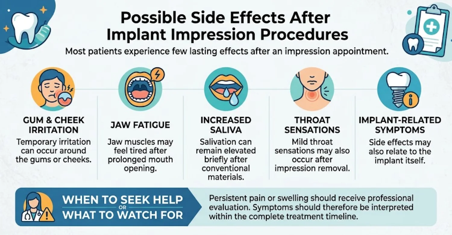
Soft tissues may feel mildly irritated after retraction or component placement. Pressure from trays can also cause temporary tenderness. Most minor irritation resolves without treatment. Patients should maintain normal oral hygiene unless instructed otherwise. Increasing pain or swelling requires clinical attention. The dentist should assess symptoms that persist beyond the expected period. Tissue health remains important before final implant restoration.
Jaw Fatigue or Mild Muscle Discomfort
Holding the mouth open can create temporary muscular fatigue. Longer scans or impressions may increase this sensation. Gentle jaw movement after treatment can feel comfortable. Persistent pain is less typical and should be assessed. Patients with existing jaw problems should mention them beforehand. Appointment breaks may improve tolerance during lengthy procedures. Comfort planning should consider the patient's individual needs.
Gagging, Salivation, or Temporary Throat Sensation
Conventional impression materials can stimulate salivation. Some patients also experience temporary gagging or throat sensations. These symptoms usually stop when the impression is removed. Digital scanning commonly reduces these reactions. Evidence shows improved patient comfort with intraoral scanning. Patients should inform the clinician if symptoms become severe. Simple breathing guidance can make the procedure easier to tolerate.
When Persistent Symptoms Should Be Discussed With a Dentist
Persistent pain should not be dismissed as a normal impression reaction. Significant swelling, bleeding, or worsening tenderness also warrants evaluation. A patient may have an unrelated dental or implant problem. Symptoms around an implant require careful clinical assessment. The timing of symptoms can help identify the cause. The dentist may inspect the soft tissues and restoration interface. Early review can prevent minor problems becoming more significant.
Medical Tips Before Choosing an Implant Impression Technique
Before choosing a technique, patients should understand the treatment objective. The number and position of implants can influence the decision. Previous gagging experiences may affect comfort considerations. The implant system determines compatible scan bodies and copings. Laboratory capabilities may also influence the workflow. Patients should ask how the final restoration will be verified. A good consultation explains Digital vs Conventional Impressions for Implant Crowns in simple terms. At Vitrin Clinic, treatment planning can integrate these considerations before fabrication.
Tell Your Dentist About Previous Difficult Impressions or Severe Gagging
Previous negative experiences are clinically relevant information. Severe gagging can make conventional impressions especially uncomfortable. Informing the dentist allows alternative techniques to be considered. Digital scanning may reduce symptoms for some patients. However, clinical suitability remains the main priority. The team can also plan breaks and positioning strategies. Patient comfort should be discussed before the appointment begins.
Ask Which Technique Is Being Recommended and Why
Patients should receive a simple explanation of the recommended technique. The reason may involve implant configuration, access, accuracy, or comfort. Asking why this method was chosen can clarify clinical reasoning. A technology should be selected because it suits the case. Patients may also ask whether another method would be equally appropriate. Understanding Digital vs Conventional Impressions for Implant Crowns can reduce anxiety before treatment. Clear communication supports truly informed consent.
Ask Whether Your Case Is a Single Implant, Multiple Implants, or Full-Arch Restoration
The restoration scope strongly influences impression complexity. Single implants generally involve shorter data capture areas. Multiple implants require more precise spatial relationships. Full-arch restorations create much larger scanning distances. Evidence shows that accuracy changes with scan length and implant arrangement. Patients should understand where their own case fits. This classification helps explain the selected impression technique.
Confirm Which Implant System and Scan Body or Impression Components Will Be Used
Implant systems have specific prosthetic connections. The scan body or coping must match the implant system. Component incompatibility can create incorrect transfer information. Patients may ask which implant brand is being restored. The laboratory must also receive the correct component library. These details are especially important for international treatment. Accurate component identification supports predictable restoration fabrication.
Ask How the Final Implant Crown Fit Will Be Verified
A reliable workflow includes checks beyond the impression itself. The dentist should assess crown seating and occlusion. Contact points should also be evaluated. Radiographic verification may be used when clinically indicated. The implant connection must engage correctly without force. Patients should understand that the final crown is still examined clinically. This step helps detect problems before definitive delivery.
Learn how ergocalciferol oral supplements support tooth strength
How Much Does an Implant Crown Cost With Digital or Conventional Impressions?
Cost depends on the implant system, crown material, clinic, and additional treatment. Impression technique may affect workflow pricing, but it is rarely the largest expense. In Digital vs Conventional Impressions for Implant Crowns, price differences are usually modest.
Current international market listings show wide price variation. Bookimed's October 2026 data lists installation of a dental implant with crown from approximately $3,600 in the United States, $1,400 in the UK, and $450 in Turkey. These are reference starting prices rather than universal national averages, and actual costs depend on clinical complexity, implant brand, restoration material, and package inclusions.
Implant Crown and Single-Implant Cost Comparison
Country | Reference Price for Implant + Crown | Pricing Context |
United States | From approximately $3,600 | Private treatment may cost more when imaging, abutment, surgery, laboratory work, or additional procedures are billed separately |
United Kingdom | From approximately $1,400 | Final private-clinic prices vary according to implant system, crown material, and treatment complexity |
Turkey | From approximately $450 | Prices vary widely by clinic, implant brand, crown material, city, and package inclusions |
Average Implant Crown and Single-Implant Cost in the US
Private implant treatment in the United States can vary substantially. Costs may include implant placement, abutment, crown, imaging, and laboratory fees. Market listings commonly show several thousand dollars for the complete procedure. Bookimed's October 2026 data gives approximately $3,600 as a reference starting figure for implant placement with a crown. Complex surgery can increase total costs considerably. Bone grafting or sinus procedures may add separate fees. Insurance coverage also differs widely between patients and plans.
US Implant Treatment Component | Possible Cost Effect |
Implant fixture | Major part of treatment cost |
Abutment | Additional restorative expense |
Implant crown | Depends on material and laboratory |
CBCT or other imaging | May be billed separately |
Bone grafting | Additional surgical cost when required |
Sinus augmentation | Additional cost in selected cases |
Temporary restoration | May add laboratory and clinical fees |
Average Implant Crown and Single-Implant Cost in the UK
UK implant prices depend on the clinic and restoration package. Private treatment can involve several separate stages and fees. Bookimed's current listings showed UK implant-with-crown prices from about $1,400. Individual clinic fees can be substantially higher. The final price depends on implant brand and crown material. Bone augmentation can increase treatment costs. Patients should request an itemized plan before treatment.
UK Implant Cost Factor | Why It Matters |
Implant system | Different brands have different component and laboratory costs |
Abutment | Stock and customized options may have different prices |
Crown material | Zirconia, porcelain, and other materials vary in cost |
Imaging | X-rays or CBCT may be charged separately |
Bone augmentation | Required only when clinically indicated |
Temporary crown | May be included or billed separately |
Average Implant Crown and Single-Implant Cost in Turkey
Turkey offers a wide range of implant treatment packages. Prices differ by implant brand, crown material, clinic, and city. Bookimed's October 2026 listings show implant-with-crown prices starting around $450, while Istanbul listings show approximately 400–630 for installation of a dental implant with crown. These figures demonstrate why advertised prices should be compared according to what is actually included.
Turkey Implant Treatment | Reference Price |
Implant + crown | From approximately $450 |
Istanbul implant + crown | Approximately 400–630 |
Premium implant systems | Usually higher than entry-level options |
Additional bone procedures | Extra cost when clinically required |
Package services | May include some imaging, transfers, or other services depending on the clinic |
At Vitrin Clinic, final treatment planning should be based on individual clinical findings rather than a headline package price.
Does the Choice of Digital or Conventional Impression Change the Final Price?
The impression method may affect workflow cost in some clinics. Digital systems require scanner investment, software, and compatible laboratory processes. Conventional methods require impression materials and physical laboratory handling. These expenses usually represent only part of the overall implant treatment.
The crown material and implant system often have greater cost effects. Digital technology may be included within a broader treatment package. Cost alone rarely settles Digital vs Conventional Impressions for Implant Crowns for patients. Patients should compare complete treatment inclusions rather than impression fees alone.
Cost Element | Digital Impression | Conventional Impression |
Impression material | Usually not required | Required |
Intraoral scanner | Required | Not required |
Physical impression shipment | Usually avoided | Usually required |
Digital laboratory workflow | Commonly integrated | May require physical model |
Separate patient fee | Clinic-dependent | Clinic-dependent |
Effect on total implant cost | Usually limited | Usually limited |
What Can Increase the Total Cost of an Implant Crown?
Treatment costs can increase when additional procedures are necessary. Bone loss may require augmentation before implant placement. Some patients need sinus-related procedures or soft-tissue treatment. Premium implant systems may also increase the restorative budget. Zirconia and other advanced materials can cost more than basic options. Imaging, temporaries, laboratory work, and follow-up can add separate charges. Every case should therefore receive an individualized estimate.
Factor | Potential Impact on Total Cost |
Implant brand | May increase component costs |
Abutment type | Customized components can cost more |
Crown material | Premium restorative materials may increase fees |
Bone grafting | Adds surgical and material costs |
Sinus augmentation | Adds a separate surgical stage |
Soft-tissue grafting | Additional periodontal procedure |
CBCT or other imaging | May be separately charged |
Temporary restoration | May create additional laboratory fees |
Laboratory work | Depends on complexity and fabrication method |
Follow-up care | May or may not be included in the package |
Implant Brand and Abutment
Implant brands differ in component design and pricing. Premium systems often cost more than economy systems. The abutment may be stock or individually customized. Certain restorations use multi-unit or specialized components. Compatibility should take priority over brand marketing.
Patients should know which implant and abutment system is planned. The chosen system affects both treatment cost and laboratory workflow.
Crown Material
Crown material strongly influences restorative cost. Zirconia, lithium disilicate, porcelain, and metal-based options have different prices. Material selection depends on location, strength requirements, and appearance. The laboratory's fabrication technology also influences cost.
A higher price does not automatically guarantee better clinical performance. Material selection should be individualized for each patient. The dentist can explain the trade-offs between strength and aesthetics.
Bone Grafting or Additional Procedures
Bone grafting can add a substantial amount to implant treatment. Sinus augmentation may also be required in selected upper-jaw cases. Soft-tissue grafting can create another additional cost.
These procedures depend on the patient's anatomy and implant plan. They are not automatically included in implant packages. Patients should ask whether preparatory surgery is included in quotes.
Imaging, Temporary Restoration, and Laboratory Fees
Imaging can include panoramic radiography or CBCT when clinically appropriate. Temporary crowns may also be required during treatment. Laboratory costs include design, manufacturing, finishing, and implant components. Complex cases may require custom components or additional verification.
Travel patients should also confirm accommodation and transportation inclusions. An itemized quote helps reduce unexpected expenses.
Why Implant Prices Should Be Treated as Planning Ranges, Not Fixed Quotes
Implant pricing is highly individualized. The same advertised package can require different additions for different patients. Clinical examination determines whether grafting or other procedures are necessary. Implant brand and restoration material also influence cost.
Current market listings show substantial differences between countries and individual clinics, reinforcing the importance of comparing treatment inclusions rather than headline prices alone.
A planning range should therefore guide budgeting rather than guarantee a final price. Patients should request a personalized treatment plan before committing.
Which Patients May Benefit From Each?
Digital
Conventional Impressions for Implant Crowns
The best impression method depends on patient anatomy and restorative goals. Digital scanning often suits single implants and short-span restorations. Conventional methods can remain valuable for challenging access or extensive implant cases. Patient comfort may favor digital workflows. Complex multi-implant transfer may require additional verification regardless of technology. Research remains mixed for full-arch accuracy. The technique should therefore be individualized. At Vitrin Clinic, the selected approach should support both clinical predictability and patient comfort.
When Digital Impressions May Be a Good Option
Digital scanning may suit single implants and short-span restorations. It can also benefit patients with strong gag reflexes. Immediate visualization allows the dentist to identify incomplete data. Electronic transmission supports rapid laboratory communication. Digital files integrate well with CAD/CAM workflows. These advantages may improve efficiency for international patients. The technique still depends on good moisture control and component visibility. Here, Digital vs Conventional Impressions for Implant Crowns often leans toward digital scanning. Digital scanning should be selected when clinical conditions support reliable capture.
When Conventional Impressions May Be a Better Option
Conventional impressions may be useful for difficult optical situations. They can also suit laboratories with strong physical-model workflows. Open-tray splinted approaches remain relevant for many multi-implant restorations. Physical impressions can sometimes provide useful tactile feedback. The technique remains a credible alternative rather than an outdated option. In these cases, Digital vs Conventional Impressions for Implant Crowns may favor conventional methods. The dentist should select it when it provides greater clinical predictability.
Why the Dentist’s Clinical Assessment Matters More Than Choosing a Technology Alone
Technology cannot replace diagnosis or restorative planning. Implant position, tissue condition, occlusion, and prosthetic requirements must be evaluated first. The dentist should determine what information the laboratory needs. Digital and conventional systems can both succeed when used correctly. A clinically inappropriate method can fail despite advanced equipment. The real answer to Digital vs Conventional Impressions for Implant Crowns comes from examination. The technique should therefore follow the case rather than dictate it.
Learn about precision and outcomes in dental implant placement accuracy
How Vitrin Clinic Selects the Right Impression for Implant Crowns
At Vitrin Clinic, impression selection should begin with individualized clinical assessment. Digital vs Conventional Impressions for Implant Crowns is considered within the broader restorative plan. The clinic can evaluate implant position, available space, soft tissues, occlusion, and restoration type. Digital scanning may be selected when suitable for the clinical situation. Conventional methods can remain an option when they provide practical advantages. Imaging may be used when clinically appropriate. The objective is accurate implant transfer and predictable crown fit. Each step should support the final restorative outcome.
Individualized Assessment Before Selecting an Implant Impression Technique
The first step is reviewing the patient's clinical condition. The dentist assesses implants, surrounding tissues, adjacent teeth, and functional relationships. Implant number and distribution are recorded carefully. Access and visibility are also considered. Patient comfort and previous impression difficulties can influence planning. Laboratory requirements should also be reviewed. This assessment helps determine the most predictable impression approach.
Digital Imaging and Implant Restoration Planning When Clinically Appropriate
Imaging can support implant planning when clinically indicated. Digital X-rays, CBCT, and three-dimensional scans may provide additional anatomical information. Imaging is not automatically required for every restoration. The clinician should balance diagnostic benefit with clinical necessity. Implant position and available restorative space are evaluated together. At Vitrin Clinic, digital information can support coordinated treatment planning when appropriate.
Using Digital Scanning Within the Implant Crown Workflow
Digital scanning can capture implant position and surrounding anatomy. The scanner creates a digital record for laboratory design. Scan-body seating should be verified before final submission. The dentist reviews the captured file for completeness. Suitable cases can then proceed through CAD/CAM fabrication. This process can reduce physical impression handling. It may also support efficient communication between clinical and laboratory teams.
Verifying the Planned Restoration Before Final Delivery
Final verification should occur before definitive placement. Contacts, seating, occlusion, and implant connection should be checked. The dentist should investigate any unexpected resistance. Radiographic assessment may be used when needed. Restoration fit must be evaluated clinically rather than assumed from digital accuracy. This verification step is essential in both digital and conventional workflows. The final objective is a safe and functional restoration.
How Vitrin Clinic Supports Your Treatment Journey
Vitrin Clinic can coordinate clinical and laboratory stages within one treatment pathway. International patients may require additional scheduling support. Previous dental records can help streamline the initial assessment. Treatment planning can include digital records and clinically appropriate imaging. Laboratory communication can be coordinated around travel timelines. Follow-up remains important after implant crown delivery. The overall process should remain individualized. Patients should receive clear expectations regarding treatment stages and timing.
Consultation and Review of Previous Dental Records
Previous radiographs and treatment records can provide valuable background information. The dentist can review implant details and previous restorative work. Existing laboratory information may also reduce unnecessary repetition. New imaging may still be needed in some cases. The consultation should identify missing clinical information. Patient concerns and goals should also be discussed openly. A complete record supports better planning.
Personalized Digital Treatment Planning
Digital treatment planning can organize clinical and restorative information. The dentist may combine scans with imaging when indicated. Virtual planning can support restoration design and communication. Digital records can also help laboratories prepare before treatment. However, digital planning should complement clinical examination. At Vitrin Clinic, the workflow can be adapted to the patient's treatment requirements.
Coordination Between Clinical and Laboratory Teams
Accurate implant restorations require strong clinician-laboratory communication. The dentist provides implant and clinical information. The laboratory uses this information for design and manufacturing. Digital workflows can accelerate communication. Conventional workflows require careful physical handling and transportation. Clear component information is essential in both systems. Good teamwork makes Digital vs Conventional Impressions for Implant Crowns work smoothly in practice. Coordination helps reduce avoidable laboratory errors.
Treatment Scheduling and Support for International Patients
International treatment may involve limited travel windows. Efficient appointment planning can therefore become important. Digital communication can reduce some laboratory delays. However, biological healing requirements cannot always be shortened. Treatment may need separate surgical and restorative visits. Travel planning should account for follow-up requirements. Vitrin Clinic can coordinate the clinical schedule around the planned treatment pathway.
Follow-Up After Implant Crown Placement
Follow-up helps assess comfort and function after restoration. The dentist can review occlusion and surrounding tissues. Patients should report pain, mobility, or unusual bite sensations. Hygiene access should also be checked. Minor occlusal adjustments may occasionally be needed. Continued implant maintenance remains important. Follow-up should be planned according to individual clinical needs.
Ready to transform your smile?
Talk to our team and get a personal FREE treatment plan.
Key Takeaways about Digital vs Conventional Impressions for Implant Crowns
Digital and conventional implant impression methods can both be clinically appropriate. Digital vs Conventional Impressions for Implant Crowns should be evaluated according to case requirements. Digital systems offer strong comfort, speed, and communication advantages. Conventional systems remain useful for selected multi-implant and challenging situations. Accuracy depends on scanner type, technique, component positioning, and implant configuration. Full-arch evidence remains mixed across different studies. The final restoration must be clinically verified. Technology should support predictable treatment rather than dictate it.
Digital and Conventional Impressions Can Both Be Appropriate for Implant Crowns
Neither technique is universally best for every patient. Digital scanning can be highly practical in suitable situations. Conventional impressions can remain reliable for complex restorative workflows. Case complexity should influence technique selection. Clinical experience also matters. Both methods require accurate component positioning. The patient's comfort should also be considered.
Accuracy Depends on the Case, Technique, Equipment, and Clinical Execution
Accuracy is influenced by several interacting factors. Implant number and spacing can affect transfer complexity. Digital systems add scanner and scan-body variables. Conventional systems add material and coping variables. Operator experience influences both techniques. Recent reviews emphasize this multifactorial nature of implant scanning accuracy.
Implant Crown Fit Requires More Than an Accurate Impression
The impression provides implant position information. Crown design determines contacts, contour, and occlusal relationships. Manufacturing affects final dimensions. Clinical seating and verification determine whether the restoration is acceptable. Soft-tissue conditions also influence the outcome. Impression accuracy should therefore not be considered the only success factor. This is why Digital vs Conventional Impressions for Implant Crowns is only one part of success.
Digital Impressions May Improve Comfort and Workflow in Suitable Cases
Digital scanning avoids most conventional impression material. Studies consistently report better patient preference for intraoral scanning. Digital files can also improve laboratory communication. Immediate scan review can identify missing information earlier. These benefits are particularly valuable for selected patients and workflows.
Conventional Impressions Remain a Relevant Option in Selected Implant Restorations
Conventional impressions remain clinically established. Open-tray splinted methods can provide reliable multi-implant transfer. They can also be useful when optical access is limited. Physical impressions remain compatible with many laboratory workflows. Their relevance continues despite the growth of digital dentistry.
When to Seek Professional Evaluation for an Implant Crown That Feels or Fits Incorrectly
Patients should seek evaluation for persistent pain or abnormal bite pressure. Mobility or unusual movement requires prompt attention. Difficulty flossing can also indicate contact problems. Swelling around an implant deserves professional assessment. The dentist can determine whether the cause is prosthetic, mechanical, or biological. Avoid making adjustments independently at home.
Discover when it is necessary to use dental fiber posts and root canal treatment
At Vitrin Clinic
Dental crown planning at Vitrin Clinic combines clinical assessment with digital and laboratory workflows. Digital vs Conventional Impressions for Implant Crowns is considered according to the crown type, tooth position, treatment complexity, and patient comfort. Digital scanning may offer practical advantages when suitable, while conventional impressions remain an option for selected cases. Each treatment plan is individualized to support an accurate, functional, and well-fitting crown.
Reference
FAQs
Dr. Rifat Alsaman has more than 5 years of clinical experience in dentistry and currently serves as the Head of the Medical Team at Vitrin Clinic. He is dedicated to providing exceptional patient care, overseeing treatment planning, and ensuring the highest clinical standards across the team. His expertise, attention to detail, and commitment to continuous professional development have helped countless patients achieve healthier, more confident smiles.


