Periodontics
September 23, 2026
What Is Dental and Periodontal Charting and How Does It Assess Gum Health?
 Dr. Rifat Alsaman
Dental and Periodontal Charting is a structured clinical record of teeth, gums, and supporting periodontal tissues. Dentists use it to identify changes that may need further evaluation over time. The record can capture probing depths, bleeding, gum recession, attachment levels, mobility, and furcation involvement. It may also note plaque, calculus, suppuration, and other findings seen during the examination. These measurements help clinicians understand periodontal health at individual sites across the mouth. Periodontal assessment matters because gum disease can progress with few noticeable symptoms. The World Health Organization estimates severe periodontal disease affects about 1 billion people globally. The American Dental Association describes periodontitis as inflammation with loss of periodontal attachment and supporting bone. Charting does not work alone, however. Dentists interpret the measurements alongside clinical examination, dental history, and appropriate imaging. This guide explains how charting works, what the numbers mean, and when further care may help.
What Is Dental and Periodontal Charting?
Dental and Periodontal Charting creates a detailed record of oral structures and periodontal findings. It helps dentists compare oral health from one visit to the next. General dental charting commonly records existing teeth, missing teeth, restorations, decay, and fractures. Periodontal charting focuses specifically on the gums and the tissues that support each tooth. A periodontal chart may contain measurements from several sites around every tooth. Full-mouth protocols commonly assess six sites per tooth for a detailed picture. The information helps establish a baseline before any treatment begins. It also supports monitoring during periodontal maintenance and long-term follow-up. Because the record is so detailed, small changes become easier to notice. Charting is therefore a clinical documentation tool, rather than a diagnosis by itself. The dentist still needs to examine the mouth and review the full history.
What Is Periodontal Charting in Dentistry?
Periodontal charting is a clinical measurement process that documents conditions around the teeth. A periodontal probe measures the distance between the gingival margin and the base of the sulcus or pocket. Dentists may record probing depth, bleeding on probing, recession, and clinical attachment level. Mobility, furcation involvement, and suppuration may also be documented where present. Six-site recording is common during comprehensive periodontal assessments. Sites include mesial, middle, and distal areas on both facial and lingual surfaces. These findings help clinicians identify areas needing closer evaluation. They also provide measurable information for comparison at future appointments.
What Is the Difference Between Dental Charting and Periodontal Charting?
Dental charting records the condition and history of individual teeth. It may include fillings, crowns, missing teeth, decay, fractures, and other findings. Periodontal charting focuses on the tissues that support those teeth. It records gum measurements, pocket depths, attachment, bleeding, recession, and tooth stability. Both records complement each other during comprehensive dental care. A tooth can carry a sound restoration while also showing periodontal concerns. Dental and Periodontal Charting brings both views together in one organized record. This matters because each condition needs different assessment methods and different treatment decisions.
Why Do Dentists Use Periodontal Charting?
Dentists use periodontal charting to document measurable periodontal findings. These records can reveal inflammation, attachment loss, recession, or deeper probing depths. Charting also establishes a baseline for monitoring. Repeated measurements show whether findings remain stable or change over time. Professional guidance recommends recording relevant periodontal findings during ongoing monitoring. For patients with periodontitis, six-point probing is commonly used during review charting. Dental and Periodontal Charting can therefore support diagnosis, treatment planning, reassessment, and maintenance. It should always be interpreted within the patient's complete clinical context.
How Dental Charting Helps Assess Oral Health
Dental charting provides an organized overview of each tooth's condition. Dentists can record restorations, missing teeth, caries, fractures, and mobility in one place. This information helps identify treatment priorities and urgent concerns. It also creates a baseline for comparison during future examinations. Dental records can support restorative, orthodontic, periodontal, and implant treatment planning. A complete chart does not replace clinical examination. Instead, it organizes findings so clinicians can evaluate oral health systematically. It also makes communication between different members of the dental team clearer.
How Periodontal Charting Helps Assess Gum Health
Periodontal charting provides measurable information about the gums and supporting tissues. Probing depth, bleeding, recession, and attachment measurements are particularly useful. Bleeding on probing can indicate periodontal inflammation at a specific site. However, one isolated bleeding site does not establish active disease by itself. Persistent bleeding with increasing probing depths may provide stronger evidence of ongoing problems. Within Dental and Periodontal Charting, these site-level details help reveal patterns across the mouth. The measurements become more meaningful when compared with clinical and radiographic findings.
Compare dental implants vs. bridges
How Does Periodontal Charting Work?
Periodontal charting usually involves systematic probing around each tooth in a set order. The clinician records measurements and other findings in a paper or digital chart. A comprehensive protocol may assess six sites around each tooth. These include mesiobuccal, midbuccal, distobuccal, mesiolingual, midlingual, and distolingual locations. The clinician may also document bleeding, recession, mobility, furcation involvement, plaque, and suppuration. Working in a consistent sequence helps prevent missed sites and recording errors. Dental and Periodontal Charting follows this structure so results can be compared reliably. The resulting chart creates a detailed periodontal baseline for the patient. It can later be compared with reassessment findings after treatment or during maintenance. This comparison is often where the real clinical value of charting appears. Digital systems can even highlight changes automatically between two visits.
How Dentists Perform Periodontal Charting
The dentist or hygienist examines the gums and supporting tissues with a periodontal probe. The probe is gently inserted alongside the tooth to assess periodontal depth. Measurements are generally recorded in millimeters at multiple sites around every tooth. Bleeding after probing can also be documented for each site. Additional findings may include recession, frucation involvement, mobility, plaque, and suppuration. The exact protocol depends on the patient's condition and the clinical purpose of the visit. An assistant or voice-activated software may record the numbers while the clinician probes.
What Happens During a Periodontal Examination?
A periodontal examination begins with visual and clinical assessment. The clinician examines the gums, teeth, plaque levels, and other oral structures. Probing measurements are then taken when clinically indicated. These measurements help assess the gingival sulcus or periodontal pocket. The clinician may also review previous records to see how findings have changed. Each step adds context to the final chart and to later treatment discussions.
Examining the Teeth and Gums
The clinician examines gum color, contour, swelling, recession, and other visible changes. Teeth are also checked for mobility, restorations, plaque, calculus, and wear. This examination provides context for the periodontal measurements taken afterward. Visual findings alone cannot determine the complete periodontal condition. For that reason, careful probing usually follows the visual assessment.
Measuring Periodontal Pockets
A periodontal probe measures the distance from the gingival margin to the base of the sulcus or pocket. Measurements are recorded in millimeters. Full-mouth examinations commonly use six sites per tooth. Careful, consistent technique helps keep readings comparable between visits. Using the same probe type over time can also improve consistency.
Checking for Bleeding on Probing
Bleeding on probing is recorded shortly after periodontal probing. It can indicate inflammation at the examined site. However, bleeding should not be interpreted independently. Repeated bleeding alongside other clinical findings provides more useful information. Tracking bleeding over several visits helps show whether inflammation is improving.
Recording Other Periodontal Findings
Clinicians may record recession, furcation involvement, mobility, plaque, calculus, and suppuration. These findings provide additional context around periodontal health. They can influence treatment planning, monitoring intervals, and home-care advice. Together, they turn simple depth numbers into a fuller clinical picture.
What Instrument Is Used for Periodontal Probing?
A periodontal probe is the standard instrument used for periodontal probing. It has calibrated markings that help clinicians estimate probing depth in millimeters. Different probe designs are available for clinical and research applications. UNC-15 probes are frequently used in periodontal research. The clinician positions the probe carefully along the tooth surface. Consistent angulation and gentle pressure help improve measurement reliability. Some clinics also use pressure-sensitive or electronic probes to support consistency.
Is Periodontal Charting Painful?
Periodontal charting is generally well tolerated by patients. Some areas may feel sensitive, particularly when gums are inflamed or recession is present. The clinician should use controlled pressure and explain each step. Gentle technique can noticeably improve patient comfort. Temporary tenderness may occur after probing. Persistent or severe discomfort should be discussed with the dental professional. Patients who feel anxious can ask for short breaks during the examination.
How Long Does Periodontal Charting Take?
The duration depends on the number of teeth examined and the complexity of the findings. A limited assessment may take less time than full-mouth charting. Six-site charting across many teeth requires careful, systematic recording. Additional examinations, photographs, or imaging can extend appointment time. Your dentist can give a more accurate estimate based on the planned examination.
Discover the relationship between oral health and overall well-being
What Measurements Are Recorded During Periodontal Charting?
Periodontal charts can contain several clinical measurements. The most common include probing depth, clinical attachment level, recession, and bleeding on probing. Additional findings can include furcation involvement, tooth mobility, plaque, calculus, and suppuration. Each measurement answers a slightly different clinical question. Probing depth describes the pocket, while attachment level describes lost support. Recession shows how far the gum margin has moved toward the root. Bleeding reflects inflammation at the time of the examination. Mobility and furcation findings describe how much support individual teeth have lost. Dental and Periodontal Charting records these values together so they can be read side by side. These measurements should always be interpreted together. No single number provides a complete periodontal diagnosis.
Periodontal Pocket Depth Measurements
Probing depth measures the distance from the gingival margin to the base of the sulcus or pocket. Measurements are recorded in millimeters. Six sites per tooth are commonly assessed during comprehensive charting. Depths should be interpreted alongside inflammation, attachment levels, recession, and bone findings. Comparing depths across visits often reveals more than a single reading.
Clinical Attachment Level
Clinical attachment level measures the position of periodontal attachment relative to the cementoenamel junction. It provides information that probing depth alone cannot show. Recession can increase attachment loss even when probing depth is not particularly deep. CAL is therefore important when assessing periodontal tissue destruction over time. It is often considered one of the most informative periodontal measurements.
Gingival Recession
Gingival recession occurs when the gingival margin moves toward the root. It can expose part of the root surface. Charting recession helps clinicians understand attachment measurements and root exposure. Recession can also contribute to tooth sensitivity. Its cause and clinical significance require individual assessment. Brushing habits, gum thickness, and periodontal disease may all play a role.
Bleeding on Probing
Bleeding on probing records whether bleeding occurs after periodontal probing. It can provide information about gingival inflammation. Absence of bleeding can be useful when monitoring periodontal stability. Persistent bleeding combined with worsening measurements deserves closer evaluation. Many charts express bleeding as a percentage of sites examined.
Furcation Involvement
Furcation involvement refers to periodontal bone loss where the roots divide. It primarily concerns multirooted teeth, such as molars. Recording furcation involvement helps clinicians assess periodontal complexity. It may influence prognosis, cleaning access, and treatment planning. Furcation areas can be harder to clean at home and professionally.
Tooth Mobility
Tooth mobility records movement beyond normal physiological movement. Loss of periodontal support can contribute to increased mobility. Mobility should be interpreted with attachment, bone levels, occlusion, and other findings. Recording it at each visit helps show whether stability is improving or declining.
Plaque and Oral Hygiene Findings
Plaque findings help clinicians assess oral hygiene and identify areas that need better cleaning. Plaque control remains important for preventing and managing periodontal disease. The CDC emphasizes regular oral hygiene and professional dental care. Plaque scores also help patients see progress in their home-care routine.
Suppuration and Other Periodontal Findings
Suppuration refers to pus discharge from a periodontal site. It can indicate localized periodontal inflammation or infection. Clinicians may also record calculus, mucogingival findings, and other abnormalities. These findings add valuable context to probing measurements. They may also influence how urgently treatment is scheduled.
Learn more about food allergies and oral health
What Do Periodontal Pocket Measurements Mean?
Periodontal pocket measurements describe the depth of the gingival sulcus or periodontal pocket. A measurement becomes clinically meaningful when read with bleeding, attachment levels, recession, and bone loss. The WHO uses a pocket depth of 6 mm or more within its population definition of severe periodontal disease. This does not mean every 6 mm site automatically establishes the same diagnosis. Population definitions serve surveys, while individual diagnosis requires a broader clinical assessment. Dental and Periodontal Charting helps by placing each pocket number beside related findings. Dentists then look for patterns across teeth rather than focusing on one site. They also compare current numbers with earlier charts whenever possible. This approach gives pocket measurements their true clinical meaning.
What Is a Periodontal Pocket?
A periodontal pocket is an abnormally deepened gingival sulcus associated with periodontal tissue changes. Healthy periodontal tissues normally have a shallow sulcus. In periodontitis, inflammation and attachment destruction can produce deeper pockets. However, inflammation can also create a pseudopocket without attachment loss. Telling these apart requires more than a single depth reading.
How Are Periodontal Pocket Depths Measured?
The clinician inserts a calibrated periodontal probe between the tooth and gingival tissue. The distance to the base of the sulcus or pocket is then recorded. Measurements are generally recorded in millimeters. Six sites per tooth are commonly used for comprehensive assessments. Consistent pressure and angulation help keep results comparable.
What Do Different Pocket Depth Measurements Indicate?
Pocket depth provides information about the gingival sulcus or periodontal pocket. Increasing depth can indicate inflammation, tissue enlargement, or periodontal attachment loss. A deeper measurement does not automatically identify the cause. Clinicians consider bleeding, recession, attachment level, and radiographic bone levels before diagnosing. Shallower readings after treatment may suggest reduced inflammation or improved attachment.
What Is Considered a Healthy Periodontal Pocket?
Healthy periodontal sulci are generally shallow and show no clinical attachment loss. A commonly referenced clinical range is approximately 1–3 mm. However, health should not be determined from depth alone. Gingival inflammation, bleeding, attachment levels, and other findings also matter. A shallow site that bleeds may still need attention.
Why Do Deep Periodontal Pockets Matter?
Deep periodontal pockets can make plaque and calculus removal more difficult. Persistent deep sites may therefore require closer monitoring or treatment. The WHO defines severe periodontal disease partly using pockets of at least 6 mm. However, depth must be interpreted with attachment loss and other findings. Deep sites can also be harder for patients to reach with home cleaning.
Periodontal Probing Depths and Charting
Probing depth is one of the central measurements in periodontal charting. It provides a numerical record for each periodontal site. Repeated measurements help identify changes over time. Dental and Periodontal Charting keeps these comparisons clear and organized. Persistent increases may warrant further evaluation.
Explore the connection between thyroid eye disease and oral health
How to Read a Periodontal Chart
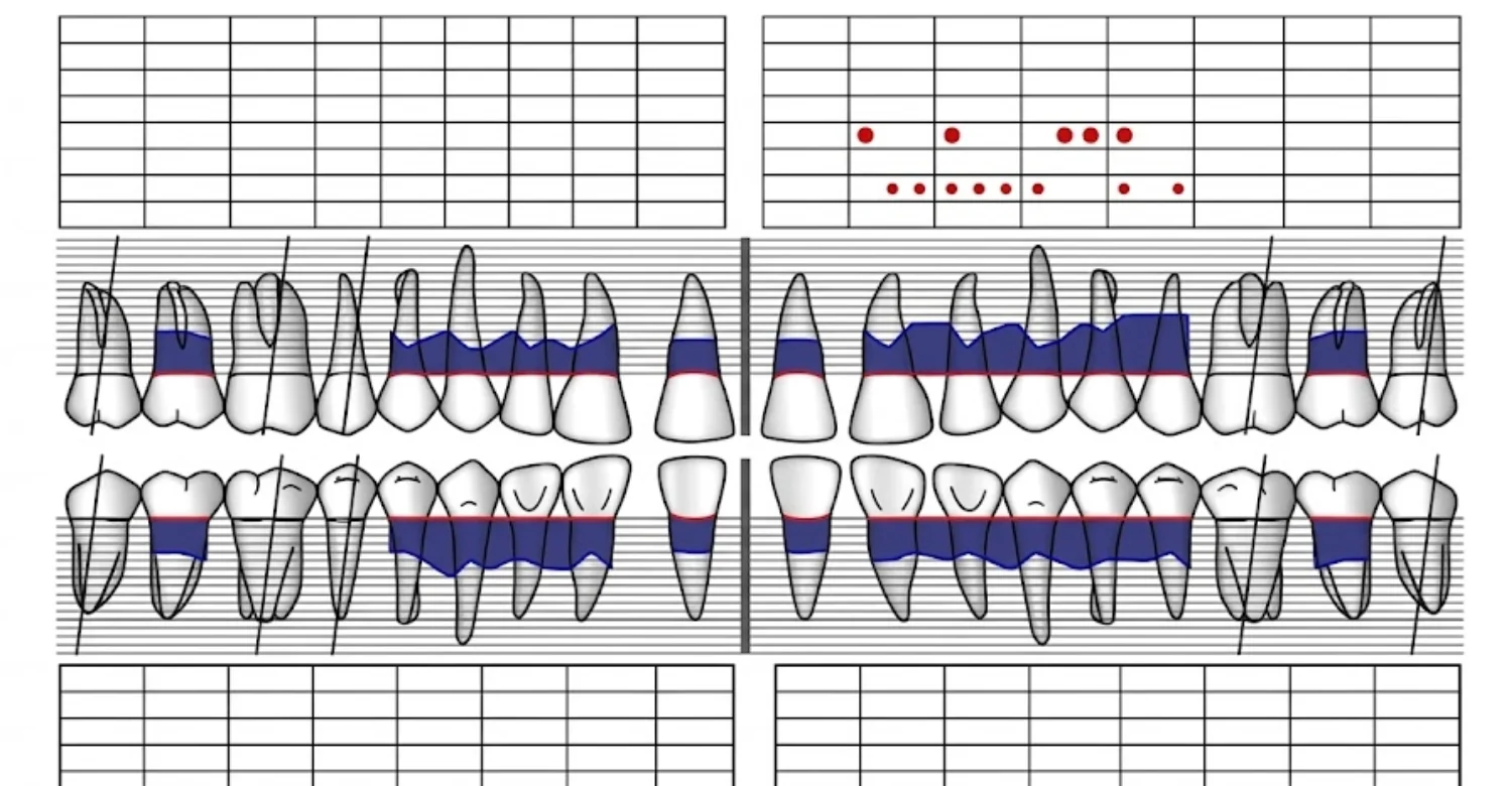
A periodontal chart converts clinical findings into an organized visual or digital record. Numbers typically represent probing depths or other measurements. Symbols or letters may identify bleeding, recession, mobility, furcation, or missing teeth. The exact layout varies between dental software systems and paper forms. Some charts show upper teeth above lower teeth, with facial and inner surfaces in separate rows. Colors are often used to highlight bleeding or deeper sites. Understanding the chart requires knowing which measurement each number represents. Dental and Periodontal Charting becomes much easier to follow once the legend is clear. Patients can always ask their dentist to walk through the chart together. A short explanation often makes the numbers far less intimidating.
Understanding the Numbers on a Periodontal Chart
Numbers commonly represent probing depth in millimeters. Other numbers may represent recession, attachment measurements, mobility, or furcation grades. The legend on each individual chart should always be checked. A single number should not be interpreted without knowing its measurement type and location. Rows are usually grouped by tooth and by surface.
How Tooth Numbers Are Organized
Dental records may use different tooth numbering systems. Common systems include the Universal system, the FDI two-digit system, and Palmer notation. The chart should clearly identify which system is being used. This prevents confusion when discussing specific teeth, especially between different clinics. International patients may notice different systems in records from different countries.
How Six-Point Periodontal Charting Works
Six-point charting records periodontal measurements at six locations around each tooth. These include three facial locations and three lingual or palatal locations. This pattern helps detect localized problems that a single reading could miss. Dental and Periodontal Charting commonly relies on this six-site approach for comprehensive records.
Mesiobuccal and Midbuccal Measurements
Mesiobuccal measurements are taken toward the front side of the facial surface. Midbuccal measurements represent the central facial surface. Both help identify localized periodontal changes on the outer side of the tooth.
Distobuccal Measurements
Distobuccal measurements are taken toward the back of the facial tooth surface. This site can reveal localized pocketing that central measurements might not show. Areas between teeth often show changes earlier than central surfaces.
Mesiolingual and Midlingual Measurements
These measurements assess the inner surface of mandibular teeth. For maxillary teeth, the corresponding surface is usually called palatal. They help complete the picture around each tooth. Plaque often collects on these inner surfaces, so they deserve careful attention.
Distolingual Measurements
Distolingual measurements assess the posterior inner surface of the tooth. Together, the six sites provide broad periodontal coverage for every tooth. Back teeth are harder to reach, so these sites can show deeper readings. Careful probing here helps avoid missing hidden problems.
How to Identify Bleeding Sites
Bleeding sites are commonly marked separately on periodontal charts. Some digital systems use colors, dots, or symbols. Bleeding indicates that bleeding occurred after probing at that site. It should be interpreted with other findings rather than treated as a standalone diagnosis. A higher number of bleeding sites usually suggests more widespread inflammation.
How to Recognize Gingival Recession
Recession measurements show how far the gingival margin has moved from its expected position. Positive or negative charting conventions can vary between systems. The clinician should explain the specific convention used in your chart. Recession is often recorded in a separate row beneath the probing depths. It is added to probing depth when attachment level is calculated.
How to Understand Clinical Attachment Level
Clinical attachment level relates periodontal attachment to the cementoenamel junction. It helps separate deeper probing from gingival enlargement versus true attachment loss. CAL therefore provides important diagnostic context within the chart. Some charts calculate this value automatically from depth and recession readings. Others require the clinician to record it directly.
What Abnormal Patterns on a Periodontal Chart May Suggest
Patterns of deep probing, bleeding, recession, mobility, or attachment loss may suggest periodontal problems. Localized findings may have different causes from generalized findings. A dentist must interpret the entire chart before determining the appropriate diagnosis. Patterns affecting specific teeth, such as molars or incisors, can also guide further assessment. Comparing patterns across visits shows whether disease is spreading or settling
Discover more about polyarteritis nodosa and oral health
.
Can Periodontal Charting Diagnose Gum Disease?
Charting is an important component of periodontal diagnosis. However, it does not independently establish every periodontal diagnosis. The ADA notes that periodontitis diagnosis considers clinical inflammation, attachment loss, and radiographic bone loss. This means periodontal charting works alongside clinical examination and imaging. Dental and Periodontal Charting supplies objective numbers, while the examination supplies clinical context. Radiographs add information about bone that probing cannot directly show. Medical and dental history can explain why certain findings appear. Together, these sources allow the dentist to reach a reliable diagnosis. A chart without that wider assessment would tell only part of the story. For this reason, patients should view charting as one important step, not the final answer.
Dental Charting for Gum Disease
Dental charting can document findings associated with gum disease. These may include tooth mobility, restorations, missing teeth, and periodontal measurements. A combined record helps clinicians understand each tooth's overall condition. It can also show how restorations and gum health affect each other.
Periodontal Charting and Gum Disease Diagnosis
Periodontal charting contributes objective measurements to the diagnostic process. Probing depth, attachment loss, bleeding, and recession can highlight areas needing further assessment. Dental and Periodontal Charting keeps these findings organized for accurate review.
Periodontal Charting for Periodontal Disease
For suspected periodontitis, full-mouth charting can show the distribution and severity of findings. The AAP and EFP classification framework uses attachment loss and bone loss within its staging approach. This helps clinicians describe disease consistently and plan care accordingly.
How Dentists Distinguish Gingivitis From Periodontitis
Gingivitis mainly involves inflammation without the attachment and bone destruction of periodontitis. Periodontitis involves periodontal attachment loss and supporting bone loss. The CDC describes gingivitis as generally reversible. Periodontitis, by contrast, involves bone loss and requires professional management.
Why Pocket Depth Alone Does Not Establish a Complete Diagnosis
Pocket depth measures the distance from the gingival margin to the pocket base. Gingival enlargement can increase this distance without corresponding attachment loss. Recession can produce attachment loss without extremely deep probing. Therefore, clinicians consider multiple measurements together.
How Clinical Attachment Loss and Bone Loss Affect Diagnosis
Clinical attachment loss indicates lost periodontal support relative to the cementoenamel junction. Radiographic bone loss provides additional information about supporting bone. The ADA states that periodontitis classification considers attachment and radiographic bone loss. Together, these findings help describe how far disease has progressed. They also guide staging within modern classification systems.
How Medical and Dental History Influences Periodontal Diagnosis
Dental history can reveal previous periodontal treatment, tooth loss, restorations, or earlier measurements. Medical history can also provide clinically relevant context. The diagnosis should consider the complete history rather than isolated measurements. For example, smoking and some general health conditions can influence periodontal risk. Sharing accurate history helps the dentist interpret the chart correctly.
Learn about HIV symptoms and oral health
What Is the Difference Between a Deep Pocket and Gingival Inflammation?
A deep probing measurement does not always mean attachment destruction. Gingival swelling can increase the distance from the gum margin to the pocket base. This can create what clinicians call a pseudopocket. True periodontal pockets, by contrast, are associated with periodontal attachment changes. The difference matters because each situation may need a different management approach. Swelling from gingivitis often improves with better plaque control and professional cleaning. True pockets may need more structured periodontal therapy and closer monitoring. Dental and Periodontal Charting helps separate these situations by recording depth, recession, and attachment together. The dentist then reads these values alongside the visual appearance of the gums.
Pseudopockets and True Periodontal Pockets
A pseudopocket occurs when gingival enlargement increases probing depth without apical migration of attachment. A true periodontal pocket involves pathological changes to periodontal attachment. Distinguishing these conditions requires careful clinical assessment. Attachment level and recession readings are especially helpful in making this distinction. Radiographs may add further information when bone loss is suspected.
How Gingival Swelling Can Increase Probing Depth
Inflamed or enlarged gums can extend farther over the tooth surface. The probe then travels a greater distance before reaching the sulcus base. This can produce a deeper reading without equivalent attachment loss. Once inflammation settles, the same site may measure noticeably shallower. This is one reason reassessment after cleaning is so useful.
How Gingival Recession Can Affect Measurements
Recession moves the gingival margin toward the root. A patient can therefore have significant attachment loss without an exceptionally deep probing depth. Recession measurements help place probing depth into context. Without recording recession, the true loss of support could be underestimated.
Why Clinical Attachment Level Provides Additional Context
Clinical attachment level accounts for the position of the gingival margin. It helps clinicians understand whether probing depth reflects attachment loss or gingival enlargement. This makes CAL a key value in every comprehensive chart.
Clinical Note: Why Pocket Measurements Should Be Interpreted With Other Findings
Pocket measurements should be interpreted alongside bleeding, recession, attachment level, mobility, and radiographic findings. This approach reduces the risk of drawing conclusions from one measurement. Dental and Periodontal Charting is most useful when every recorded value is read in context.
Explore the science behind ferroptosis in periodontitis
What We Notice Clinically
Clinicians often see periodontal findings that vary considerably between teeth and sites. A patient may have healthy areas alongside isolated deeper pockets. Another patient may show mild inflammation spread evenly across the mouth. This variation makes site-specific charting valuable in everyday practice. Dental and Periodontal Charting captures these differences instead of averaging them away. It allows the dental team to focus attention exactly where it is needed. It also helps explain findings to patients using their own measurements. Seeing specific numbers often motivates patients more than general advice does. Over time, repeated charts reveal whether care is working as planned.
Common Patterns Dentists See During Periodontal Charting
Common patterns include localized bleeding, generalized inflammation, recession, deeper pockets, and isolated furcation involvement. Patterns help clinicians decide whether findings appear localized or generalized. This distinction can shape both diagnosis and treatment.
Why One Isolated Measurement Does Not Tell the Whole Story
One measurement represents one site at one point in time. Its meaning depends on surrounding measurements and clinical findings. Repeated measurements can provide stronger evidence of real change. Small differences can also reflect normal variation in probing technique. Trends across several visits are usually more reliable than single readings.
Why Bleeding on Probing Matters
Bleeding can indicate inflammation at the examined periodontal site. Persistent bleeding combined with increasing pocket depth may signal greater concern. Reduced bleeding over time often suggests improving gum health. It is also one of the easiest findings for patients to notice at home.
Why Recession and Pocket Depth Should Be Interpreted Together
Recession can make attachment loss look different from probing depth. Recording both measurements gives the clinician a more complete picture of periodontal support. A shallow pocket with marked recession may hide significant attachment loss. Reading both values together prevents this kind of misunderstanding.
Common Patient Misconceptions About Periodontal Charting
Patients sometimes assume every deep number means severe gum disease. Others believe bleeding automatically confirms periodontitis. Both assumptions are incomplete. Diagnosis requires multiple clinical findings interpreted together by a dental professional.
Dr. Rifat Alsaman’s Clinical Opinion on Periodontal Charting
Dr. Rifat Alsaman is Head of the Medical Team at Vitrin Clinic and a cosmetic dentist. Dr. Alsaman considers periodontal measurements an important part of comprehensive dental assessment. The clinical value comes from interpreting measurements alongside tooth condition, gum appearance, bite, and diagnostics. In Dr. Alsaman's view, charting highlights areas needing attention before restorative or implant treatment.
Find out more about sialadenitis and salivary gland inflammation
What Happens After Periodontal Charting?
After charting, the dentist reviews the measurements and other clinical findings. The next step depends on the patient's periodontal status. Some patients may need only routine preventive care and regular check-ups. Others may require periodontal treatment or evaluation by a specialist. The dentist usually explains the findings and discusses the options available. Dental and Periodontal Charting results also become the baseline for future comparisons. Keeping this baseline accurate helps measure the effect of any treatment. Patients can ask for a copy or explanation of their chart. Understanding the results helps patients take an active role in their care.
How Dentists Use Charting Results to Plan Treatment
Charting identifies sites requiring monitoring, professional cleaning, periodontal therapy, or further evaluation. Treatment should reflect the patient's overall findings rather than single numbers. The results also help decide how often a patient should return for review. Clear records make it easier to explain each recommendation to the patient.
When Additional Dental X-Rays May Be Needed
Radiographs may be recommended when bone levels or other structures require assessment. X-rays are not automatically necessary at every charting appointment. Clinical indication should guide imaging decisions.
When Periodontal Treatment May Be Recommended
Treatment may be considered when periodontal inflammation or tissue destruction is identified. Options depend on disease severity, distribution, risk factors, and patient needs. The dentist should explain the goals, expected stages, and likely follow-up of any treatment.
When a Patient May Be Referred to a Periodontist
Referral may suit complex periodontal disease, persistent deep sites, advanced bone loss, or significant recession. Surgical needs may also prompt referral. The decision depends on clinical findings.
How Periodontal Charting Supports Treatment Monitoring
Repeat charting allows clinicians to compare new measurements with baseline findings. This helps assess whether periodontal conditions are stable or changing. Stable readings suggest that current care is working well. Worsening readings may prompt a change in treatment or maintenance intervals.
Periodontal Charting Before Dental Treatment
Periodontal assessment can be particularly important before complex restorative or implant treatment. Healthy supporting tissues provide a stronger foundation for comprehensive treatment planning.
Discover the possible causes of loss of taste
Periodontal Charting and Gum Disease Treatment
Charting provides measurable information that supports periodontal treatment planning. The treatment approach depends on the diagnosis, severity, and distribution of disease. Mild inflammation may respond to professional cleaning and improved home care. More advanced disease may need nonsurgical periodontal therapy or, occasionally, surgery. Dental and Periodontal Charting shows which sites need attention and which appear stable. It also sets clear starting values against which healing can be measured. After treatment, repeat charting shows how each site has responded. This cycle of measuring, treating, and remeasuring supports long-term periodontal stability. Patient home care remains essential throughout every stage of treatment.
How Charting Can Guide Periodontal Treatment
Measurements help identify areas requiring professional intervention. They also show which sites need closer monitoring after treatment. This helps focus professional time on the areas that need it most. It also avoids unnecessary treatment in healthy sites.
When Scaling and Root Planing May Be Considered
Scaling and root planing may be considered for sites with calculus, inflammation, and periodontal pocketing. The decision should follow careful clinical assessment. The ADA describes nonsurgical periodontal therapy as professional removal of plaque, biofilm, and calculus. Appropriate patient home care supports this treatment.
How Charting Can Help Monitor Treatment Response
Post-treatment measurements can be compared with baseline findings. Changes in pocket depth, bleeding, and other parameters help assess response. Reduced bleeding and shallower pockets often suggest a favorable response. Sites that do not improve may need further evaluation.
When Surgical Periodontal Treatment May Be Considered
Surgery may be considered when problems persist despite appropriate nonsurgical management. The exact procedure depends on anatomy and disease characteristics. A periodontist usually assesses whether surgery is appropriate. Charting results help support that decision.
Why Periodontal Reassessment Matters
Reassessment helps determine whether treatment achieved periodontal stability. It also identifies sites that may need additional care. Dental and Periodontal Charting at reassessment provides the evidence for these decisions.
Note : All images used are for editorial and illustrative purposes only and may not originate from the original news provider or associated company.
Learn what can cause a yellow tongue
How Often Should Periodontal Charting Be Done?
There is no single interval appropriate for every patient. Frequency depends on periodontal history, disease status, risk factors, treatment response, and clinical findings. Patients with healthy gums may need less detailed monitoring than those treated for periodontitis. People with a history of disease often benefit from more regular review. Changes in health, medication, or habits can also influence the interval. Dental and Periodontal Charting should follow a schedule the dentist tailors to each patient. Regular records make it easier to detect small changes early. Patients can ask their dentist when their next full chart is due.
Periodontal Charting for Patients With Healthy Gums
Healthy patients may receive periodontal assessment during routine dental examinations. The exact frequency depends on individual risk and clinical findings. Basic screening can show whether a more detailed chart is needed. Healthy baseline records also help detect future changes early.
Periodontal Charting for Patients With Gingivitis
Patients with gingivitis may need monitoring alongside professional preventive care. The dentist decides whether comprehensive charting is appropriate. Improved home care often helps gingivitis settle. Follow-up measurements can confirm whether bleeding has reduced.
Periodontal Charting for Patients With Periodontitis
Patients with periodontitis generally require structured periodontal monitoring. Guidance recommends six-point measurements during review charting. These records help track stability during long-term maintenance. They also show early signs of recurrence.
How Previous Periodontal Disease Changes Monitoring
Previous periodontitis increases the importance of long-term periodontal monitoring. Historical charts provide valuable comparison points. Patients treated in the past can remain at higher risk of recurrence. Regular review helps catch any return of disease early.
Why Charting Frequency Depends on Individual Findings
Periodontal disease does not progress identically in every patient. Monitoring should therefore reflect clinical risk and previous findings. Smoking, diabetes, and previous disease may justify closer follow-up. Low-risk patients may need less frequent detailed charting.
Explore salivary gland stones and their symptoms
Periodontal Charting Before Dental Treatment
Periodontal health can influence restorative and implant treatment planning. A comprehensive dental plan should consider the condition of supporting tissues. Crowns, veneers, bridges, and implants all depend on stable gums and bone. Untreated inflammation can affect impressions, margins, and long-term results. Dental and Periodontal Charting before treatment helps identify these risks early. Where needed, periodontal care can be completed before restorative work begins. This sequencing can support more predictable and durable outcomes. It also helps patients understand why certain steps come first.
Why Gum Health Should Be Assessed Before Certain Dental Procedures
Inflamed or unstable periodontal tissues can affect treatment planning. Assessment helps clinicians identify issues before extensive procedures begin. Treating inflammation first can create more stable conditions for later dental work.
Periodontal Charting Before Crowns and Veneers
Gum health can influence the appearance and stability of restorative margins. Periodontal assessment may be useful before extensive crown or veneer treatment. Stable gum margins can help restorations look natural over the long term.
Periodontal Assessment Before Dental Implants
Implant planning requires evaluation of the supporting oral environment. Periodontal disease should be identified and managed appropriately before implant placement. A history of periodontitis may influence implant risk and maintenance needs. Careful assessment supports safer planning.
Periodontal Charting Before Full-Mouth Rehabilitation
Full-mouth rehabilitation involves multiple teeth and treatment stages. Periodontal assessment helps establish whether supporting tissues suit the planned treatment. It can also help decide which teeth have a reliable long-term outlook.
How Gum Health Can Influence a Personalized Treatment Plan
Gum condition may influence treatment sequencing, restorative design, hygiene planning, and follow-up. This supports a more individualized approach. Plans built on healthy foundations tend to be easier to maintain.
Dr. Rifat Alsaman’s Opinion on Periodontal Assessment Before Dental Treatment
Dr. Rifat Alsaman emphasizes reviewing periodontal findings before complex dental treatment. At Vitrin Clinic, clinical findings help guide personalized treatment sequencing. Dental and Periodontal Charting is especially relevant when restorative or implant procedures are planned.
Discover how the salivary glands work
Tips for Patients
Good periodontal care begins with consistent home care and professional monitoring. Patients should also understand their own periodontal measurements. Knowing what the numbers mean makes dental visits more productive. It also helps patients notice when something changes between appointments. Dental and Periodontal Charting works best when patients and clinicians share information openly. The following tips can help you prepare and stay involved in your care.
How to Prepare for a Periodontal Examination
Continue your normal oral hygiene before the appointment. Bring previous dental records when available. Tell your dentist about recent changes in gum bleeding, sensitivity, or tooth mobility.
Tell Your Dentist About Gum Bleeding, Swelling, or Sensitivity
Bleeding, swelling, tenderness, and sensitivity provide useful clinical information. Reporting changes helps the dentist decide whether further assessment is needed.
Maintain Regular Brushing and Interdental Cleaning
Daily plaque control supports periodontal health. The CDC recommends brushing twice daily and flossing daily as part of oral hygiene.
Bring Previous Dental and Periodontal Records When Available
Previous charts help clinicians compare current findings with earlier measurements. This can make changes easier to identify. Radiographs and treatment summaries from other clinics can also be helpful.
Ask Your Dentist What Your Measurements Mean
Ask which numbers represent probing depth, recession, attachment, or bleeding. Understanding the chart makes treatment discussions clearer. You can also ask which sites need the most attention at home.
Do Not Diagnose Gum Disease From Pocket Depth Alone
A single pocket measurement cannot establish the complete periodontal diagnosis. Attachment, bleeding, recession, bone levels, and other findings may be necessary.
Medical Tips for Maintaining Periodontal Health
Avoid tobacco, maintain daily plaque control, and attend recommended dental appointments. Tell your dental team about relevant medical history. WHO identifies tobacco use and poor oral hygiene among important periodontal risk factors.
Find out what can cause a white tongue
Periodontal Charting Relief, Comfort, and Possible Side Effects
Periodontal probing is generally a brief clinical procedure. Some patients experience temporary sensitivity afterward, especially when gums are inflamed. Most discomfort settles quickly without any special treatment. Clinicians can adjust their technique for patients with sensitive gums. Letting the team know about anxiety or sensitivity beforehand can help. Dental and Periodontal Charting should feel manageable for most patients. The sections below explain what to expect and when to seek advice.
What Can Patients Expect During and After Probing?
You may feel gentle pressure during probing. Some areas can feel more sensitive than others. Mild temporary tenderness can occur afterward. The clinician can pause if any area feels uncomfortable.
Can the Gums Feel Sensitive After Periodontal Probing?
Yes, temporary sensitivity can occur after probing. It may be more noticeable in inflamed or recessed areas. Sensitivity usually fades within a short time without special care.
How to Reduce Temporary Gum Discomfort
Continue gentle oral hygiene unless your dentist advises otherwise. Avoid aggressive brushing around sensitive areas. Your dentist can recommend suitable measures when sensitivity persists.
When Discomfort After Periodontal Examination Should Be Reported
Contact your dental professional if pain becomes severe, persistent, or progressively worse. Significant swelling or other unexpected symptoms should also be reported.
Possible Side Effects of Periodontal Probing
Possible short-term effects include mild tenderness or minor bleeding. These effects are generally temporary. Your dentist can explain what to expect after your examination.
When Should You See a Dentist About Your Gum Health?
Early assessment can help identify periodontal problems before they become more advanced. WHO estimates severe periodontal disease affects about 19% of people older than 15 years globally. Many people notice only mild signs, or none at all, in the early stages. Regular examinations are therefore important even when your mouth feels comfortable. Dental and Periodontal Charting can detect changes that are not yet visible or painful. The warning signs below should prompt a dental visit rather than self-diagnosis. Acting early usually means simpler care and better long-term outcomes.
Persistent Gum Bleeding
Repeated bleeding during brushing or professional probing deserves dental assessment. Bleeding can indicate gingival inflammation. Bleeding that returns often should not be ignored, even without pain.
Swollen, Red, or Tender Gums
Swelling, redness, and tenderness may indicate gingival inflammation. A dental examination can identify possible causes. Early treatment can often help inflamed gums recover more quickly.
Persistent Bad Breath or Bad Taste
Persistent bad breath or an unpleasant taste can accompany periodontal problems. These symptoms should be assessed rather than self-diagnosed.
Loose or Shifting Teeth
Tooth mobility can occur for several reasons. Periodontal support loss is one possible cause. A dentist should evaluate persistent or increasing mobility.
Gum Recession or Increasing Tooth Sensitivity
Recession can expose tooth roots and increase sensitivity. It may also affect clinical attachment measurements. Your dentist can suggest ways to protect exposed root surfaces.
Deep or Increasing Periodontal Pocket Measurements
Increasing pocket depths may indicate changing periodontal conditions. Repeat charting helps determine whether changes are clinically meaningful.
When Urgent Dental Evaluation May Be Needed
Rapid swelling, severe pain, significant bleeding, fever, or difficulty swallowing requires prompt professional evaluation. These symptoms may indicate a spreading infection. Seek care promptly rather than waiting for a routine appointment.
How Much Does Dental and Periodontal Charting Cost?
There is no universal price for Dental and Periodontal Charting. Costs vary by country, clinic, clinician, examination complexity, and imaging. Price also depends on whether charting is part of a broader periodontal consultation. In the UK, published 2026 private examples show full periodontal assessments commonly costing about £85 to £160. Some specialist assessments cost more when radiographs and additional diagnostics are included. Patients should compare what each quote includes, not just the headline price. The sections below outline typical ranges and the factors behind them.
Average Periodontal Charting Cost in the United States
Pricing in the United States varies substantially by provider and insurance arrangement. A comprehensive periodontal evaluation may be billed separately from routine examination or treatment. Published negotiated-rate data should not be confused with a patient's actual out-of-pocket price. Patients should request an itemized estimate before treatment.
Average Periodontal Charting Cost in the United Kingdom
Private UK prices vary by clinic and scope. Current published examples include approximately £85–£160 for full periodontal assessment or charting. Specialist consultations can cost more when imaging and treatment planning are included.
Average Periodontal Charting Cost in Turkey
Turkey has no single nationally fixed private price for periodontal charting. Published 2026 Turkish clinic guides report periodontal examination prices around ₺800–₺2,500. Prices depend on location and scope. These figures are indicative rather than a Vitrin Clinic quotation.
What Is Usually Included in the Cost?
A periodontal assessment may include clinical examination, probing measurements, bleeding assessment, and periodontal charting. Some clinics also include photographs or radiographs. Always confirm what the quoted fee covers.
What Factors Can Affect the Price?
Cost can depend on appointment length, clinician expertise, examination complexity, and imaging. Specialist periodontal assessment may also raise the fee. Location can affect private dental fees as well.
Does the Cost Include Dental X-Rays?
Not necessarily. Some clinics include radiographs within periodontal assessments, while others charge separately. For example, some UK specialist assessments explicitly include X-rays. Other fee schedules list radiographs as separate items.
Charting Cost vs. Periodontal Treatment Cost
Dental and Periodontal Charting is an assessment procedure, not periodontal treatment. Treatment can include professional cleaning, nonsurgical periodontal therapy, maintenance, or surgery. Treatment costs therefore differ substantially from charting fees.
Comprehensive Examination of Teeth, Gums, and Oral Tissues
Clinical examination helps identify visible periodontal and dental findings. The dentist assesses gum appearance, tooth condition, bite, and oral structures. These findings form the starting point for any recommended treatment.
Digital Dental Diagnostics When Clinically Appropriate
Digital diagnostics may support treatment planning when clinically indicated. Technology is selected according to each patient's clinical needs.
Digital X-Rays and CBCT When Indicated
Digital X-rays or CBCT may be recommended when additional structural information is clinically necessary. Imaging is not automatically required for every periodontal examination.
3D Intraoral Scanning When Clinically Appropriate
Three-dimensional intraoral scanning can support digital dental workflows. Its use depends on the planned treatment and clinical requirements.
Personalized Treatment Planning Based on Clinical Findings
Vitrin Clinic develops treatment plans according to clinical findings and individual goals. Periodontal considerations can influence treatment sequencing.
Periodontal Assessment Before Restorative or Implant Treatment
Gum and supporting-tissue health can matter before crowns, veneers, implants, and extensive rehabilitation. Assessment helps identify concerns before treatment begins.
How Vitrin Clinic Supports Your Treatment Journey
Vitrin Clinic combines clinical assessment with coordinated digital treatment planning. This approach helps international patients understand their proposed treatment pathway. Each stage is explained before it begins, so patients know what to expect. Periodontal findings are considered alongside restorative and cosmetic goals. The team also plans appointments around travel and follow-up needs. The steps below outline how this support typically works.
Initial Dental Consultation and Clinical Assessment
The initial consultation reviews the patient's dental concerns and clinical findings. Additional assessment is performed when indicated. Patients can share their goals, concerns, and previous treatment experiences at this stage.
Reviewing Existing Dental and Periodontal Records
Previous records provide useful information about treatment history. They may also help clinicians compare historical periodontal findings.
Coordinating Digital Diagnostics With Treatment Planning
Relevant digital diagnostics can be incorporated into treatment planning. The exact technologies depend on the patient's clinical needs.
Personalized Treatment Sequencing
Complex dental treatment may require several stages. Sequencing follows clinical priorities and treatment requirements. Periodontal care may come first when gum health needs attention.
Explaining Treatment Options and Expected Stages
Patients can discuss proposed treatment stages with the dental team. The final plan depends on examination findings. Questions are encouraged so patients can make informed decisions.
Follow-Up and Aftercare Planning
Aftercare supports continued monitoring following treatment. The follow-up schedule depends on the procedure and clinical findings.
Support for International Dental Patients
Vitrin Clinic provides coordinated support for international patients throughout treatment. Planning considers appointments, clinical requirements, and follow-up needs.
Key Takeaways About Dental and Periodontal Charting
Dental and Periodontal Charting provides structured information about teeth, gums, and supporting tissues. It documents probing depth, bleeding, recession, attachment, mobility, and other findings. The record also creates a baseline for future comparison. Its value grows when measurements are repeated and compared over time. The points below summarize what patients should remember.
What Dental and Periodontal Charting Tells Your Dentist
Charting provides site-specific clinical measurements. These help dentists understand periodontal conditions and establish a reliable baseline. They also highlight which sites need closer attention.
Why Periodontal Pocket Measurements Matter
Pocket measurements show the depth between the gingival margin and the sulcus or pocket base. Changes help identify sites requiring further evaluation.
Why Charting Should Be Interpreted With Other Diagnostic Findings
Pocket depth alone cannot establish every periodontal diagnosis. Attachment, bleeding, recession, bone levels, and history provide additional context.
When Periodontal Charting May Lead to Further Evaluation or Treatment
Abnormal patterns may lead to more examination, radiographs, periodontal therapy, or specialist referral. The next step depends on the overall clinical picture.
When to Seek Professional Dental Care for Gum Problems
Seek dental evaluation for persistent bleeding, swelling, recession, mobility, sensitivity, or worsening findings. Early professional assessment can help identify treatable periodontal problems.
At Vitrin Clinic
At Vitrin Clinic, periodontal assessment forms part of comprehensive dental evaluation when clinically appropriate. The team evaluates teeth, gums, oral tissues, bite, and relevant supporting structures. Findings are recorded carefully so they can guide treatment decisions. Dental and Periodontal Charting may be included when the clinical situation calls for it. Digital tools are used where they add genuine diagnostic value. The aim is a clear, personalized plan based on each patient's findings.
Reference
FAQs
Dr. Rifat Alsaman has more than 5 years of clinical experience in dentistry and currently serves as the Head of the Medical Team at Vitrin Clinic. He is dedicated to providing exceptional patient care, overseeing treatment planning, and ensuring the highest clinical standards across the team. His expertise, attention to detail, and commitment to continuous professional development have helped countless patients achieve healthier, more confident smiles.


