
An oral mucocele is a fluid-filled swelling linked to a salivary gland or its duct. It commonly develops after trauma damages a small salivary duct, and the leaked mucus collects beneath the oral lining. Most lesions are benign and painless, and they often appear on the lower lip. Some develop inside the cheek or beneath the tongue instead. The appearance can vary with depth and location. Superficial lesions may look bluish or translucent, while deeper lesions can resemble normal pink oral tissue. Many resolve without invasive treatment. Persistent or repeatedly returning lesions may require professional evaluation. A large Brazilian study reviewed 1,901 cases and found the lower lip represented 67.4% of affected sites, while the floor of the mouth accounted for 10%.
What Is an Oral Mucocele?
An oral mucocele is a benign, mucus-filled swelling that forms when a small salivary duct is damaged or blocked. It is not an infection and not a form of cancer. Most cases are painless and many resolve on their own, though some need dental treatment to prevent recurrence.
Oral Mucocele Definition
This is a localized collection of mucus within oral tissues. It usually develops after disruption of a minor salivary gland duct, and trauma can allow mucus-like saliva to escape into surrounding tissue. Less commonly, duct blockage causes mucus retention instead. These lesions are generally benign and appear as soft, smooth, fluctuant swellings. Many are painless during daily activities, and color can range from normal pink to translucent blue. Small lesions may disappear after spontaneous rupture, and the leaked contents can then be reabsorbed. However, the lesion may return if the underlying duct remains affected. Persistent lesions need assessment by a dentist or oral healthcare professional.
What Does an Oral Mucocele Look Like?
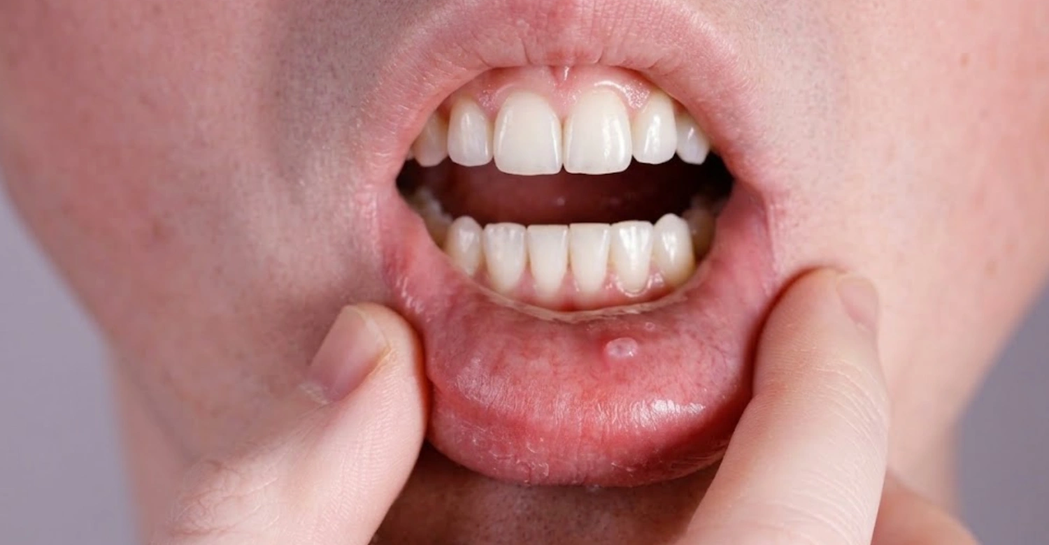
The appearance depends on depth, size, and location. A superficial lesion may look like a small, rounded, bluish bubble, since mucus sits close to the surface. Deeper lesions can share the color of surrounding oral tissue and instead look like a smooth, soft lump. Most are not painful when gently examined. Clinical studies describe these lesions as dome-shaped, fluctuant, and usually non-tender, with reported sizes ranging from very small up to roughly four centimeters. A changing oral lump should not automatically be assumed to be this condition, since other cysts, tumors, vascular lesions, and inflammatory conditions can look similar. Professional examination can establish the correct diagnosis.
Where Do Oral Mucoceles Usually Develop?
These lesions can develop wherever minor salivary glands are present, though certain locations are far more common. The lower lip is the classic site, and the inner cheek, floor of the mouth, and tongue can also be affected. A large multicenter study found the lower lip involved in 67.4% of cases, while the floor of the mouth accounted for 10%. Location can offer an important diagnostic clue, but location alone cannot confirm the diagnosis.
Oral Mucocele on the Lower Lip
The lower lip is the most frequently affected location. Repeated biting or accidental trauma can damage minor salivary gland ducts, allowing mucus to escape beneath the mucosal surface. The swelling may look smooth, rounded, and slightly bluish. Some lesions stay small and unnoticed, while others become obvious while eating or speaking. A Brazilian multicenter study of 1,901 cases found that 67.4% occurred on the lower lip. Recurrent lip trauma can contribute to repeated lesions, so avoiding biting and irritation may help reduce further injury.
Oral Mucocele Inside the Cheek
The inner cheek contains minor salivary glands, so trauma here can also produce a mucus-filled swelling. Cheek lesions may follow accidental biting or occur when teeth repeatedly rub against the mucosa. These lesions may keep a normal pink color, though superficial ones can look translucent or bluish. The cheek is affected less often than the lower lip, but published studies confirm the buccal mucosa can be involved. Persistent cheek swellings deserve professional assessment, since several oral conditions can look similar.
Oral Mucocele Under the Tongue
A swelling beneath the tongue needs careful evaluation, since lesions here can involve different glands and structures. A lesion from the sublingual region may represent a ranula, which is related to major salivary glands and differs from a typical minor-gland lesion. Larger swellings can interfere with speech or swallowing, and very large ones can occasionally affect breathing. A persistent swelling under the tongue should therefore be assessed professionally, since this location makes accurate differentiation especially important.
Mucocele Lip: Why Does It Commonly Affect the Lower Lip?
A mucocele lip is most commonly associated with the lower lip because this area contains numerous minor salivary glands and is frequently exposed to accidental biting or other trauma. Damage to a small salivary duct can allow mucus to collect beneath the oral lining, creating a soft, rounded swelling that may appear translucent or slightly bluish. Most lesions are painless, but persistent or recurrent swelling should be assessed by a dental professional.
Clinical Note
Clinical examination usually provides the initial basis for diagnosis. Imaging is not routinely necessary for straightforward lesions, though it can help when a lesion is unusual or extensive. A dentist considers location, consistency, color, duration, and history, and previous trauma offers an additional clue. However, trauma is not always reported, since some patients develop lesions without remembering an injury. At Vitrin Clinic, persistent oral swellings are assessed according to their clinical appearance and history, and the right diagnostic path depends on individual findings.
Discover why chewing pens can be bad for your teeth
What Causes an Oral Mucocele?
An oral mucocele usually develops when a minor salivary gland or its duct is damaged or blocked, causing mucus to collect in the surrounding tissue. Accidental lip or cheek biting is a common trigger, while other forms of trauma or irritation may also contribute. Some mucoceles develop when the salivary duct becomes obstructed rather than directly injured. They commonly occur on the lower lip but can appear elsewhere in the mouth. Repeated trauma or habits such as biting the lips may increase the risk of recurrence. A dental examination can help confirm the cause and distinguish the lesion from other types of oral swelling.
Salivary Duct Injury or Blockage
The most common mechanism involves disruption of a salivary duct. Trauma can damage the duct and let mucus escape into surrounding tissue, a process commonly called mucus extravasation. Another mechanism involves obstruction of the duct, which lets mucus accumulate within the ductal system; this type is generally called a retention phenomenon. Possible causes of obstruction include scarring and other local structural changes, and less commonly other lesions can contribute. Understanding the mechanism helps guide management. A small lesion may resolve naturally, while a persistent one may need removal of the affected tissue.
Lip Biting and Other Oral Trauma
Repeated trauma is an important contributor to these lesions. Lip biting matters most because the lower lip contains many minor salivary glands. Accidental biting can injure a small duct, and repeated irritation may prevent normal healing. Other trauma can also contribute, including sharp dental edges, oral habits, or previous procedures. Not every patient remembers a traumatic event, so absence of reported trauma does not rule out the diagnosis. Reducing repeated irritation can help protect the tissue, and professional evaluation remains important when swelling persists.
Why Can Oral Mucoceles Develop Repeatedly?
Recurrence can occur when the affected salivary gland or duct remains involved. Simply releasing the accumulated mucus may not correct the underlying source. This explains why aspiration alone is generally linked with higher recurrence. Definitive treatment may involve removing the lesion and its associated minor salivary gland. A 2022 multicenter study reported recurrence in 6.2% of diagnosed cases. Recurrence can also reflect continued trauma, since repeated biting may keep damaging nearby salivary ducts. Treatment decisions should therefore weigh both the lesion itself and any contributing habits.
Are Some People More Likely to Develop an Oral Muscle?
These lesions can occur at many ages, but they are frequently reported among younger patients. A large Brazilian study found a mean patient age of 19.8 years, with a reported range from one to 87 years. Children and adolescents may face increased exposure to accidental lip biting, and sports injuries and oral habits can also contribute. However, age alone does not establish risk, since adults can develop these lesions too. A persistent swelling should be evaluated regardless of age, because clinical characteristics matter more than age when choosing a diagnostic approach.
Explore the relationship between tick-borne diseases and oral health
What Are the Symptoms of an Oral Mucocele?
Most cases cause a painless, soft swelling that a patient notices while eating, speaking, or brushing. Some feel a small fluid-filled bubble, while others notice a firmer bump if the lesion sits deeper in the tissue. Size and depth largely determine which symptoms appear.
Common Oral Mucocele Symptoms
Most present as painless, localized oral swellings that may feel soft or fluctuant when examined. Some patients notice a small bubble inside the lip, while others discover it accidentally while brushing. Superficial lesions can look bluish or translucent, and deeper lesions may appear pink and resemble surrounding tissue. Larger lesions can occasionally interfere with speaking, chewing, or swallowing, and these symptoms matter more when the lesion sits beneath the tongue. Symptoms alone cannot confirm the diagnosis, since several oral conditions can produce similar lumps.
Small or Tiny Oral Mucoceles
Tiny lesions may be hard to notice and can appear as small bumps within the lip or cheek. Some disappear without treatment after spontaneous rupture, and the mucus is then absorbed by surrounding tissue. A small size does not automatically mean treatment is needed, and observation may be appropriate when the diagnosis is clear and symptoms are minimal. However, persistent growth should prompt professional assessment, since a small lesion that keeps returning may need definitive treatment.
Is an Oral Mucocele Painful?
Most lesions are painless, and patients usually notice swelling rather than pain. Tenderness can develop if the lesion is repeatedly traumatized, and accidental biting may cause irritation or bleeding. Large lesions in sensitive spots can also interfere with normal oral function, and under-tongue lesions may affect swallowing or speech once they grow substantial. Significant pain should not automatically be blamed on this condition, since infection, trauma, ulceration, or another oral issue may explain painful symptoms. Professional assessment is appropriate when pain persists or steadily worsens.
Can an Oral Mucocele Come and Go?
Yes. Some lesions rupture spontaneously and temporarily disappear, and the mucus is then reabsorbed by surrounding tissues. The lesion can return later if the affected duct remains disrupted, which can make the swelling seem intermittent. Clinical references describe courses ranging from several days to several weeks, though rare lesions may persist much longer. Repeated disappearance and reappearance is therefore not necessarily reassuring, since recurrence can indicate that the underlying salivary duct is still affected.
Learn about H5N1 bird flu and oral health
How Long Does an Oral Mucocele Last?
Most small lesions last from a few days to several weeks before resolving on their own. Persistent or recurring lesions can last much longer without proper treatment, especially if the underlying duct problem is never corrected.
Can an Oral Mucocele Go Away on Its Own?
Many small lesions resolve without invasive treatment. Spontaneous rupture can release the accumulated mucus, and surrounding tissue can then reabsorb the contents. Observation can therefore be appropriate for selected small lesions, though the diagnosis should still be reasonably established. A lesion that persists, enlarges, or repeatedly returns deserves professional assessment.
How Long Do Oral Mucoceles Usually Take to Heal?
The clinical course varies between individuals. StatPearls reports durations ranging from a few days to several weeks, and some uncommon lesions can persist much longer. Healing also depends on whether trauma continues, since repeated biting can interfere with normal recovery. After surgical treatment, healing depends on lesion size and the procedure used, and following postoperative instructions can support uncomplicated recovery.
Why Some Mucoceles Keep Coming Back
Repeated recurrence can happen when the underlying salivary gland stays affected. Repeated trauma can also contribute to this pattern. Aspiration may temporarily reduce swelling, but it does not necessarily remove the source, and StatPearls notes that aspiration carries a higher recurrence rate. For persistent lesions, complete removal of the lesion and associated gland may reduce recurrence. Treatment decisions should therefore consider both the lesion and any ongoing habits.
Oral Mucocele Monitoring for Changes or Improvement
Monitoring involves watching size, color, consistency, symptoms, and how often the lesion returns. Patients should avoid repeatedly squeezing or traumatizing it, and photographs can help document changes between appointments. A rapidly changing lesion deserves professional evaluation, and bleeding, persistent ulceration, or unexplained pain also warrants assessment. At Vitrin Clinic, clinicians can evaluate persistent oral lesions and determine whether monitoring or further investigation fits.
Dive deeper into the oral-gut microbiome axis
Are Oral Mucoceles Dangerous or Cancerous?
A typical lesion is benign and is not considered dangerous or cancerous on its own. However, persistent or unusual oral lumps should still be checked by a professional, since a few other conditions can look similar at first glance.
Are Oral Mucoceles Cancerous?
A typical lesion is a benign salivary-related swelling and is not considered oral cancer. However, not every oral lump is this condition, since other benign and malignant conditions can look similar. Persistent or unusual lesions should therefore receive professional evaluation, and a clinician may recommend a biopsy when the diagnosis stays uncertain.
Oral Mucocele vs Cancer
These lesions and oral cancers are different clinical conditions. A typical lesion is usually soft, fluctuant, and painless. Oral malignancies can present in many different ways, and some may cause persistent ulcers, firm masses, bleeding, or unexplained tissue changes. Appearance alone cannot reliably rule out every alternative diagnosis, so persistent lesions require careful clinical judgment. A biopsy can help separate benign lesions from malignant disease when clinically indicated.
Can an Oral Mucocele Cause Cancer?
A typical lesion does not cause oral cancer. It results from salivary duct disruption or obstruction, and there is no established mechanism by which it transforms into cancer. The important distinction is diagnostic, since another lesion may initially resemble this one. Persistent or atypical findings should be investigated rather than assumed to be harmless.
When Does an Oral Bump Need Further Investigation?
An oral bump deserves evaluation when it persists or repeatedly returns, and rapid enlargement also warrants assessment. Additional warning features include unexplained bleeding, ulceration, significant pain, firmness, or major color change. A lesion beneath the tongue deserves particular attention, since several conditions can develop there. Clinical evaluation helps determine whether observation, imaging, or biopsy fits best.
What We Notice Clinically
Clinicians usually assess several features together, including location, size, color, consistency, duration, and recurrence. A classic lower-lip lesion may have a straightforward presentation, while an unusual one may need further investigation. A large multicenter study found that 78% of diagnosed cases received excisional biopsy. This statistic does not mean every patient needs surgery, since treatment selection depends on individual circumstances. At Vitrin Clinic, assessment focuses on identifying the likely cause before recommending treatment.
Find out more about Alexander disease and oral health
Can You Pop an Oral Mucocele?
Popping a mucocele at home is not recommended and rarely fixes the underlying problem. It can cause bleeding, infection, and a higher chance of the swelling coming back.
Can I Pop an Oral Mucocele at Home?
Trying to puncture the lesion at home is not recommended, since a needle or sharp object can injure oral tissue. Self-treatment can introduce bacteria and cause bleeding, and it can also make diagnosis harder later. Even if the swelling disappears, the underlying duct problem may remain, so the lesion can return. Professional assessment is safer when a lesion persists or causes concern.
Why Popping an Oral Mucocele Is Not Recommended
Popping only releases the accumulated fluid and does not necessarily remove the affected gland or correct duct disruption. StatPearls specifically notes that aspiration carries a higher recurrence rate. Home puncturing also creates unnecessary tissue trauma, and infection and delayed healing are additional concerns. A dentist can determine whether observation or definitive treatment fits better.
What Can Happen After Oral Mucocele Popping?
The swelling may temporarily shrink, but it can refill if the underlying source remains. Bleeding and local irritation can also occur, and infection is another possible complication after inappropriate tissue manipulation. Repeated rupture can create a cycle of swelling and healing. Persistent recurrence should prompt professional assessment rather than repeated self-treatment.
What Should You Do Instead?
Avoid squeezing, cutting, or puncturing the lesion, and try to prevent biting or rubbing the area. Maintain normal oral hygiene without aggressively brushing the swelling. If the lesion persists, grows, or keeps returning, arrange a dental assessment, since a clinician can determine whether observation or treatment fits.
Explore antibiotic stewardship in dentistry
How Is an Oral Mucocele Diagnosed?
Diagnosis is usually made through a clinical oral exam, based on the lesion's appearance, location, and history. Additional tests such as imaging or biopsy are only needed for unusual or uncertain cases.
Clinical Examination of the Mouth
Diagnosis usually begins with an oral examination, evaluating the lesion's location, appearance, consistency, and duration. A history of lip biting or other trauma can provide useful information. Most uncomplicated lesions can be recognized clinically, and imaging is generally unnecessary for typical presentations. However, unusual lesions may require further evaluation.
How Dentists Identify a Typical Mucocele
A typical lesion is usually soft, smooth, and fluctuant, and superficial ones may appear translucent or bluish. The lower lip is particularly characteristic, and the lesion is often painless and localized. These findings can make clinical diagnosis fairly straightforward. However, dentists must still consider alternative diagnoses when features look atypical.
When Is Additional Testing Needed?
Additional testing may be considered when a lesion is unusually large or deep, or when the diagnosis stays uncertain. Ultrasound may help identify cystic structures or other abnormalities, while CT or MRI is generally reserved for select extensive lesions. The specific test chosen depends on location and clinical findings.
When Might a Biopsy Be Considered?
A biopsy may be considered when a lesion cannot be confidently diagnosed clinically. Histopathology can distinguish a benign lesion from other conditions, so persistent, unusual, or recurrent lesions may need tissue evaluation. Biopsy decisions should be individualized, based on examination findings and the clinician's differential diagnosis.
Dr. Rifat Alsaman’s Clinical Perspective
Dr. Rifat Alsaman, Head of the Medical Team at Vitrin Clinic and a cosmetic dentist, emphasizes correct diagnosis before treatment. A visible oral lump should not be treated based on appearance alone, since location, duration, recurrence, and clinical features all matter. When a lesion presents unusually, additional investigation may be appropriate. The goal is always identifying the cause before choosing a treatment approach.
Learn why lip biting can affect your teeth
How Is an Oral Mucocele Treated?
Treatment ranges from simple observation for small lesions to surgical removal for persistent or recurring ones. The right approach depends on size, location, symptoms, and how often the lesion returns.
Monitoring Small Oral Mucoceles
Small, uncomplicated lesions may sometimes just be monitored, since spontaneous resolution is common. Observation can avoid unnecessary intervention when the diagnosis is clear. However, monitoring should include attention to growth and recurrence, since persistent lesions may eventually need treatment.
Oral Mucocele Surgical Removal
Surgical excision is commonly used for persistent, recurrent, or symptomatic lesions, and the lesion is removed under appropriate local anesthesia. The associated minor salivary gland may also be removed to help reduce recurrence. Treatment should be individualized according to lesion location and size.
Removing the Associated Minor Salivary Gland When Appropriate
Removing the involved gland can help address the source of repeated mucus leakage, and this approach is particularly relevant for persistent or recurrent lesions. Complete excision generally carries a lower recurrence risk than simple aspiration. The clinician must also consider nearby anatomical structures during treatment.
Cryotherapy for Oral Mucocele
Cryotherapy uses controlled freezing to treat selected superficial lesions and has been studied as an alternative to conventional excision. A clinical study evaluated 24 patients treated with cryosurgery, and two later required surgical excision after the initial treatment failed. This approach can be considered in select cases, depending on lesion characteristics and clinical expertise.
Other Oral Mucocele Treatment Options
Other approaches include laser ablation, electrocautery, marsupialization, and select sclerotherapy techniques. Evidence varies between procedures and lesion types, and some options are better established for specific locations. Laser and cryotherapy have been reported for superficial lesions. Treatment should be selected according to individual clinical findings.
Oral Mucocele Supportive Therapy
Supportive care focuses on protecting the affected tissue, and patients should avoid biting or repeatedly irritating the lesion. Soft foods may help after certain procedures, and oral hygiene should continue per postoperative instructions. Supportive care does not necessarily fix the underlying salivary duct problem, so professional treatment may still be needed for persistent lesions.
Discover what chewing ice can do to your teeth
What Is the Oral Mucocele Removal Healing Process?
Healing after removal typically involves mild swelling for a few days, followed by gradual improvement over one to two weeks. Recovery time depends on the size of the lesion and the procedure used.
What to Expect After Oral Mucocele Removal
Mild swelling and tenderness can occur after removal, and the treated area may need several days of careful protection. The exact recovery period depends on the procedure and lesion size. Patients should follow instructions regarding diet, oral hygiene, and physical activity.
How Long Does an Oral Mucocele Take to Heal?
Healing varies between individuals, and minor procedures may need only a short recovery period. Larger procedures can require longer tissue healing. Patients should follow the treating clinician's instructions rather than comparing recovery with another patient.
Eating and Oral Hygiene During Healing
Soft foods may be recommended temporarily, and very hot, hard, or irritating foods can increase discomfort. Oral hygiene remains important during healing, though patients should avoid directly traumatizing the surgical site. Specific instructions depend on the procedure performed.
Monitoring the Treatment Area
Monitor the area for increasing swelling, bleeding, severe pain, or signs of infection. A small amount of postoperative discomfort can be expected. Unexpected worsening should be reported to the treating clinician right away.
What Happens if the Mucocele Comes Back?
Recurrence may indicate persistent involvement of the affected salivary gland or duct. The clinician may reassess the diagnosis and previous treatment. Further treatment can sometimes provide more definitive, lasting management.
Can You Treat an Oral Mucocele at Home?
Home care cannot cure a mucocele, but it can support healing and reduce further irritation. Definitive treatment for persistent or recurring lesions still requires a dental professional.
Oral Mucocele Home Treatment
Home care should focus on avoiding additional trauma, and patients should not puncture, cut, or squeeze the lesion. Good oral hygiene should continue, and gentle care can reduce unnecessary irritation. Home measures cannot reliably remove an affected salivary gland or damaged duct, so persistent lesions should still receive professional assessment.
Home Care for Oral Discomfort
If mild irritation occurs, avoiding spicy or abrasive foods may help, and patients should also avoid biting the affected area. Any medication should be used according to professional advice. Severe or persistent pain requires clinical evaluation.
Why Home Remedies Cannot Always Remove the Cause
Home remedies cannot reliably correct salivary duct disruption. The swelling may temporarily decrease after spontaneous rupture, but it can return afterward. Definitive treatment may become necessary when recurrence continues.
When Professional Treatment Is Needed
Professional evaluation is appropriate for persistent or recurrent lesions. Assessment matters especially when the lesion changes rapidly or develops concerning symptoms.
Find out how nail biting can affect the jaw
Can Oral Mucoceles Be Prevented?
Prevention mainly means reducing lip and cheek trauma, since most lesions follow accidental biting. Good oral hygiene and addressing sharp dental edges can also lower the chance of recurrence.
Avoiding Lip and Cheek Biting
Reducing accidental biting can protect minor salivary gland ducts. Patients with frequent biting should discuss possible causes with their dentist.
Protecting Oral Tissues From Repeated Trauma
Sharp dental surfaces and irritating habits can traumatize oral tissues. Identifying and reducing these sources can support healthier oral mucosa.
Maintaining Good Oral Hygiene
Good oral hygiene supports general oral health, though it does not directly prevent every case. Regular dental care can help identify persistent oral lesions early.
Monitoring Recurrent Oral Lesions
Repeated lesions should not simply be ignored. Recurrence may indicate ongoing trauma or persistent salivary duct involvement.
Note : All images used are for editorial and illustrative purposes only and may not originate from the original news provider or associated company.
Is an Oral Mucocele Contagious?
This condition is not contagious and cannot spread through kissing, sharing utensils, or casual contact. It is not related to any sexually transmitted infection or to HIV.
Can an Oral Mucocele Spread?
This is not considered a contagious infection and does not spread from person to person through ordinary contact.
Is an Oral Mucocele an STD?
A typical lesion is not a sexually transmitted infection. Oral lesions linked to sexually transmitted infections can look different, and a professional exam can distinguish the possible causes.
Does an Oral Mucocele Mean HIV?
A typical lesion does not indicate HIV infection. Many unrelated conditions can produce oral swellings, so any oral lump should be assessed according to its actual clinical features.
Understanding the Difference Between a Mucocele and an Infection
This condition results from salivary mucus accumulation, while infection involves microorganisms and inflammatory responses. Fever, pus, significant redness, or worsening pain can suggest an infectious process, and these symptoms require professional assessment.
When Should You See a Dentist About an Oral Mucocele?
See a dentist if the swelling persists beyond a few weeks, keeps coming back, or changes in size, color, or texture. Pain, bleeding, or ulceration should also prompt a prompt evaluation.
A Mucocele That Does Not Improve
A persistent lesion should be examined professionally. Observation may be appropriate initially, but continued persistence changes the clinical decision.
A Lump That Keeps Coming Back
Repeated recurrence can indicate an unresolved salivary duct problem, and further assessment may help identify the source.
A Lesion That Changes in Size, Color, or Appearance
Significant changes deserve professional evaluation. A changing lesion should not automatically be assumed to remain this condition.
Pain, Bleeding, Ulceration, or Other Concerning Changes
These symptoms can have several causes, and professional assessment can determine whether further testing is necessary.
A Persistent Oral Bump With an Unclear Cause
Any unexplained persistent oral lump deserves proper examination. A biopsy may sometimes be recommended when the diagnosis is uncertain.
Medical and Dental Tips for Oral Mucocele
If you develop an oral mucosal, avoid biting, squeezing, or puncturing the swelling because this can cause additional tissue injury, infection, or recurrence. Maintain good oral hygiene and try to avoid habits that repeatedly irritate the affected area. Monitor the lesion for changes in size, color, pain, or duration. If it persists, repeatedly returns, becomes painful, or interferes with eating or speaking, seek professional dental assessment. Do not attempt to remove the lesion yourself, as proper diagnosis is important before deciding whether observation or treatment is appropriate.
Do Not Try to Pop the Lesion
Avoid needles, blades, and other sharp objects. Self-treatment can cause bleeding, infection, and additional tissue injury.
Avoid Repeated Trauma to the Area
Try not to bite or repeatedly touch the lesion. Reducing trauma can protect the affected tissue.
Monitor Changes in the Lesion
Observe changes in size, color, discomfort, and recurrence. Documenting changes can help during a dental consultation.
Seek Professional Assessment for Persistent Lesions
Persistent lesions may require treatment or additional investigation.
Tips for Patients
Patients with an oral mucocele should avoid popping or biting the lesion and should protect the affected area from further trauma. Keep up regular oral hygiene and monitor whether the swelling changes or resolves naturally. If you frequently bite your lip or cheek, addressing this habit may help reduce repeated irritation. Seek dental advice if the lesion persists, grows, returns after disappearing, bleeds, becomes painful, or affects normal mouth function. Professional evaluation can confirm whether the swelling is an oral mucocele or another type of oral lesion requiring different management.
Avoid Biting or Irritating the Mucocele
Protect the area from repeated trauma. This matters most when lesions occur on the lower lip.
Keep Good Oral Hygiene
Continue gentle brushing and routine oral hygiene. Avoid directly traumatizing the affected area.
Do Not Attempt Surgical Removal at Home
Professional treatment is safer and allows for a proper diagnosis.
Follow Your Dentist’s Aftercare Instructions
Post-treatment care depends on the procedure performed. Following instructions can support healing and reduce avoidable complications.
At Vitrin Clinic: Oral Mucocele Assessment
At Vitrin Clinic, an oral mucocele can be assessed through a structured examination of the lesion and surrounding oral tissues. Our dental team reviews factors such as the location, appearance, duration, symptoms, and history of trauma or repeated irritation. Based on the clinical findings, the lesion may be monitored or discussed for appropriate treatment when necessary. If the appearance is unusual or the diagnosis is uncertain, further evaluation may be recommended. Treatment decisions are based on the patient's individual clinical findings rather than appearance alone.
Clinical Examination of Oral Lesions
Vitrin Clinic can assess persistent oral lesions through clinical examination. The assessment considers location, appearance, duration, symptoms, and recurrence.
Identifying Possible Dental and Oral Causes
A detailed history can help identify possible trauma or local irritation. The clinician can also consider alternative explanations for persistent lesions.
Personalized Evaluation and Treatment Planning
Treatment planning depends on the individual lesion. Some lesions may be monitored, while others may require removal or further investigation.
How Vitrin Clinic Supports Your Treatment Journey
At Vitrin Clinic, our approach to an oral mucocele begins with accurate assessment and individualized treatment planning. Our dental team can evaluate the lesion, discuss its possible cause, and explain whether observation or professional treatment may be appropriate. When intervention is indicated, available treatment options can be discussed according to the lesion's size, location, symptoms, and recurrence. Patients also receive guidance on avoiding further trauma and maintaining appropriate oral care. Follow-up recommendations can be provided based on the clinical response and individual treatment needs.
Digital Diagnostic Workflow
Vitrin Clinic uses a digital diagnostic workflow when clinically appropriate. Digital imaging and documentation can support broader dental treatment planning, though not every case requires advanced imaging.
Individualized Treatment Planning
Treatment recommendations should reflect the lesion's location, size, recurrence, and symptoms. The objective is appropriate care rather than unnecessary intervention.
Follow-Up and Aftercare
Follow-up allows clinicians to monitor healing and identify recurrence. Aftercare instructions depend on the treatment selected.
Key Takeaways About Oral Mucocele
An oral mucocele is generally a benign, mucus-filled swelling associated with a damaged or blocked minor salivary gland or duct. Trauma, particularly accidental lip or cheek biting, is a common contributing factor. Some lesions resolve naturally, while persistent or recurrent cases may require professional treatment. Patients should avoid puncturing or squeezing the swelling and should seek dental assessment if it persists, changes, or causes discomfort. Accurate diagnosis is important because other oral lesions can have a similar appearance and may require different management.
Oral Mucoceles Are Usually Related to Salivary Gland Ducts
Most develop after disruption or obstruction involving salivary ducts, and trauma is a common contributor.
Many Small Mucoceles May Resolve Without Invasive Treatment
Spontaneous resolution is common, so observation can suit selected lesions.
Persistent or Recurrent Lesions Should Be Evaluated
Recurrence can indicate ongoing duct involvement.
Avoid Popping or Cutting an Oral Mucocele Yourself
Self-treatment can cause unnecessary injury and infection.
The Appropriate Treatment Depends on the Lesion and Its Cause
Options include observation, excision, cryotherapy, laser treatment, and other selected approaches.
Reference
FAQs

Dr. Rifat Alsaman has more than 5 years of clinical experience in dentistry and currently serves as the Head of the Medical Team at Vitrin Clinic. He is dedicated to providing exceptional patient care, overseeing treatment planning, and ensuring the highest clinical standards across the team. His expertise, attention to detail, and commitment to continuous professional development have helped countless patients achieve healthier, more confident smiles.


