General Dentistry
September 20, 2026
Tooth Luxation: What are The Types, Symptoms, Treatment, and Recovery?
 Dr. Rifat Alsaman
Tooth luxation is a dental injury where trauma loosens or displaces a tooth without necessarily breaking it. It commonly follows falls, sports injuries, accidents, or direct blows to the mouth. A tooth that looks only slightly out of place can still have damage to the periodontal ligament, the surrounding bone, or the dental pulp. The type and severity of displacement decide which treatment is appropriate. Prompt dental assessment can help protect the tooth and reduce the risk of complications. This guide explains the types, symptoms, treatment, and recovery in clear terms.
Tooth Luxation: What Is a Luxated Tooth?
Tooth luxation is an injury to the tissues that hold a tooth in its socket. Trauma forces the tooth out of its normal position, so it may feel loose, look crooked, or sit too high or too low. The tooth luxation definition is simple: the tooth stays in the socket, but its supporting structures are damaged. These structures include the periodontal ligament, the alveolar bone, and the gum. Understanding this injury helps patients react quickly and protect the tooth from further harm.
What Is a Luxated Tooth?
A luxated tooth is a tooth that has been displaced or loosened by trauma but remains inside its socket. This is what separates it from a knocked-out tooth, which leaves the socket completely. The injury affects the ligament fibers, blood vessels, and bone that anchor the tooth. So, when people ask what is a luxated tooth, the answer is a tooth that has moved but not fallen out. Even so, it needs a dentist's evaluation as soon as possible to protect the tooth.
How Does Tooth Luxation Happen?
Tooth luxation happens when a strong force hits the mouth or face. Falls are a leading cause, especially in children learning to walk or run. Sports injuries, road or vehicle accidents, and direct impact to the mouth also play a role. Accidental blows during physical activity, such as an elbow or a ball, can loosen a tooth too. The direction of the force determines whether the tooth moves sideways, outward, or deeper into the bone. Each direction leads to a different injury.
Why Can a Luxated Tooth Be More Serious Than It Looks?
A luxated tooth can look almost normal while hiding real damage. The periodontal ligament may be torn or crushed, which affects how the tooth heals. The pulp and its blood supply can be injured, sometimes leading to color change later. The alveolar bone and gum tissue may also be bruised or fractured. Finally, tooth position and stability may change over time. Because these problems are not always visible, an examination and often an X-ray are essential. Do not judge the injury by appearance alone.
Learn more about nail biting and teeth
Tooth Luxation Types and How They Differ
Tooth luxation types are grouped by the direction and severity of displacement. Dentists describe five main patterns: concussion, subluxation, lateral luxation, extrusive luxation, and intrusive luxation. Each one affects the tooth and its supporting tissues differently. Some cause only tenderness, while others move the tooth clearly out of line. Knowing the type guides the treatment plan and the expected healing. The sections below explain each pattern in simple terms and compare luxation with avulsion, so you can follow your dentist's explanation.
Concussion and Subluxation
In a concussion, the tooth is tender when touched but is not loose or displaced. The supporting tissues are bruised, yet the tooth stays firmly in position. In subluxation, the tooth has increased mobility but still sits in its normal place. There may be gum bleeding around the tooth. Neither injury moves the tooth, but both can affect the pulp. For this reason, dentists still monitor these teeth carefully after the injury. Mild injuries can still develop later problems.
Lateral Luxation of Tooth
Lateral luxation of tooth means the tooth is pushed sideways out of its normal alignment. The crown often tilts toward the tongue or the lip. The tooth may feel locked in its new position. Because the root moves against the socket wall, the surrounding bone and periodontal ligament are often affected. The injury can also involve a fracture of the socket wall. A dentist usually needs to reposition the tooth carefully and check the bone and pulp. Early care gives the tissues the best chance to heal.
Extrusive Luxation of Tooth
Extrusive luxation of tooth causes the tooth to look partially pulled out of its socket. It often appears longer than the neighboring teeth. The tooth is usually very loose, and the patient may feel that the bite is off. The periodontal ligament is torn, and the blood supply to the pulp may be disrupted. Treatment often involves gently moving the tooth back into place and stabilizing it. Prompt care improves the chance of good healing. Do not try to push the tooth back yourself.
Intrusive Luxation of Tooth
Intrusive luxation of tooth pushes the tooth deeper into the socket and the surrounding bone. The tooth may look shorter than its neighbors, or it may seem to have disappeared. This injury often damages the pulp and the bone around the root. It also needs careful assessment with imaging. In some young patients, the tooth may re-erupt on its own, while other cases need active repositioning. Only a dentist can decide which approach is safe. Delays can make treatment harder.
How Is Tooth Luxation Different From Tooth Avulsion?
Tooth luxation keeps the tooth inside the socket, even when it is loose or displaced. Tooth avulsion means the tooth has been completely knocked out. Avulsion is a dental emergency, and the tooth needs to be handled carefully and replanted quickly. Luxation needs prompt care too, but the tooth is still attached to some supporting tissue. Both injuries can damage the ligament and pulp. The right treatment depends on which one has occurred. A dentist can tell them apart after a quick examination.
Explore common brushing and flossing mistakes
Tooth Luxation Symptoms: What Patients May Notice
Tooth luxation symptoms vary with the type and severity of the injury. Some patients feel only mild tenderness, while others notice a loose or shifted tooth right away. Symptoms can appear immediately after the trauma or develop over the following days. Pain, bleeding, and a change in the bite are common signs. Noticing these signals early helps patients seek care sooner. The sections below list the common symptoms and the warning signs that need prompt attention. Do not ignore any change after trauma.
Common Tooth Luxation Symptoms
Common tooth luxation symptoms include tooth mobility, pain, and tenderness when touching the tooth. Many patients feel sensitivity to biting or chewing. The gum may bleed, and the area can swell. The tooth may look displaced, or the teeth may no longer meet in the usual way. Some people also notice sensitivity to hot or cold. Not every patient has all of these signs, and mild symptoms should still be checked by a dentist. Early checks prevent bigger problems.
Signs That Need Prompt Dental Assessment
Seek a dental assessment if the tooth is significantly displaced, very loose, or painful. Ongoing bleeding is another warning sign. A sudden change in tooth color also matters, because it may signal pulp injury. Patients should also come in if the bite feels wrong or the tooth looks longer or shorter than before. Waiting can reduce the chance of successful treatment. When in doubt, contact a dental professional the same day rather than watching the tooth at home.
Can a Luxated Tooth Die?
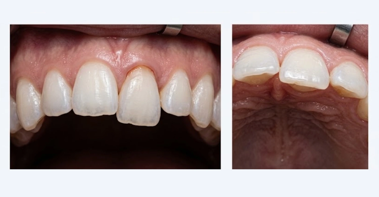
Yes, a luxated tooth can lose its vitality, but this does not happen to every injured tooth. Trauma can damage the blood vessels and nerves inside the pulp. Some teeth recover, while others slowly develop pulp necrosis. These changes may appear weeks or months later. For this reason, dentists test the tooth's response at follow-up visits. A darkening tooth or a new gum bump should be reported quickly. Early detection allows timely treatment and better protection of the tooth.
Note: All images used are for editorial and illustrative purposes only and may not originate from the original news provider or associated company.
Discover important information about aortic dissection and dental treatment
How Is Tooth Luxation Diagnosed?
A dentist diagnoses tooth luxation by combining questions, examination, and imaging. First, the dentist asks how the injury happened and when. Then, the mouth and teeth are checked for movement, position, and bite changes. X-rays show the root and bone. Sometimes 3D imaging adds more detail. The goal is to identify the type of luxation and any related injuries. Only then can the dentist choose the most suitable treatment for each patient. A careful diagnosis also sets the plan for follow-up.
Clinical Examination
During the clinical examination, the dentist checks tooth mobility and the position of the displaced tooth. They also assess the bite, or occlusion, to see whether the teeth meet normally. The gum and periodontal tissues are examined for tears and bleeding. The dentist notes pain and tenderness, often by gentle tapping. Pulp response tests may also be used, although results can be unreliable right after trauma. Each finding helps build a full picture of the injury. This picture guides every later decision.
Dental X-Rays and 3D Imaging
Dental X-rays are often needed to evaluate the root, the surrounding bone, and the exact position of the tooth. They can show whether the tooth has been pushed into the bone or whether a root fracture is present. Three-dimensional imaging, such as CBCT, may be recommended when a standard X-ray is not enough. It gives a detailed view of the bone and the root. The dentist uses imaging only when it is clinically justified. This approach keeps diagnosis accurate and safe.
Why Follow-Up Examinations Matter
An initial examination that looks reassuring does not always rule out later problems. Pulp damage and periodontal complications can develop slowly. A tooth may seem stable at first and then change color or become tender weeks later. Follow-up visits allow the dentist to repeat tests, take new X-rays, and compare results over time. This monitoring helps catch complications early. Patients should attend every scheduled visit, even when the tooth feels fine. Skipping visits can allow hidden problems to grow.
Learn more about medical diagnostic technology
Tooth Luxation Treatment: What Are the Options?
Tooth luxation treatment depends on the type of injury, the amount of displacement, and the stability of the tooth. The patient's age, the stage of root development, and the condition of the pulp also matter. A mild injury may need only observation. A displaced tooth may need repositioning and splinting. Some cases need further pulp treatment later. The dentist chooses the plan after a full assessment. The next sections describe the main options in simple terms.
Repositioning the Luxated Tooth
A displaced luxated tooth may need to be gently repositioned by a dental professional. The dentist usually numbs the area first to keep the patient comfortable. Then the tooth is guided back into its proper place, with care to protect the root and bone. Timing matters, because early repositioning supports better healing. Not every case needs this step. Intruded teeth, for example, may be managed differently, depending on age and root development. Never attempt this at home.
Stabilizing the Tooth With a Splint
A temporary splint may be recommended after selected luxation injuries. It is a thin, flexible wire or band attached to the injured tooth and its healthy neighbors. The splint holds the tooth steady while the periodontal ligament and bone recover. It also protects the tooth from accidental movement during eating. Splints are usually worn for a limited period, which the dentist decides. Patients should keep the splint clean and report any looseness or discomfort. Careful splint care supports healing.
Monitoring the Dental Pulp
Vitality monitoring is a key part of care after trauma. The dentist checks whether the pulp still responds normally, and repeats these checks over time. A tooth may start well and later show signs of damage. If the pulp stops responding or an infection appears, the treatment plan can change. Monitoring also includes watching tooth color and looking for gum swelling. Careful follow-up helps the dentist act at the right time. Patients play a role by reporting any changes early.
Root Canal Treatment When Indicated
Root canal treatment may be required when the pulp dies or when another documented complication appears. It is not automatic for every luxated tooth. Many teeth heal without it, especially when the injury is mild. The dentist decides based on test results, X-rays, and symptoms over time. When it is needed, the procedure removes damaged pulp tissue, cleans the canal, and seals it. This can help save a tooth that would otherwise be lost. Timing is decided case by case.
Treatment for Associated Gum or Bone Injuries
Luxation often occurs with other injuries. Gum lacerations may need cleaning and sometimes stitches. Damage to the alveolar bone may require extra stabilization, and healing is monitored closely. Other teeth may also be injured, such as chipped or fractured crowns. The dentist treats these problems together with the luxated tooth. A complete plan helps protect all the affected tissues. Where multiple injuries exist, the sequence of treatment is chosen carefully. Soft-tissue healing usually takes days, while bone healing takes longer.
Explore cosmetic dentistry in Turkey
What Happens After Tooth Luxation Treatment?
After tooth luxation treatment, recovery follows a gradual path. The tooth needs time to settle, and the supporting tissues heal in stages. Each patient recovers differently, so no exact outcome can be promised. The dentist explains what to expect, how to protect the tooth, and when to return. Following this guidance improves the chances of a smooth recovery. The next sections cover the first days, eating habits, and follow-up care. Patience is important during this period. Healing is gradual.
What to Expect in the First Few Days
In the first few days, the tooth may feel tender, and biting can feel different. Mild swelling is possible. The gum may look sore or bruised. Protect the injured area by avoiding pressure and touching. Over-the-counter pain relief may help, if the dentist agrees. Contact the dentist if pain increases, swelling grows, or the tooth becomes looser. These signs could point to a complication that needs attention. Rest and gentle care help the area settle during this early stage.
Eating and Chewing During Recovery
Softer foods are often recommended during recovery. Yogurt, mashed vegetables, soup, and eggs put little pressure on the injured tooth. Avoid biting directly into hard or sticky foods, such as apples, nuts, or caramel. Cut food into small pieces and chew on the opposite side. How long this lasts depends on the injury and the dentist's instructions. Hot drinks may also feel uncomfortable at first. Return to normal eating only when the dentist says it is safe. This protects the healing tissues.
Dental Follow-Up and Monitoring
Follow-up appointments are essential after tooth luxation. The dentist checks tooth stability, pulp health, and periodontal healing at planned intervals. Changes in tooth color are also recorded and compared over time. X-rays may be repeated to look for problems in the root or bone. Monitoring can continue for months, and sometimes longer. Regular visits allow small issues to be treated before they become serious. Keep every appointment, even if you feel fine. Consistent care gives the tooth its best chance.
Tips for Patients
Protect the injured tooth: Avoid unnecessary pressure, hard foods, and activities that could cause another impact.
Keep your mouth clean: Continue gentle brushing and oral hygiene, especially around the injured area.
Follow your dentist’s plan: Attend follow-up appointments and follow any specific instructions for your tooth.
Watch for changes: Pay attention to increasing pain, swelling, mobility, sensitivity, or tooth discoloration.
Report concerns early: Contact your dentist if you notice new or worsening symptoms.
Choose foods carefully: Follow any dietary recommendations and avoid foods that place excessive pressure on the injured tooth.
Brush gently: Clean the area carefully without putting unnecessary force on the affected tooth.
Ask questions: If any part of your treatment or recovery instructions is unclear, ask your dentist for guidance.
Protect the Injured Tooth
Avoid unnecessary pressure on the injured tooth. Do not bite hard foods, and do not use the tooth to open packages or chew pens. Avoid touching or wiggling the tooth with your tongue or fingers. Postpone contact sports and other activities that could cause another impact. A second blow can undo early healing and worsen the damage. If sport is unavoidable, ask the dentist about a protective mouthguard. Extra care in the first weeks makes a real difference.
Maintain Gentle Oral Hygiene
Keep cleaning your mouth, but be gentle. Use a soft toothbrush and brush the injured area lightly. Avoid scrubbing the gum near the tooth, especially if it is sore or stitched. A dentist may suggest a mild rinse to keep the area clean. Follow that advice and do not swish forcefully. Good hygiene lowers the risk of infection and supports gum healing. Clean the rest of the mouth as usual, but avoid flossing the injured tooth until the dentist agrees.
Follow the Dentist's Instructions
Follow every instruction your dentist gives. This includes splint care, follow-up visits, dietary recommendations, and any prescribed treatment. Take medication exactly as directed. Do not remove the splint yourself, and do not skip appointments. If something is unclear, ask the dental team to explain it again. Clear communication prevents mistakes. Patients who follow the plan closely usually help their teeth heal in the best possible way, although results still vary from person to person. Your effort matters.
Watch for Changes
Report any changes rather than waiting for the next appointment. Warning signs include increasing pain, swelling, more mobility, discoloration, or a change in the bite. A pimple-like bump on the gum can also signal infection. These changes may mean the pulp or supporting tissues are struggling. Early action often makes treatment simpler. Call your dentist promptly, and describe exactly what you have noticed. Photos can help the dental team understand how the tooth has changed. Never assume a symptom will fade by itself.
Clinical Note
A tooth can appear relatively intact after trauma while still having damage to its supporting tissues or pulp. The absence of a visible fracture does not automatically mean the injury is minor. Clinical examination and appropriate imaging help determine the extent of trauma. They also show whether monitoring or active treatment is required. Because hidden damage may show later, patients should keep follow-up appointments and report any change in the tooth. A calm, early check is always safer than guessing.
A Luxated Tooth May Change Over Time
A luxated tooth can change after the first examination. Pulp vitality may improve, stay stable, or decline. Tooth color can shift from normal to yellow, gray, or dark. Mobility may reduce as the ligament heals, or it may increase if a problem develops. Symptoms can also come and go. Because of this, one normal check does not close the case. Ongoing monitoring gives the dentist a clear view of how the tooth is really healing. Patience and regular visits are key.
Do Not Self-Reposition a Displaced Tooth
Never try to push a displaced tooth back into position yourself. Repositioning must be done by a dental professional. Forcing the tooth can crush the ligament, damage the bone, or break the root. It can also cause more pain and bleeding. If the tooth has moved, keep your hands away and avoid biting on it. Contact a dentist right away. Gentle cold compresses on the cheek may reduce swelling while you arrange care. Professional care is always the safest route.
When to Seek Urgent Dental Care
Seek urgent dental care if the tooth is significantly displaced or extremely mobile. Severe pain and uncontrolled bleeding also need immediate attention. Facial trauma or a suspected jaw injury is an emergency, too. If you feel dizzy, confused, or have trouble breathing, go to an emergency department first. Otherwise, call a dentist as soon as possible. Quick action can protect the tooth and prevent complications. Do not wait to see if the problem improves on its own. Time matters after trauma.
Explore cosmetic dentistry in Turkey
Can Tooth Luxation Be Prevented?
Tooth luxation cannot always be prevented, but the risk can be reduced. Most injuries come from sport, falls, and accidents, so protection and safety habits matter. A mouthguard, safer play areas, and proper protective equipment all help. Awareness also plays a role, since previously injured teeth are more fragile. The sections below explain realistic steps that families, athletes, and adults can take. Even small changes can lower the chance of a serious dental injury. Prevention is easier than repair.
Use a Mouthguard During Contact Sports
A properly fitted mouthguard can reduce the risk of dental trauma in contact sports. It absorbs part of the force from a blow and spreads it over several teeth. Football, boxing, hockey, martial arts, and basketball are common examples. Custom mouthguards made by a dentist usually fit better than boxed versions. A comfortable guard is more likely to be worn consistently. Replace it when it becomes worn, or when a child's mouth grows. Wear it in training, not only in matches.
Address Environmental and Accident Risks
Many dental injuries happen at home or on the road. Reduce fall hazards by fixing loose rugs, using safety gates, and keeping floors dry. Wear a helmet when cycling or riding a scooter. Use seat belts and child car seats in vehicles. Choose protective equipment that suits high-risk activities, such as skating or climbing. Supervise young children near stairs and playgrounds. These practical habits cannot remove all risk, yet they prevent many avoidable injuries. Small safety steps protect smiles.
Do Not Ignore Previous Dental Trauma
A tooth that was injured in the past may still need attention. Old trauma can lead to delayed pulp changes, root problems, or slow color change. Many patients forget minor injuries from childhood or sport. Tell your dentist about any earlier blow to the mouth, even if it seemed small. The dentist may recommend periodic check-ups and X-rays. This monitoring helps detect silent problems before they cause pain or infection. A previously injured tooth also deserves extra protection during future activities.
Find out more about opioids and dental pain
What We Notice Clinically
Dr. Rifat Alsaman is the Head of the Medical Team at Vitrin Clinic and a cosmetic dentist. The key message from Dr. Rifat Alsaman is that a luxated tooth should not be judged only by how it looks right after trauma. Tooth position, mobility, periodontal tissues, pulp response, root condition, and surrounding bone all contribute to treatment planning. A tooth that looks acceptable may still need monitoring, while a dramatic displacement may respond well to prompt care. This is why a full assessment of tooth luxation comes first.
Vitrin Clinic's Approach to Dental Trauma and Tooth Care
Vitrin Clinic approaches dental trauma with careful assessment and clear communication. Patients receive an explanation of the findings, the available options, and the reasons behind each recommendation. Care aims to protect natural teeth whenever it is clinically appropriate. Care is planned around the patient's needs, rather than a fixed routine. The sections below describe how assessment, technology, coordination, and restorative planning fit together after a dental injury. Details always depend on the individual case. Each plan begins with listening to the patient.
Individualized Dental Assessment
An individualized dental assessment looks at the complete oral condition before any restorative or cosmetic treatment is planned. After trauma, the dentist reviews the injured tooth, neighboring teeth, gums, bite, and existing dental work. This broader view helps identify risks and priorities. Treatment for the injury comes first, and cosmetic decisions follow only when the tooth is stable. Taking this order avoids rushed choices. Each patient's history and goals are considered. This keeps care safe and realistic.
Digital Dental Technology When Clinically Indicated
Digital technology can support diagnosis and planning when it is clinically indicated. CBCT or 3D imaging may show the root, the bone, and the position of a displaced tooth in detail. Intraoral scanning can record the teeth and bite accurately without traditional impressions. These tools help the dentist plan restorative evaluation after injury. They are not used for every patient. The dentist recommends them only when the findings justify the added information. Technology supports judgment; it never replaces it.
Coordinated Care When Multiple Dental Needs Are Present
Some injuries involve more than one dental need. A luxated tooth may occur with fractured teeth, gum damage, bite problems, or older restorations. In these cases, coordination between relevant dental disciplines can help. Depending on the injury, this may include gum care, root treatment, orthodontic input, or restorative planning. The team shares findings so that treatment steps follow a logical order. This approach reduces repeated procedures and keeps care organized for the patient. Clear coordination also improves communication.
From Trauma Management to Restorative Care
Once the injured tooth has been assessed and stabilized, future needs can be reviewed. Some teeth need no further work. Others may need a filling, crown, veneer, or other restoration if the shape or color has changed. The dentist decides on these options according to the patient's individual condition. Restorative care should wait until the tooth is stable and healing is clear. This step-by-step path protects health first and appearance second. Patients can then choose treatments with confidence.
Discover the connection between heartburn and jaw pain
References:
FAQs
Dr. Rifat Alsaman has more than 5 years of clinical experience in dentistry and currently serves as the Head of the Medical Team at Vitrin Clinic. He is dedicated to providing exceptional patient care, overseeing treatment planning, and ensuring the highest clinical standards across the team. His expertise, attention to detail, and commitment to continuous professional development have helped countless patients achieve healthier, more confident smiles.


