General Dentistry
September 20, 2026
Impacted Tooth: How It Happens and When It Should Be Removed?
 Dr. Rifat Alsaman
Impacted tooth is a tooth that does not fully erupt into its expected position because bone, gum tissue, another tooth, or an abnormal eruption path blocks it. Wisdom teeth are the most common culprits, since they arrive last and often find little room. Some impactions cause no symptoms, while others lead to pain, infection, swelling, or damage to neighboring teeth. Early dental assessment helps decide between monitoring, preventive care, and extraction. This practical guide covers diagnosis, treatment, recovery, and impacted wisdom tooth symptoms.
Impacted Tooth: What Is an Impacted Tooth and What Does It Mean?
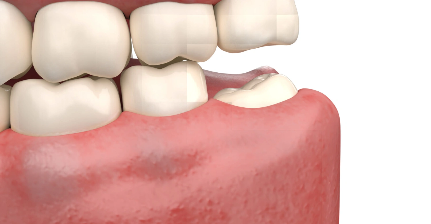
Impacted tooth refers to any tooth that stays trapped under the gum or inside the jawbone instead of emerging normally. The term describes a mechanical problem, not a disease by itself. Patients often ask what this condition means because it can stay hidden for years. Understanding the definition helps you recognize when a routine finding becomes a concern. The sections below explain the definition, everyday meaning, common causes, and the teeth most often affected by impaction.
What Is an Impacted Tooth?
This condition occurs when a tooth cannot finish erupting because it becomes trapped beneath the gum or bone. Dentists call this incomplete eruption. The tooth may stay fully buried, or it may emerge only partly. Because it sits out of sight, many patients feel nothing at first. Impaction does not always cause immediate pain, which is why routine X-rays often discover it by chance. Clear clinical definitions help patients understand why monitoring and timely follow-up matter.
Impacted Tooth Meaning in Everyday Terms
In everyday terms, the meaning is simple: the tooth is stuck. It is not loose, broken, or missing. It is present in the jaw but blocked from reaching its normal spot in the mouth. Many people carry one for years without realizing it. Discomfort, gum swelling, or a routine dental X-ray often reveals the problem. Recognizing this difference prevents confusion and helps patients describe their concerns accurately during a dental visit. Early understanding keeps more options open.
Why Do Teeth Become Impacted?
Teeth become impacted when something blocks their path. The most common reason is a lack of space in the dental arch. Crowding, an abnormal eruption angle, or a tooth positioned awkwardly can also trap it. Retained baby teeth sometimes hold the way closed. Developmental factors play a role too, and another tooth or dense bone may act as a barrier. In most cases, several factors combine, so the cause differs from one patient to the next. An X-ray usually shows the main cause.
Which Teeth Are Most Commonly Impacted?
Wisdom teeth are the most commonly impacted teeth, because they erupt last and often lack room. Canines come next, since they have a long eruption path. Less commonly, other permanent teeth, such as premolars, can become trapped. The location matters greatly. A wisdom tooth may cause gum infections, while an impacted canine can disturb the bite and alignment. Each position affects symptoms, risks, and treatment choices differently, so dentists tailor decisions to the specific tooth. This is why a proper examination always comes first.
Find out how stress and oral habits may affect your mouth
Impacted Tooth Symptoms: What Patients May Notice
Signs of impaction range from mild pressure to severe pain, depending on the tooth and surrounding tissue. Some people notice swollen gums or a bad taste, while others feel nothing at all. Symptoms often appear when a partly erupted tooth traps bacteria and food. They may come and go, which makes them easy to dismiss. Knowing what to watch for helps you seek care before an infection or damage develops. The sections below outline common signs and warning signals.
Common Impacted Tooth Symptoms
Common signs include pain or pressure around the affected area and swollen, tender gum tissue. Chewing may become difficult. Gum inflammation is frequent, and food often becomes trapped around a partially erupted tooth. Some patients notice bad breath or an unpleasant taste. In certain cases, discomfort spreads to the head or jaw. These symptoms can be mild at first and then worsen, so persistent changes deserve a professional examination rather than home guesswork. Early attention protects surrounding teeth and gums.
Impacted Wisdom Tooth Symptoms
Impacted wisdom tooth symptoms usually appear at the back of the mouth. Patients may feel pain, notice swelling around the tooth, and find the gums tender or inflamed. Chewing can hurt, and opening the mouth may feel difficult. Recurrent gum infections around a partially erupted tooth are a classic sign. Not every case follows this pattern, since symptoms depend on the angle and depth of the tooth. A dental examination confirms whether the wisdom tooth is the true source.
Can an Impacted Tooth Cause No Symptoms?
Yes, many impactions stay silent. Dentists often discover them by chance on routine X-rays, long before the patient feels anything. Still, a symptom-free tooth is not automatically risk-free. Hidden decay, gum problems, or pressure on a neighboring tooth can develop quietly. That is why an apparently harmless impaction still needs periodic monitoring. Regular checkups and follow-up imaging allow the dentist to act early if the situation changes, instead of waiting for pain to appear. Silence does not equal safety.
When Is Impacted Wisdom Tooth Pain a Warning Sign?
Impacted wisdom tooth pain becomes a warning sign when it is persistent or worsening. Facial swelling, fever, or other signs of infection also matter. Difficulty swallowing or opening the mouth should never be ignored. Pain accompanied by a bad taste or pus suggests an active infection. In these situations, urgent dental assessment is appropriate. Breathing difficulty or rapidly spreading swelling requires emergency care. Do not wait for symptoms to settle on their own, because infections can progress quickly.
Note: All images used are for editorial and illustrative purposes only and may not originate from the original news provider or associated company.
Discover the connection between sugar and bacteria
Types of Wisdom Tooth Impaction and How They Differ
Types of wisdom tooth impaction are classified by the tooth's angle and by how deeply it sits in the jaw. The main categories are mesial, distal, vertical, and horizontal, plus soft-tissue and bony impaction. Each type behaves differently. Some press against the second molar, while others sit upright but remain trapped. Knowing the type helps the dentist predict complications and plan removal. Not all types need the same treatment. The following sections explain each type and offer practical tips for patients.
Mesial Impaction
Mesial impaction means the wisdom tooth tilts forward, toward the second molar. It is one of the most common orientations, because the tooth often grows toward the available space in front of it. The angled crown can press against the neighboring molar. Over time, this pressure may trap food and plaque, which raises the risk of decay or gum problems in the second molar. Dentists evaluate the angle carefully on X-rays when deciding whether monitoring or removal is better.
Distal Impaction
Distal impaction describes a wisdom tooth angled backward, toward the back of the jaw. This position can limit eruption because there is little room behind it. It also affects surgical access, since the tooth sits farther from where the dentist works. Distal impactions are less common than mesial ones. Because the tooth points away from the second molar, it may press less on that neighbor. However, it can still become trapped and cause discomfort or infection over time.
Vertical Impaction
Vertical impaction occurs when the wisdom tooth points straight up, much like a normal tooth, yet stays trapped. The orientation looks favorable, but the tooth still cannot emerge if bone or gum tissue blocks it. Available space remains the deciding factor. A vertically positioned tooth may erupt partially, leaving a flap of gum that collects debris. This can lead to recurring inflammation. Orientation alone never tells the whole story. X-rays help the dentist judge whether enough room exists for healthy eruption.
Horizontal Impaction
Horizontal impaction places the wisdom tooth on its side, with the crown pointing toward the second molar. This position often causes the greatest pressure against the neighboring tooth. The pressure can lead to decay, root damage, or gum problems that are hard to clean. Horizontal teeth are often deeply positioned. As a result, removal can be more involved. Some horizontal teeth cause no symptoms for years. Careful imaging helps the dentist plan the safest approach for each patient.
Soft-Tissue vs. Bony Impaction
Soft-tissue impaction means the tooth is covered mainly by gum tissue, while bony impaction means part of the tooth is held within the jawbone. Depth matters. A tooth under gum only is usually simpler to remove, whereas a tooth locked in bone often needs bone removal or sectioning. The type and depth of impaction therefore influence the complexity, duration, and recovery of the procedure. Both types can still become infected. Imaging confirms which category applies before treatment is planned.
Tips for Patients
Keep the area around the impacted tooth clean with careful, consistent oral hygiene.
Do not ignore repeated pain, swelling, or discomfort.
Avoid guessing the cause of jaw pain, as several dental conditions can cause similar symptoms.
Attend every follow-up appointment recommended by your dentist.
Continue monitoring the impacted tooth when removal is not currently planned.
Learn the warning signs that require urgent dental care.
Learn why your teeth may feel tightFollow your dentist’s instructions to reduce the risk of complications.
Protect the neighboring teeth by addressing changes early.
Take small, consistent steps to maintain comfort and oral health.
What Causes an Impacted Wisdom Tooth?
An impacted wisdom tooth usually results from a mismatch between tooth size and jaw space. Wisdom teeth are the last to erupt, typically in the late teens or early twenties, when the jaw has little room left. Abnormal angles, crowding, and obstruction from gum or bone can worsen the problem. Each cause blocks the normal eruption path in a different way. The following sections explain the main causes, so you can understand why wisdom teeth are so often affected.
Limited Space Behind the Second Molar
The jaw often lacks enough room behind the second molar for a wisdom tooth to emerge. Wisdom teeth are the final teeth to develop, so the space may already be filled. When the jaw is too small for the tooth, the crown cannot reach its normal position. The tooth then remains partly or fully trapped. This relationship between jaw space and eruption is the most common reason wisdom teeth become impacted, and X-rays help measure the available room.
Abnormal Eruption Direction
A wisdom tooth does not always grow straight up. It may tilt forward, backward, or sideways, or it may rotate as it develops. An angled or rotated tooth can hit the second molar or the surrounding bone and stop moving. Once blocked, it cannot correct its path by itself. The direction of eruption therefore strongly influences whether the tooth becomes trapped, which impaction type develops, and how difficult removal may be later. Early X-rays can reveal these angles.
Tooth Crowding and Jaw Development
Crowded teeth leave little space for late-arriving molars. Jaw size and growth patterns vary between people, and a smaller jaw offers less room. If the dental arch is already tightly packed, the wisdom tooth has even fewer options. Genetics often influence these features, so they are not the patient's fault. Understanding how space, crowding, and jaw development interact explains why some people have no trouble with wisdom teeth while others develop repeated impaction problems. Dentists can assess this early.
Gum or Bone Obstruction
Sometimes the barrier is tissue rather than space. Thick gum tissue can cover the crown and slow eruption, while dense bone can hold the tooth in place. In other cases, the tooth is partly through the gum but blocked by bone beneath it. These obstructions explain why some impactions are classed as soft-tissue and others as bony. Identifying the barrier guides treatment, because removing gum tissue is very different from removing bone around a buried tooth. Imaging shows which barrier is present.
Find out why teeth can hurt when you have the flu
How Is an Impacted Tooth Diagnosed?
Diagnosis combines a clinical examination with imaging. The dentist checks symptoms, gum condition, and tooth position, then uses X-rays to see what lies beneath the surface. Three-dimensional imaging may be added when the tooth is complex or close to important structures. This step-by-step approach ensures that every case is evaluated accurately before treatment is chosen. Each step answers a different question. The sections below explain the examination, standard X-rays, and CBCT scanning when it is clinically indicated.
Dental Examination
The dentist reviews your symptoms, checks the gums, and examines the tooth's visible position. Signs of infection, such as redness, swelling, or pus, are noted. Neighboring teeth are checked for decay or sensitivity. However, a clinical examination alone cannot show the full position of a buried tooth. The crown, roots, and surrounding bone remain hidden beneath the gum. That is why examination is only the first step, and imaging is needed to complete the picture. Your medical history is reviewed too.
Dental X-Rays
Panoramic X-rays are the standard tool for evaluating an impaction. A single image shows both jaws, all teeth, and the surrounding structures. It reveals the tooth's angle, depth, and relationship to neighboring teeth and the lower jaw nerve canal. Smaller periapical films can add detail for a specific area. These images help the dentist classify the impaction, spot decay or cysts, and decide whether monitoring or removal is the most suitable option. This overview guides every later decision.
CBCT 3D Imaging When Clinically Indicated
CBCT, or cone beam computed tomography, provides a three-dimensional view that a flat X-ray cannot. It is valuable when the tooth lies close to nerves or other important structures, or when the position is unclear. The scan helps the dentist plan the safest surgical approach. However, advanced imaging should match clinical need rather than being used routinely for every patient. Your dentist selects the right level of imaging after examining you, balancing the benefits of extra detail with radiation exposure.
Explore the process of chewing step by step
What We Notice Clinically
Dr. Rifat Alsaman, Head of the Medical Team at Vitrin Clinic and a cosmetic dentist, emphasizes assessing the position and condition of an impacted tooth before deciding on removal. Symptoms, infection risk, tooth position, neighboring teeth, and imaging findings can all influence the treatment plan. Not every impaction needs immediate extraction. Some can be monitored safely, while others need timely treatment. The decision follows the evidence gathered during examination, not assumptions or fixed rules. This measured approach avoids unnecessary surgery.
Why Individual Assessment Matters
Two patients with impacted wisdom teeth may need completely different care. One may be young with a symptom-free tooth, while another has repeated infections and decay in the neighboring molar. Age, tooth position, symptoms, infection history, anatomical relationships, and overall oral health all matter. Treatment should follow clinical findings rather than symptoms alone, because pain does not always reflect the true condition. A personalized assessment ensures that each patient receives care that fits their situation. There is no one-size-fits-all answer.
Learn how thumb sucking can affect teeth
When Does an Impacted Tooth Need Treatment?
An impaction needs treatment when it causes problems or carries a foreseeable risk. If the tooth is symptom-free and healthy, monitoring may be enough. If it leads to infection, pain, or damage, intervention becomes more important. The decision depends on professional assessment, not on the mere presence of impaction. Every case deserves an individual evaluation. The sections below explain when watchful waiting is reasonable, when treatment becomes more important, and what may happen if a problematic tooth is left alone.
When Monitoring May Be Appropriate
Monitoring may be appropriate when the tooth causes no symptoms and no significant pathological findings appear on examination or imaging. In these cases, regular clinical and radiographic follow-up allows the dentist to watch for changes. The tooth is not ignored; it is observed. Monitoring should always rest on a professional assessment rather than personal guesswork. If symptoms, decay, or gum problems develop later, the plan can change quickly and the tooth can be treated before complications grow. Patience can be wise.
When Treatment Becomes More Important
Treatment becomes more important when problems appear. Recurrent infection, persistent pain, and significant gum disease are common triggers. Damage or decay affecting the neighboring tooth also matters, as do cystic or other pathological changes around the impaction. Sometimes the tooth position itself predicts a foreseeable complication, even before symptoms begin. In these cases, dentists weigh the risks of leaving the tooth against the risks of surgery and recommend the option that best protects long-term oral health. Timely care often means simpler treatment.
What Happens If an Impacted Tooth Is Left Untreated?
Not every problematic impaction causes complications, but some do. Possible outcomes include recurrent infection, periodontal problems, and decay in the adjacent tooth. In selected cases, cysts or other pathology may form around the tooth. These risks do not occur in every patient, and the likelihood varies with the tooth's position and surrounding anatomy. That is why professional evaluation is essential before deciding to leave it untreated or to remove it. Understanding these possibilities helps you make an informed choice.
Impacted Wisdom Tooth Removal: What Are the Treatment Options?
Impacted wisdom tooth removal is only one of several options. Depending on the case, a dentist may recommend monitoring with preventive care, surgical exposure, simple or surgical extraction, or a more complex surgical removal. The right choice depends on the tooth involved, its depth, and your symptoms. No option suits everyone, so a good dentist explains the benefits and limits of each. The sections below describe each approach, so you can understand what a dentist may propose and why.
Monitoring and Preventive Care
Observation may be chosen when the tooth is healthy, symptom-free, and not causing damage. During this period, careful oral hygiene around a partially erupted tooth is essential, because trapped plaque can trigger inflammation. Scheduled reviews allow the dentist to track changes on X-rays and clinical examination. Monitoring is an active plan, not neglect. Reviews keep this approach safe. If new symptoms or findings appear, the treatment approach can shift promptly, and the tooth can be addressed before problems become more serious.
Surgical Exposure
Surgical exposure is considered in selected cases, particularly for non-wisdom impacted teeth such as canines. The dentist lifts the gum and exposes the crown, so the tooth can be guided into place, often with orthodontic support. This approach aims to save a valuable tooth rather than remove it. It is rarely used for wisdom teeth. Coordination between specialists can improve results. Treatment depends heavily on the tooth involved and its position, which is why a full assessment is essential first.
Impacted Tooth Extraction
Extraction is the definitive treatment for some impacted teeth, especially when infection, damage, or pathology is present. It is usually performed under local anesthesia and, where appropriate, sedation for added comfort. The surgical approach varies with the tooth's depth and position. A tooth covered only by gum may need a small incision, while one within bone may require bone removal. Removing the tooth resolves the problem at its source. Your dentist explains the plan, risks, and expected recovery beforehand.
Complex Surgical Removal
Deeply positioned or anatomically challenging teeth may require a more involved surgical approach. The dentist may section the tooth into smaller pieces when clinically appropriate, which allows removal through a smaller opening and protects surrounding bone. Proximity to nerves or other structures also affects planning, so advanced imaging is often reviewed carefully beforehand. Complex cases demand experience, precise technique, and thorough preparation. With proper planning, risks can be reduced and managed carefully. Every step aims to protect nearby nerves and teeth.
Discover what can cause a tight jaw
What to Expect Before, During, and After Impacted Tooth Extraction
Knowing what to expect before, during, and after surgical removal reduces anxiety and helps you prepare. The process begins with assessment and imaging, continues with the procedure itself, and ends with careful recovery at home. Each stage has its own instructions. Every patient's experience differs, depending on the tooth and the technique. Being informed makes the whole experience calmer and more manageable. The sections below walk through the stages, along with clinical guidance and advice on timing.
Before the Procedure
Preparation starts with a clinical assessment and imaging to understand the tooth's position. The dentist reviews your medical and dental history, including medications and allergies. Anesthesia or sedation options are discussed where appropriate. You also receive a personalized explanation of risks, expected recovery, and aftercare. Ask questions freely, and share any health changes. Follow any fasting instructions if sedation is planned. Clear communication before treatment helps the team plan safely and helps you feel confident about what will happen on the day.
During Impacted Tooth Removal
The general sequence varies, because no two procedures are identical. After the area is numbed, the dentist may open the gum to reach the tooth. Bone removal may be needed, depending on the impaction. In selected cases, the tooth is sectioned and removed in pieces. The site is then cleaned, and sutures may be placed when appropriate. Time varies with complexity. You should feel pressure but not sharp pain, and you can tell the team if you are uncomfortable.
After Extraction
Expect some swelling and tenderness during the first days. Following the postoperative instructions is essential for smooth healing. Keep oral hygiene gentle, avoiding vigorous rinsing early on, and follow the guidance on brushing near the site. Choose soft, cool foods at first and avoid hard or hot items. Do not disturb the blood clot. Rest well during the first day. Attend follow-up visits when recommended, and call the clinic promptly if pain, swelling, or bleeding worsens instead of improving.
Before-and-After Expectations
The immediate goal is to remove the problematic tooth and manage the surrounding tissues. When the tooth was the source of pain or infection, patients can expect symptoms to reduce as the area heals. Healing time varies according to the procedure and individual factors such as depth, age, and general health. Some people return to routine activities quickly, while others need more rest. Patience during recovery pays off. Your dentist will give a realistic timeline based on your specific case.
Clinical Note
Clinical decisions about impacted teeth work best when they rest on the whole picture, not one finding. Symptoms, imaging, gum health, and the condition of neighboring teeth are reviewed together. Extraction is neither automatic nor avoided by default. It is chosen when the benefits outweigh the risks. Good care is transparent care. This note introduces the guidance of Dr. Rifat Alsaman, and it also reminds patients that every recommendation should be explained clearly and tailored to their own mouth.
A Dentist’s Guidance on Impacted Teeth
Dr. Rifat Alsaman, Head of the Medical Team at Vitrin Clinic and a cosmetic dentist, considers the full oral environment when evaluating impacted teeth. The evaluation checks whether the tooth affects neighboring teeth, gum health, or overall oral function. Diagnostic imaging is selected according to the clinical situation. Treatment planning balances the risks of leaving the tooth untreated against the risks and benefits of intervention, so every recommendation reflects the patient's real needs. This balanced view prevents both overtreatment and delay.
Why Timing Should Be Individualized
No universal age or timeline applies to every case. Timing depends on symptoms, anatomy, infection history, and radiographic findings. A young patient with a developing wisdom tooth may be monitored, while an adult with recurrent infection may need prompt removal. Waiting too long can complicate treatment in some cases, but rushing is not always necessary either. An individualized schedule, set by your dentist after examination, offers the best balance of safety and comfort. Your case is judged on its own merits.
Read more about tingling of the mouth
How Can Impacted Teeth Be Prevented?
Prevention is limited, because many causes are developmental and cannot be controlled. Even so, early dental examinations can identify eruption problems before they become complicated. Regular monitoring of developing wisdom teeth and other unusual eruption patterns gives your dentist time to plan. Protecting neighboring teeth from decay and gum problems is also important. Awareness and regular checkups remain your strongest tools against avoidable complications. Small habits add up. The sections below explain what prevention realistically means and how routine care helps.
Can an Impacted Tooth Actually Be Prevented?
In most cases, no. Many developmental factors, such as jaw size, tooth position, and genetics, cannot be controlled. Brushing well does not stop a tooth from becoming trapped. What you can do is detect problems early. Early dental examinations can identify eruption problems before they become complicated, giving the dentist more options. Prevention therefore means early awareness, not stopping impaction itself. Timely X-rays during the teenage years often reveal how wisdom teeth are developing. Ask your dentist when to begin screening.
Regular Dental Monitoring
Routine examinations and appropriate imaging let the dentist follow how teeth are developing. Monitoring developing wisdom teeth, and any teeth with abnormal eruption patterns, helps identify trouble early. Not every visit needs an X-ray; the schedule depends on your age, risk, and findings. Consistent checkups also catch decay and gum disease that might otherwise go unnoticed. Over time, these records show whether a tooth is moving into position or staying stuck, guiding timely decisions. Consistency is what makes monitoring effective.
Protecting the Neighboring Teeth
Early identification helps protect neighboring teeth. An impaction can press on the adjacent molar or create a pocket where bacteria collect. Regular checks detect decay, gum problems, or pressure-related changes before they cause lasting damage. Good brushing and flossing next to the affected area also help. If the dentist finds early signs of harm, treatment can be planned sooner and the healthy teeth around the impaction are better preserved for the long term. Protection is a key goal of care.
Vitrin Clinic’s Approach to Impacted Tooth Assessment and Treatment
Vitrin Clinic is a dental clinic in Istanbul, Turkey, that plans each case around the patient's clinical findings. The approach combines a thorough examination, suitable imaging, and coordination between dental disciplines when needed. International patients receive clear explanations and a structured plan. Every recommendation is based on what your examination and images actually show, not on a standard template. The sections below describe individualized assessment, diagnostic technology, multidisciplinary care, and how to book your consultation.
Individualized Dental Assessment
Vitrin Clinic in Istanbul, Turkey, follows a patient-centered approach to assessment and treatment planning. Each visit begins by listening to your symptoms and reviewing your history. Treatment is planned according to your clinical findings rather than using the same approach for every case. Some patients need monitoring, others need extraction, and some need coordinated care. This individualized method aims to deliver the right treatment, at the right time, for the right reasons. Your goals and comfort matter throughout.
Diagnostic Technology When Clinically Indicated
Vitrin Clinic uses a comprehensive dental examination, digital intraoral scanning where relevant, and dental X-rays. CBCT 3D imaging is added when extra anatomical information is clinically necessary, such as when a tooth lies near a nerve. Imaging supports treatment planning by showing depth, angle, and neighboring structures accurately. Technology is a tool, not a routine for every patient. Each option is selected according to clinical need, so you receive only the diagnostics that truly help. Clear images make clear decisions possible.
Multidisciplinary Dental Care
An impaction can affect surrounding teeth, periodontal health, or a broader restorative plan, so several dental disciplines may be involved. Oral surgery, periodontal care, orthodontics, and restorative dentistry may each contribute when needed. Dr. Rifat Alsaman, Head of the Medical Team at Vitrin Clinic and a cosmetic dentist, is involved in coordinating individualized treatment planning. This teamwork ensures that solving one problem does not create another and that the final result supports function and appearance. Coordination keeps the plan consistent.
Book Your FREE Treatment Plan
If you have impacted wisdom tooth symptoms, persistent pain, swelling, or recurrent infection, seek a professional dental assessment. An examination is the only way to determine whether monitoring, extraction, or another approach suits your situation. Waiting rarely makes a complicated problem simpler. Do not let discomfort linger. Vitrin Clinic will review your findings, explain your options clearly, and help you choose the next step with confidence, wherever in the world you are traveling from.
Explore the connection between tinnitus and teeth
References:
FAQs
Dr. Rifat Alsaman has more than 5 years of clinical experience in dentistry and currently serves as the Head of the Medical Team at Vitrin Clinic. He is dedicated to providing exceptional patient care, overseeing treatment planning, and ensuring the highest clinical standards across the team. His expertise, attention to detail, and commitment to continuous professional development have helped countless patients achieve healthier, more confident smiles.


.webp&w=3840&q=75)


