General Dentistry
October 1, 2026
Chewing Tobacco Teeth Damage: Is it Really That Bad?
 Dr. Rifat Alsaman
Chewing tobacco teeth damage often starts quietly, long before any pain appears. Chewing tobacco and other smokeless tobacco products affect the teeth, the gums, and the soft tissues lining the mouth. The main concerns are tooth discoloration, gum irritation, recession, tooth decay, and tooth loss. Some users also develop more serious oral health problems. How severe the damage becomes depends on the product type, how often it is used, and for how long. Oral hygiene and individual dental health also play a role. From a dental perspective, stopping tobacco exposure and attending regular examinations help reduce ongoing harm and identify changes early.
Chewing Tobacco Teeth Damage: How Does It Affect Your Teeth and Gums?
Chewing tobacco cause teeth damage begins with direct contact. A pinch or plug is usually held against the teeth and gums for long periods, often several times a day. This repeated chemical and physical exposure affects the enamel, the gingival tissues, saliva, and the wider oral environment. The chewing tobacco effects on teeth may look purely cosmetic at first, such as light staining. Over time, however, smokeless tobacco teeth damage can become clinically significant, involving recession, decay, or changes in the soft tissues.
Chewing Tobacco and Tooth Discoloration
Regular tobacco exposure leaves pigments that cling to the enamel and settle into tiny surface irregularities. Over months, this creates yellow or brown staining. Chewing tobacco stained teeth are one of the most common concerns patients raise. External stains sit on the tooth surface and often respond well to professional cleaning. Deeper discoloration is different. It may involve changes within the tooth structure or exposed root surfaces. This is why cleaning improves many cases of chewing tobacco teeth damage but cannot reverse every type of discoloration.
Can Chewing Tobacco Cause Tooth Decay?
Tobacco use can influence several factors linked to cavities. It may affect saliva, encourage plaque accumulation, and coincide with weaker oral hygiene habits. Some products also contain added sweeteners. Still, the link between chewing tobacco and tooth decay is not simple, because cavities always have more than one cause. Gum recession adds another layer of risk. Exposed root surfaces lack protective enamel, so they decay more easily. Fluoride toothpaste and regular dental examinations help protect these vulnerable areas and detect early decay.
Chewing Tobacco Gum Damage and Gum Recession
Tobacco held in the same spot repeatedly irritates the gum tissue beneath it. Over time, this irritation can cause the gum line to pull away from the teeth. This chewing tobacco gum damage is usually most visible where the product is habitually placed. As the gum recedes, the tooth root becomes exposed. Patients then notice sensitivity to cold, sweet, or acidic foods. Recession is one of the clearest forms of chewing tobaccocause teeth damage, and it rarely reverses on its own.
Chewing Tobacco and Gum Disease
Tobacco use affects periodontal health in several ways. Plaque hardens into tartar, which irritates the gums and keeps inflammation active. If this continues, periodontal pockets form between the gums and teeth. Eventually, the bone supporting the teeth may be lost. Chewing tobacco gum disease can be hard to notice early. Tobacco may reduce bleeding and redness, masking the usual warning signs. This is why tobacco users benefit from regular periodontal checks, even when their gums feel healthy.
Find out what causes and treats White Spots on Teeth
Note: All images used are for editorial and illustrative purposes only and may not originate from the original news provider or associated company.
What Are the Other Chewing Tobacco Mouth Problems?
Chewing tobacco teeth damage is only part of the picture. The soft tissues of the mouth, including the cheeks, lips, and tongue, are exposed too. Common chewing tobacco mouth problems include irritation, soreness, and visible tissue changes. Some users develop persistent lesions where the tobacco rests. These chewing tobacco side effects on mouth tissues vary widely in appearance. Persistent or unusual changes require professional examination, because self-diagnosis can miss important findings. A complete picture of chewing tobacco oral health effects always requires a dental assessment.
Changes Where the Tobacco Is Usually Placed
Most users place tobacco in the same spot every time. Repeated contact with one area can cause localized tissue changes. The lining may become white, wrinkled, or thickened. Some patches look harmless, while others need closer monitoring. A dentist should evaluate any white or thickened area, especially if it persists. Persistent lesions should never simply be assumed to be harmless irritation. Early assessment allows the dentist to decide whether monitoring, further investigation, or referral is the right next step.
Does Chewing Tobacco Increase the Risk of Oral Cancer?
There is an established association between smokeless tobacco and an increased risk of oral cancer. Commonly affected areas include the mouth, lips, cheeks, gums, tongue, and throat. This does not mean every user will develop cancer. However, early assessment matters. Seek prompt evaluation for any sore that does not heal within about two weeks. The same applies to a lump, a bleeding area, or an unexplained tissue change. Early detection generally offers more treatment options and better outcomes.
Learn about managing Amelogenesis Imperfecta
What Are the Signs of Smokeless Tobacco Oral Health Damage?
Recognizing problems early makes a real difference. Many signs of chewing tobacco teeth damage develop slowly and without pain, so they are easy to dismiss. Some signs relate to the teeth, while others affect the gums or soft tissues. Smokeless tobacco oral health problems often progress quietly between dental visits. The lists below separate early warning signs from more advanced changes. Noticing any of them is a good reason to book a dental examination rather than waiting.
Early Signs to Watch For
Early changes are often subtle. Watch for:
Tooth staining: yellow or brown discoloration, especially near where tobacco rests.
Tooth sensitivity: discomfort with cold, hot, or sweet foods.
Gum irritation: tenderness or redness along the gum line.
Gum recession: teeth that look longer than before.
Bad breath: a persistent odor despite brushing.
Localized soreness: discomfort in the usual placement area.
Changes in the tissues where tobacco is placed: white, wrinkled, or rough patches.
More Advanced Signs
As damage progresses, the signs become harder to ignore:
Persistent gum bleeding or swelling: a possible sign of active gum disease.
Increasing tooth mobility: loose teeth may have lost bone support.
Root exposure: visible root surfaces, often with marked sensitivity.
Persistent oral sores or lesions: areas that do not heal within two weeks.
Changes in the color or texture of oral tissues: red, white, or thickened patches.
Pain that does not resolve: ongoing discomfort needs prompt assessment.
Discover when pocket reduction is needed in Osseous Surgery
Tips for Patients
Small daily habits can limit chewing tobacco teeth damage and protect long-term oral health:
Stop using chewing tobacco and other smokeless tobacco products.
Brush twice daily with fluoride toothpaste.
Clean between the teeth every day.
Maintain regular professional dental examinations.
Do not ignore persistent mouth sores or tissue changes.
Avoid repeatedly placing tobacco against the same area.
Ask a dentist about sensitivity, recession, staining, or bleeding gums.
Follow recommended periodontal maintenance if gum disease is present.
Consider professional support for tobacco cessation when needed.
How Is Chewing Tobacco Teeth Damage Diagnosed?
Diagnosing chewing tobacco teeth damage involves more than a quick look at stained enamel. A dentist assesses the teeth, gums, supporting bone, and soft tissues together. The examination usually combines visual inspection, periodontal measurements, and soft-tissue screening. Imaging is added when deeper problems are suspected. An accurate history matters as well. Telling your dentist how long and how often you use tobacco helps them interpret the findings correctly and plan safe, appropriate treatment.
Dental and Gum Examination
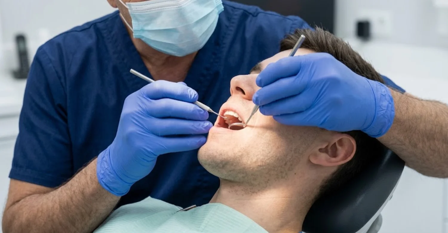
The dentist examines each tooth surface for staining, wear, decay, and damaged restorations. The gum margins are checked for recession, inflammation, plaque, and tartar. A small probe measures the periodontal pockets around each tooth, which can reveal hidden gum disease. Particular attention goes to the areas where tobacco is regularly placed. These sites often show the earliest changes. Together, these findings show whether the problems are mainly cosmetic or whether active disease needs treatment first.
Oral Soft-Tissue Examination
A soft-tissue examination covers the cheeks, lips, tongue, floor of the mouth, palate, and throat. The dentist looks for unusual white, red, thickened, ulcerated, or persistent areas. Some changes may simply be monitored over a few weeks. Others call for additional investigation, such as a biopsy, or referral to a specialist. This decision depends on the appearance, location, and duration of the change. The patient's tobacco history also guides how closely an area is followed.
Dental Imaging When Needed
Some problems cannot be seen during a visual examination. X-rays or other dental imaging may be recommended when the dentist needs to evaluate bone levels, root surfaces, or decay between the teeth. Imaging also helps assess existing restorations and possible infections. In chewing tobacco cause teeth damage, imaging can reveal bone loss caused by gum disease. Imaging is not automatic for every patient. It is recommended when it is clinically justified and genuinely guides treatment planning.
How Is Chewing Tobacco Teeth Damage Treated?
No single procedure treats chewing tobacco teeth damage. Treatment is condition-specific, so it depends on what the examination finds. One patient may only need a cleaning, while another requires periodontal care and restorations. Stopping tobacco exposure is an important part of every plan. Without it, new damage keeps developing even as existing problems are treated. The sections below explain the most common treatment options and when each one may be appropriate.
Professional Dental Cleaning
Professional dental cleaning removes plaque and hardened tartar that brushing cannot reach. It also lifts much of the surface staining caused by tobacco. Patients usually notice smoother, brighter-looking teeth afterward. However, expectations should be realistic. Cleaning does not change the natural shade of the teeth. It cannot remove deeper discoloration within the tooth structure either. For many patients, cleaning is the first step, because it allows the dentist to assess the teeth and gums clearly.
Fluoride and Preventive Decay Management
Fluoride strengthens enamel and helps protect against early decay. Daily fluoride toothpaste is the foundation of prevention. When the risk is higher, the dentist may apply professional fluoride treatments during visits. Exposed root surfaces need special attention, since they decay faster than enamel. The dentist will monitor these vulnerable areas over time. Catching early weakening allows it to be managed before a cavity forms and a restoration becomes necessary.
Treatment for Gum Disease
Gum disease treatment begins with professional periodontal cleaning. When pockets are deeper, the dentist may recommend scaling and root planing. This deep cleaning removes tartar below the gum line and smooths the root surfaces. Smoother roots make it easier for the gums to heal and tighten around the teeth. Patients with ongoing gum concerns usually need periodontal maintenance. These visits are scheduled more often than routine cleanings. They help control inflammation and reduce the risk of further bone loss.
Treatment for Gum Recession
Treatment for recession depends on its severity and the symptoms it causes. Mild recession may only need monitoring, a gentler brushing technique, and desensitizing products. A periodontal evaluation helps determine whether the recession is stable or progressing. In selected cases, gum-grafting procedures may cover exposed roots and reduce sensitivity. Grafting works best when tobacco exposure has stopped. Continued use at the same site can undermine healing and worsen chewing tobacco teeth damage over time.
Restorative Treatment for Damaged Teeth
When decay or wear has compromised tooth structure, restorative treatment rebuilds the tooth. Options include fillings, bonding, crowns, and other restorations. The right choice depends on how much healthy tooth structure remains and on overall oral health. Small cavities may only need a filling. Larger defects may require a crown for strength and protection. Restorations repair existing chewing tobacco teeth damage, but they still need healthy gums and good hygiene to last.
Cosmetic Treatment for Tobacco-Stained Teeth
Professional whitening can improve tobacco-stained teeth when the teeth and gums are healthy enough for treatment. Active gum disease or untreated decay should be managed first. When whitening is not effective, veneers or other cosmetic restorations may be considered, but only when clinically appropriate. Patients should understand one key point. Cosmetic procedures improve appearance, yet they do not remove the underlying risks of continued tobacco use. Stains may also return if use continues.
Understand the role of Gutta Percha Solvents in Root Canal Retreatment
Can Teeth Recover After Stopping Chewing Tobacco?
Some changes improve after stopping, while others need treatment. Surface staining may improve with professional cleaning or whitening. Gum inflammation often settles once the irritant is removed and oral hygiene improves. However, established gum recession does not simply grow back. Significant loss of tooth structure also requires restoration. Quitting cannot undo all chewing tobacco teeth damage, but it prevents additional exposure-related harm. It also gives any treatment a much better chance of lasting.
Clinical Note
Tobacco-related dental problems do not look the same in every patient. One person may have heavily stained teeth with relatively healthy periodontal tissues. Another may show recession, periodontal disease, root exposure, or localized mucosal changes. Clinical examination is therefore more informative than appearance alone. Dr. Rifat Alsaman, Head of the Medical Team at Vitrin Clinic and a cosmetic dentist, emphasizes identifying active oral disease before elective cosmetic dentistry. Recommendations for chewing tobacco teeth damage depend on examination findings and, when indicated, imaging or periodontal assessment.
Learn how to prevent enamel loss from Sports Drinks and Dental Erosion
How Can You Prevent Further Smokeless Tobacco Oral Health Damage?
Preventing further harm depends on two things: removing the cause and protecting what remains. Treatment alone cannot stop chewing tobacco teeth damage if exposure continues. Prevention combines tobacco cessation, preventive dental care, and careful self-monitoring. Each element supports the others. Regular visits catch new problems early, while good home care slows their development. The steps below outline how patients can protect their smokeless tobacco oral health over the long term.
Make Tobacco Cessation the Priority
Eliminating tobacco exposure protects the mouth more effectively than treating visible symptoms. Every new pinch brings fresh contact with the gums and soft tissues. Even after cleaning, whitening, or restorations, continued use can cause new chewing tobacco teeth damage. Quitting can be difficult, and many people benefit from support. A doctor, pharmacist, or cessation program can offer practical help. Telling your dentist about your plans also helps them adjust your care.
Maintain Preventive Dental Care
Preventive dental care stops small problems from becoming larger ones. Key steps include:
Regular examinations and professional cleaning, at intervals your dentist recommends based on your risk.
Fluoride protection, through daily fluoride toothpaste and professional applications when advised.
Daily interdental cleaning with floss or interdental brushes to remove plaque between the teeth.
Together, these habits lower the risk of decay and gum disease. They also give your dentist regular opportunities to track any changes.
Monitor Changes in the Mouth
Patients play an important role in early detection. Check your mouth regularly in good light, especially where you used to place tobacco. Report any persistent sore, lump, or color change to your dentist. Unexplained bleeding and areas that do not heal also deserve attention. As a general guide, any change lasting longer than two weeks should be examined. Prompt reporting allows investigation early, when more options are usually available.
Understand the connection between Circulatory System Diseases and Periodontal Disease
What Can Patients Expect Before and After Treatment?
Knowing what happens before and after treatment helps patients feel prepared. Every plan starts with a thorough assessment, since treatment depends on the problems found. Afterward, results depend on the extent of existing damage and on whether tobacco use continues. Some improvements are visible quickly, such as cleaner teeth. Others, like healthier gums, develop gradually. The sections below describe the typical steps and the outcomes patients can realistically expect.
Before Treatment
Treatment begins with a comprehensive examination of the teeth, gums, and soft tissues. The dentist identifies any active gum disease, decay, recession, staining, or soft-tissue changes. Imaging may be added when needed. These findings shape an individualized treatment plan. Health problems are addressed first, followed by restorative and then cosmetic care. This order ensures that chewing tobacco teeth damage is managed safely, rather than simply covered up.
After Treatment
After professional hygiene treatment, tooth surfaces feel cleaner and smoother. Gum inflammation can be better managed when the underlying cause is addressed. If appropriate cosmetic treatment is performed, the appearance of the teeth may also improve. Continued monitoring and preventive care help maintain these results. Outcomes vary according to the extent of existing damage. They also depend heavily on whether tobacco exposure continues after treatment ends.
Learn about the causes and solutions for Habitual Jaw Clenching and Tooth Wear
What We Notice Clinically
Dr. Rifat Alsaman, Head of the Medical Team at Vitrin Clinic and a cosmetic dentist, notes that patients often focus on visible tobacco staining. Meanwhile, they may overlook gum recession or changes in the surrounding tissues. Dr. Rifat Alsaman recommends evaluating underlying oral health before considering cosmetic procedures. He may assess gum condition, tooth structure, recession, existing restorations, and overall oral health first. Only then are whitening, veneers, or crowns considered. Cosmetic treatment should address genuine dental concerns, not mask ongoing tobacco exposure.
Find out how thick saliva impacts recovery in Salivary Viscosity and Root Canal Retreatment Healing
Why Choose Vitrin Clinic for Tobacco-Related Dental Concerns?
Vitrin Clinic in Istanbul is a multidisciplinary dental clinic where treatment planning addresses both oral health and cosmetic concerns. Gum and tooth health are assessed before any cosmetic procedure begins. Each patient receives an individualized examination and treatment plan. Where clinically indicated, digital scans, intraoral scanning, dental imaging, and multidisciplinary assessment support accurate decisions. Dr. Rifat Alsaman, Head of the Medical Team at Vitrin Clinic and a cosmetic dentist, oversees individualized planning and coordinates care across specialties. For chewing tobacco teeth damage, the focus stays on realistic expectations, not promised results.
.jpg)
.jpg)
.jpg)
Ready to transform your smile?
Talk to our team and get a FREE treatment plan.
References:
FAQs
Dr. Rifat Alsaman has more than 5 years of clinical experience in dentistry and currently serves as the Head of the Medical Team at Vitrin Clinic. He is dedicated to providing exceptional patient care, overseeing treatment planning, and ensuring the highest clinical standards across the team. His expertise, attention to detail, and commitment to continuous professional development have helped countless patients achieve healthier, more confident smiles.


