Hollywood Smile
September 13, 2026
Drill Free Tooth Decay Treatment : What Is Tooth Decay Treatment Without Drilling ?
 Dr. Rifat Alsaman
Drill Free Tooth Decay Treatment focuses on managing early tooth decay without removing healthy tooth structure. It is mainly suitable when the lesion has not formed a true cavity. Early enamel changes can sometimes be controlled through remineralization, fluoride, sealants, or resin infiltration. The appropriate approach depends on the lesion's depth, location, activity, and progression risk. A dentist must first determine whether the enamel surface remains intact. Once a cavity forms, restorative treatment may become necessary. The American Dental Association recognizes several nonrestorative approaches for selected carious lesions. Vitrin Clinic approaches diagnosis before treatment to determine the most conservative suitable option. Modern digital diagnostics can help clinicians assess tooth structure more accurately. The goal is not simply avoiding a drill. The goal is preserving as much healthy natural tooth structure as clinically possible.
What Is Drill Free Tooth Decay Treatment?
This approach describes Drill Free Tooth Decay Treatment for selected stages of dental caries. It does not mean every cavity can be treated without conventional restoration. Noninvasive care is most relevant to early, noncavitated lesions. Treatment can include professional fluoride applications, improved fluoride exposure, dietary changes, sealants, and resin infiltration. These methods target disease activity rather than immediately removing tooth structure. Clinical assessment remains essential because different lesions behave differently. A white spot may represent early mineral loss rather than a conventional cavity. A dark area may also have several possible explanations. Therefore, treatment should follow diagnosis rather than appearance alone. The ADA provides recommendations according to lesion type, tooth surface, dentition, and caries risk. This individualized approach supports conservative dentistry while reducing unnecessary intervention.
How Does Tooth Decay Develop?
Tooth decay develops through repeated interactions between dental plaque, dietary carbohydrates, saliva, and tooth minerals. Bacteria in plaque metabolize fermentable carbohydrates and produce acids. Frequent acid attacks can lower the local pH around enamel. Mineral begins leaving the tooth during repeated periods of demineralization. Saliva can help buffer acids and supply minerals for repair. Fluoride can strengthen the remineralization process and improve resistance to future acid attacks. When these processes remain balanced, early lesions may stay stable. When mineral loss exceeds repair, the lesion can progress deeper. Eventually, structural breakdown may produce a cavity. The World Health Organization defines untreated caries using clinical signs such as unmistakable cavitation or softened tooth structure. Understanding this progression explains why early diagnosis can change treatment choices.
Can Early Tooth Decay Be Treated Without Drilling?
Yes, selected early lesions can be managed without conventional drilling. The key factor is whether the tooth surface remains intact. Noncavitated enamel lesions can sometimes respond to fluoride and improved plaque control. Dental sealants can protect susceptible pits and fissures from plaque accumulation. Resin infiltration can penetrate porous noncavitated lesions and create a physical diffusion barrier. Evidence supports resin infiltration for selected proximal noncavitated lesions. A 2019 meta-analysis found substantially lower progression risk in infiltrated permanent-tooth lesions. However, treatment suitability cannot be determined from symptoms alone. Dentists consider lesion activity, depth, location, hygiene, diet, saliva, and overall caries risk. Drill Free Tooth Decay Treatment therefore does not mean automatically choosing one intervention. It means selecting the least invasive effective approach for the specific lesion.
Why Early Detection Matters
Early detection can preserve tooth structure and expand Drill Free Tooth Decay Treatment options. Enamel lesions may remain treatable before structural breakdown occurs. Once a cavity develops, damaged tooth tissue cannot simply be rebuilt with fluoride. The treatment objective then changes from controlling mineral loss to restoring lost structure. Early diagnosis can also identify patients with elevated caries risk. Dentists can address dietary frequency, oral hygiene, fluoride exposure, dry mouth, and other contributing factors. This prevents treatment from focusing only on one visible lesion. WHO data demonstrate the substantial global burden of untreated dental caries. Early assessment can therefore support both individual treatment and long-term prevention. Regular examinations are particularly important when previous decay, orthodontic appliances, dry mouth, or frequent sugar exposure increases risk.
Signs of Early Tooth Decay
Early decay may appear as a chalky, matte, or white area on enamel. These changes are often called white spot lesions. Some early lesions produce no pain or obvious sensitivity. That absence of symptoms does not necessarily mean the tooth is healthy. Lesions between teeth may also remain difficult to see during routine visual inspection. Dentists may use bitewing radiographs when appropriate to evaluate hidden areas. Surface texture, plaque retention, lesion location, and changes over time can provide additional information. Some discoloration is inactive or unrelated to active decay. Therefore, patients should not diagnose cavities solely from tooth color. A professional examination helps distinguish active mineral loss from staining or other enamel changes. Early recognition is valuable because Drill Free Tooth Decay Treatment is most relevant before substantial structural damage develops.
Dr. Rifat Alsaman's Clinical Opinion
Dr. Rifat Alsaman, Head of the Medical Team at Vitrin Clinic and cosmetic dentist, emphasizes diagnosis before intervention. Early caries management should focus on protecting healthy tooth structure whenever clinically appropriate. Avoiding unnecessary tooth preparation can be beneficial because natural enamel and dentin cannot be replaced biologically. However, Drill Free Tooth Decay Treatment must never mean ignoring progressive disease. A lesion should be assessed according to its activity, depth, location, and risk factors. When a lesion remains noncavitated, preventive or minimally invasive approaches may be considered. When structural breakdown is present, restorative treatment may provide a safer long-term solution. This distinction is central to modern minimally invasive dentistry. The objective is not to avoid treatment at all costs. The objective is to deliver the smallest effective intervention while maintaining oral health.
Discover more about drill free tooth decay treatment
Can Cavities Be Treated Without Drilling?
Some early carious lesions can be managed without drilling, but established cavities are different. A true cavity represents physical loss of tooth structure. Fluoride can support mineral processes but cannot recreate a missing wall of enamel or dentin. Treatment therefore depends heavily on whether the lesion is cavitated. Noncavitated lesions may be managed with nonrestorative or minimally invasive techniques. Cavitated lesions may require restoration depending on their depth, cleansability, symptoms, and progression risk. The ADA specifically distinguishes noncavitated and cavitated lesions within its caries-management recommendations. This distinction prevents unrealistic expectations about Drill Free Tooth Decay Treatment. Patients should therefore ask whether their lesion is noncavitated or cavitated. That single clinical distinction can substantially influence the treatment pathway.
When Is Cavity Treatment Without Drilling Possible?
Treatment without drilling is most plausible when mineral loss has occurred without significant structural collapse. The dentist may identify an intact enamel surface despite visible discoloration or a white spot. Fluoride can support remineralization and reduce future demineralization. Sealants can protect susceptible occlusal surfaces when appropriate. Resin infiltration may be considered for selected noncavitated lesions. Treatment also includes controlling the factors that drive disease progression. These factors can include frequent sugar intake, inadequate fluoride exposure, plaque accumulation, and reduced saliva. The goal is to change the oral environment while controlling the lesion. Patient cooperation is therefore an important part of Drill Free Tooth Decay Treatment. Without effective daily plaque control and risk management, even professionally treated early lesions may remain vulnerable.
When Does a Cavity Require a Filling?
A filling becomes more likely when decay has caused irreversible structural loss. A cavity can trap plaque and become difficult for patients to clean effectively. The dentist may also consider lesion depth, symptoms, function, and risk of further progression. Deep lesions require particularly careful assessment because disease may approach the pulp. Restorative treatment can replace missing tooth structure and improve cleansability. However, not every dark or stained area automatically requires restoration. Diagnosis should establish whether active caries and structural breakdown are actually present. Modern dentistry increasingly emphasizes preserving sound tissue whenever possible. This means restoration should be based on clinical need rather than appearance alone. Patients should discuss conservative alternatives when a lesion is noncavitated or uncertain.
Decay That Has Reached the Dentin
Dentin is softer and structurally different from enamel. Once decay extends into dentin, progression can become more concerning. Treatment decisions depend on lesion depth and whether the surface has collapsed. Some noncavitated dentinal lesions can be managed conservatively in carefully selected situations. However, deeper or active lesions may require restorative treatment. Radiographs can help estimate lesion depth when decay lies between teeth. Dentists also evaluate whether the lesion can remain cleanable. A lesion that traps plaque may have a greater risk of progression. Therefore, the word "dentin" alone does not automatically determine treatment. The complete clinical picture matters. Patients should avoid assuming that every dentinal lesion can safely be treated only with home care.
Decay That Has Created a Cavity
Once tooth structure collapses, the clinical situation changes significantly. A cavity creates an area where plaque and food can accumulate. The patient may notice sensitivity, food trapping, roughness, or visible discoloration. Some cavities remain symptom-free despite progressing deeper. This is why absence of pain does not guarantee that treatment can be postponed. The dentist evaluates whether the lesion can be cleaned and controlled without restoration. If structural support is compromised, restorative treatment may become necessary. The Drill Free Tooth Decay Treatment objective is then to control disease while rebuilding function and protecting remaining tooth structure. Delaying treatment simply because drilling is undesirable can allow disease to progress. Conservative dentistry means minimizing unnecessary removal, not refusing necessary restoration.
Clinical Note: Why the Stage of Decay Matters
The stage of caries strongly influences the available treatment options. Early demineralization can sometimes be arrested or remineralized. Noncavitated lesions may also be suitable for sealants or resin infiltration. Cavitated lesions are more difficult to manage without restoring the lost structure. Deep lesions require additional consideration because the pulp may be involved. This staged approach reflects modern caries management. The ADA's evidence-based guidance separates recommendations according to lesion characteristics. Therefore, patients should ask their dentist about lesion stage rather than simply asking whether drilling can be avoided. The most appropriate treatment depends on disease biology and tooth structure. Early diagnosis provides the greatest opportunity for Drill Free Tooth Decay Treatment.
Explore the connection between oral health and systemic disease
How to Treat Cavities Without Drilling
Drill Free Tooth Decay Treatment depends on whether the lesion remains suitable for nonrestorative management. Fluoride is one of the most established approaches for controlling early caries activity. Remineralization strategies support the natural repair cycle of enamel. Sealants can protect susceptible surfaces from plaque and dietary acids. Resin infiltration can treat selected noncavitated lesions by penetrating porous enamel. Dietary modification and plaque control are also essential components. No single method works for every patient or every lesion. The dentist must consider caries risk, lesion location, depth, activity, and cleansability. Professional monitoring is also important because treatment success requires evidence that the lesion remains stable. Conservative dentistry therefore combines clinical treatment with long-term disease control.
Fluoride Treatment for Early Cavities
Fluoride supports enamel remineralization and can make enamel more resistant to acid challenges. It is available through toothpaste, professional varnishes, gels, rinses, and other formulations. The appropriate product depends on age, caries risk, and clinical circumstances. Cochrane evidence supports fluoride toothpaste for reducing dental caries compared with non-fluoride toothpaste. Professional fluoride treatments may provide additional protection for patients with elevated risk. However, fluoride cannot rebuild a large cavity. Its role is strongest when tooth structure remains largely intact. Patients should use fluoride products according to professional advice. Children require particular attention to age-appropriate fluoride exposure. Excessive ingestion during tooth development can increase fluorosis risk. Therefore, stronger fluoride does not automatically mean better treatment for every patient.
Remineralization for Tooth Decay
Remineralization occurs when minerals return to previously demineralized tooth surfaces. Saliva naturally contributes calcium, phosphate, and buffering capacity. Fluoride can enhance the repair process and improve mineral stability. Remineralization is most relevant to early enamel lesions. It cannot replace substantial tooth structure that has already been lost. Successful management also requires reducing the frequency of acid challenges. Frequent sugary snacks can repeatedly lower plaque pH and limit opportunities for recovery. Good brushing removes plaque and improves fluoride contact with enamel. Adequate saliva is also important because dry mouth can increase caries risk. Therefore, remineralization is better understood as a biological process supported by clinical habits. It is not a quick method for rebuilding an established cavity.
Dental Sealants for Early Tooth Decay
Dental sealants create a protective barrier over susceptible pits and fissures. They are particularly useful when grooves are deep and difficult to clean effectively. Sealants can prevent plaque and food from entering protected areas. They may also be used for selected early noncavitated lesions. The ADA includes sealants among evidence-based caries-management options. Sealants do not remove a large cavity or repair major structural loss. Their usefulness depends on accurate diagnosis and appropriate isolation during placement. Dentists also monitor sealed surfaces over time. A sealant that becomes damaged or partially lost may need repair or replacement. For suitable patients, this Drill Free Tooth Decay Treatment can protect vulnerable tooth anatomy while preserving natural structure.
Resin Infiltration for Non-Invasive Cavity Treatment
Resin infiltration is designed for selected noncavitated carious lesions. The technique uses a low-viscosity resin that penetrates porous enamel. Once infiltrated, the resin can reduce pathways for acid and mineral movement. Research has shown reduced progression of selected proximal lesions. A systematic review found that resin infiltration could inhibit progression in noncavitated lesions. A later meta-analysis also found significantly lower progression risk in permanent proximal lesions. Suitability depends on lesion depth, surface integrity, location, and clinical accessibility. Resin infiltration is not a universal substitute for fillings. It is a targeted minimally invasive option for appropriate lesions. Long-term monitoring remains important after treatment.
How Resin Infiltration Works
The tooth surface is first prepared according to the clinical protocol. The lesion is then conditioned to allow resin penetration. A low-viscosity resin is applied to the porous lesion. The material penetrates microscopic spaces within the affected enamel. Light curing then hardens the resin within those spaces. This process creates a barrier that can slow lesion progression. The procedure is generally considered minimally invasive because it does not require conventional cavity preparation. Its effectiveness depends on proper diagnosis and technique. Resin infiltration is especially studied for proximal noncavitated lesions. Evidence from systematic reviews supports its ability to reduce progression in selected cases. Patients should still maintain fluoride exposure and good oral hygiene afterward.
Who May Benefit From Resin Infiltration?
Patients with suitable noncavitated lesions may benefit from resin infiltration. Proximal lesions between teeth are among the most studied indications. Early enamel lesions can sometimes be difficult to manage using brushing alone. Infiltration can provide an additional lesion-specific intervention. It may be particularly useful when the lesion shows progression risk despite preventive measures. However, not every white spot or radiographic lesion qualifies. The dentist must assess lesion depth and surface integrity. Deep cavitated lesions require a different treatment strategy. Patient expectations also matter because infiltration controls disease rather than replacing missing tooth structure. Follow-up remains necessary to assess stability. Evidence indicates meaningful benefits for selected proximal lesions, but treatment should always be individualized.
Dr. Rifat Alsaman's Opinion on Minimally Invasive Treatment
Dr. Rifat Alsaman considers minimally invasive dentistry an important part of contemporary caries management. Preserving sound enamel can protect the tooth from unnecessary structural loss. The first question should always be whether intervention is actually required. If intervention is required, the next question should concern the least invasive effective option. This approach requires accurate diagnosis and appropriate monitoring. Preventive measures should accompany any lesion-specific treatment. Patients should understand that Drill Free Tooth Decay Treatment is not synonymous with avoiding all procedures. Sometimes the safest approach still requires restoration. Vitrin Clinic emphasizes personalized assessment before selecting a treatment pathway.
Learn more about electrolyte drinks and dental health
The objective is predictable disease control while preserving natural tooth structure whenever clinically possible.
Can Tooth Decay Be Reversed Without Drilling?
Early enamel mineral loss can sometimes be arrested or remineralized. This process is different from reversing a large established cavity. A white spot lesion may retain an intact surface despite mineral changes underneath. Fluoride and saliva can support mineral recovery under favorable conditions. Reducing plaque and sugar exposure also helps create a less cariogenic environment. Once significant structural loss occurs, biological remineralization cannot recreate the missing tooth wall. This distinction is critical for realistic patient expectations. Early management therefore focuses on arresting progression before cavitation develops. Professional examination is necessary to determine whether a lesion remains suitable. Drill Free Tooth Decay Treatment works best when disease is identified before irreversible structural damage becomes extensive.
What Does Reversing Tooth Decay Mean?
"Reversing" early decay generally means reducing mineral loss and promoting lesion arrest. It does not mean regrowing a missing piece of tooth. A developing enamel lesion may become harder and less active over time. Its appearance may also change after successful mineral recovery. The biological process involves mineral exchange between tooth tissues, saliva, and the surrounding environment. Fluoride can enhance mineral stability and reduce future acid damage. Good plaque control decreases the bacterial biofilm responsible for repeated acid production. Dietary changes reduce the frequency of acid challenges. Therefore, successful early management requires multiple factors working together. Patients should avoid products claiming to regenerate large cavities without professional treatment. Established structural damage requires clinical assessment and potentially restorative care.
Can Enamel Remineralize?
Yes, enamel can undergo remineralization when mineral conditions become favorable. Saliva supplies minerals that can return to previously demineralized areas. Fluoride can promote formation of more acid-resistant mineral phases. The process is most effective during early stages before cavitation. Regular exposure to fluoride toothpaste supports ongoing caries prevention. Cochrane evidence demonstrates that fluoride toothpaste reduces new decay compared with non-fluoride toothpaste. Remineralization is therefore an important component of preventive dentistry. However, it should not be confused with regeneration of lost enamel. Once enamel has physically collapsed, remineralization cannot recreate its original shape. Professional monitoring helps determine whether an early lesion is stable or progressing.
Which Early Tooth Decay Can Be Reversed?
Early noncavitated enamel lesions are the primary candidates for remineralization. These lesions may appear as white or chalky areas. Some can become inactive after successful control of disease activity. The exact prognosis depends on location, depth, plaque exposure, and patient risk. Proximal lesions may require additional imaging because their progression is difficult to observe directly. Occlusal lesions can sometimes be protected with sealants. Resin infiltration can provide another option for selected noncavitated lesions. Patients with frequent sugar exposure may need more intensive preventive management. Dry mouth can also make control more challenging. The earlier the lesion is identified, the greater the opportunity to preserve intact structure. This is why routine dental examinations remain important even without pain.
White Spot Lesions
White spot lesions often represent areas of enamel demineralization. Their chalky appearance results from changes in enamel structure and light scattering. Some lesions remain inactive while others continue progressing. Active lesions may feel rougher and accumulate plaque more easily. Treatment can include fluoride, improved hygiene, dietary changes, sealants, or resin infiltration. The appropriate option depends on lesion characteristics. White spots can also result from causes unrelated to active caries. Developmental enamel defects, fluorosis, and other conditions can produce similar appearances. Therefore, appearance alone cannot establish a diagnosis. Dentists consider history, location, surface texture, and progression. Early assessment can prevent unnecessary treatment and identify lesions that require active management.
Early Enamel Demineralization
Demineralization begins when acidic conditions cause minerals to leave enamel. Repeated episodes can gradually weaken the affected area. Saliva and fluoride can help restore mineral balance between acid challenges. The process can remain reversible while the enamel surface stays intact. Once structural collapse occurs, the lesion enters a different clinical stage. Patients can reduce risk by brushing with fluoride toothpaste and limiting frequent sugar exposure. Regular professional examinations can identify early changes before they become symptomatic. The objective is to maintain a favorable balance between demineralization and remineralization. This biological approach is central to preventive and Drill Free Tooth Decay Treatment. It also explains why early intervention can reduce the need for conventional restorative treatment.
What Happens When Tooth Decay Progresses?
Progressive decay can move from enamel toward deeper tooth tissues. As the lesion advances, mineral loss becomes increasingly difficult to control biologically. Structural collapse may eventually produce a visible cavity. Deeper disease can cause sensitivity when dentin becomes exposed. If decay approaches the pulp, spontaneous pain or prolonged sensitivity may develop. Severe progression can result in infection and potentially tooth loss. The timing varies between individuals because caries activity depends on multiple risk factors. Frequent sugar consumption, inadequate fluoride exposure, poor plaque control, and dry mouth can accelerate progression. Early treatment aims to interrupt this cycle before substantial tissue destruction occurs. Patients should seek assessment when they notice persistent sensitivity, food trapping, discoloration, or visible defects.
Find out more about chickenpox in mouth
What Are the Modern Alternatives to Dental Fillings?
Modern caries management includes several approaches beyond conventional restorations. These options are mainly relevant to noncavitated or carefully selected lesions. Fluoride therapy supports remineralization and caries control. Sealants protect vulnerable pits and fissures. Resin infiltration can arrest selected noncavitated proximal lesions. Dietary counseling and plaque control address the underlying disease process. Some clinical guidelines also include additional nonrestorative options for particular situations. The correct option depends on lesion characteristics and patient risk. Alternatives should therefore be discussed with a dentist rather than selected from online recommendations. The goal remains disease control with maximum preservation of healthy tissue.
Fluoride Therapy
Fluoride therapy is one of the most established methods for caries prevention and early lesion management. It can strengthen enamel and support remineralization after acid exposure. Professional fluoride varnish may be recommended for patients with increased caries risk. Home fluoride toothpaste provides regular exposure during daily oral hygiene. Evidence from large systematic reviews supports fluoride toothpaste for reducing caries development. Treatment concentration and frequency should follow professional recommendations. Children require age-appropriate guidance because excessive fluoride ingestion can contribute to fluorosis. Fluoride cannot restore missing tooth structure. Its greatest value comes from preventing progression and supporting early lesion control. It is often combined with dietary and behavioral measures for better long-term results.
Dental Sealants
Sealants cover pits and fissures where plaque can accumulate. They are especially useful when tooth anatomy makes effective brushing difficult. A sealed surface becomes less accessible to food particles and bacterial biofilm. Sealants can be used preventively or for selected early noncavitated lesions. ADA recommendations include sealants among evidence-based caries-management approaches. Their success depends on correct case selection and moisture control. Dentists should periodically inspect sealed teeth because sealants can wear or partially detach. Sealants are not designed to replace restorations in large cavities. They are better viewed as a protective strategy for susceptible surfaces. When combined with fluoride exposure and good hygiene, they can support a comprehensive preventive plan.
Resin Infiltration
Resin infiltration targets porous noncavitated enamel lesions. Unlike a filling, it does not require conventional cavity preparation. The resin penetrates the lesion and creates a barrier within the porous structure. Clinical research supports reduced progression for selected proximal lesions. A 2023 systematic review found firm evidence for arresting proximal caries lesions in included scenarios. Earlier systematic evidence reached similar conclusions for noncavitated lesions. However, treatment selection requires careful diagnosis. Lesion depth and surface integrity influence suitability. Resin infiltration is therefore a specialized minimally invasive option rather than a universal cavity treatment. Continued preventive care remains necessary after infiltration.
Minimally Invasive Cavity Treatment
Minimally invasive dentistry aims to remove as little healthy structure as possible. It combines prevention, monitoring, nonrestorative therapy, and conservative restoration. The approach recognizes dental caries as a disease process rather than simply a hole. This perspective encourages dentists to control the causes of disease. It also supports early intervention before extensive structural damage develops. Depending on the case, treatment can involve fluoride, sealants, infiltration, or conservative restoration. Digital diagnostics can assist clinicians in evaluating lesion location and progression. The Drill Free Tooth Decay Treatment plan should be based on disease activity rather than appearance alone. Patients benefit when preventive care continues after any procedure. Modern dentistry therefore emphasizes preserving natural tissue whenever this remains clinically safe.
When Are Alternatives to Fillings Appropriate?
Alternatives are most appropriate when tooth structure remains sufficiently intact. The lesion should generally be noncavitated or otherwise suitable for conservative management. The dentist also considers whether the area can remain cleanable. Active lesions with high progression risk may require targeted treatment. Patient risk factors influence the decision as well. Someone with frequent sugar exposure may require stronger preventive support. Someone with dry mouth may need additional fluoride strategies. Lesion location also matters because some surfaces are easier to monitor than others. A personalized assessment prevents both overtreatment and undertreatment. Drill Free Tooth Decay Treatment should always include follow-up because disease activity can change. The objective is long-term control rather than simply postponing a restoration.
Discover more about veneers and bad breath
Is Drill Free Dental Treatment Painless?
Many conservative procedures are comfortable because they may require little or no tooth preparation. Fluoride applications are generally straightforward and do not require drilling. Sealant placement is usually focused on cleaning and conditioning the tooth surface. Resin infiltration also avoids conventional cavity preparation. However, comfort varies between patients and procedures. Sensitivity may already exist because of the underlying disease. Anxiety can also influence how patients perceive dental treatment. Anesthesia may not be necessary for many Drill Free Tooth Decay Treatment procedures. Still, the dentist should determine the appropriate pain-control approach for each patient. Avoiding drilling does not guarantee that every dental visit will be completely sensation-free. Clinical diagnosis remains more important than promising a particular experience.
Why Drill-Free Treatment May Be More Comfortable
Drill Free Tooth Decay Treatment can involve less mechanical manipulation of the tooth. There may be no high-speed handpiece or extensive preparation. This can reduce vibration, pressure, and some common sources of dental anxiety. Procedures such as fluoride application can be particularly simple. Resin infiltration also avoids conventional cavity preparation. Patients may therefore experience a more comfortable appointment. However, comfort depends on the specific procedure and existing tooth sensitivity. Inflamed or damaged tissues can remain sensitive even without drilling. Anxiety can also amplify normal dental sensations. Vitrin Clinic focuses on explaining treatment steps so patients understand what will happen. A clear treatment plan can help make the dental experience more predictable.
Is Anesthesia Needed?
Local anesthesia is often unnecessary for purely Drill Free Tooth Decay Treatment. Fluoride applications and many sealant procedures generally do not involve drilling. Resin infiltration also usually avoids conventional cavity preparation. However, anesthesia decisions depend on the actual treatment being performed. If a lesion requires restorative preparation, local anesthesia may become appropriate. Existing sensitivity or patient anxiety may also influence comfort planning. Dentists should discuss anesthesia before beginning treatment. Patients should not assume that avoiding a drill automatically eliminates every possible sensation. The clinician can select appropriate comfort measures according to the procedure. The safest approach combines disease control with appropriate patient-centered care.
What Affects Treatment Comfort?
Treatment comfort depends on procedure type, tooth sensitivity, anxiety, lesion depth, and individual tolerance. A simple fluoride application may feel very different from restorative treatment. Resin infiltration can be comfortable because it avoids conventional cavity preparation. Sensitive teeth may still respond to air, temperature, or pressure. Dental anxiety can increase perceived discomfort even during simple procedures. Communication can help patients understand expected sensations. Dentists can also pause treatment when necessary. Vitrin Clinic's patient-centered approach can help international patients understand each stage before treatment begins. Comfort should therefore be considered during treatment planning. However, clinical effectiveness must remain the primary objective.
Clinical Note: Why Treatment Suitability Matters
A comfortable treatment is not necessarily an appropriate treatment. A patient may strongly prefer avoiding a filling, but the tooth may still require restoration. Conversely, a patient may expect drilling despite having a lesion suitable for Drill Free Tooth Decay Treatment. Accurate diagnosis resolves this conflict. Dentists evaluate lesion activity, depth, cavitation, and progression risk. The treatment should then match the disease stage. This principle protects patients from both unnecessary procedures and delayed care. Conservative dentistry works best when treatment is selected scientifically. Patients should therefore ask what the diagnosis shows before asking which procedure feels easiest. The safest approach is the one that controls disease while preserving healthy structure.
Learn more about ADHD and gum disease
How to Stop Tooth Decay Without Drilling
Stopping progression requires controlling the biological factors that drive caries. Fluoride exposure is central to prevention and early lesion management. Daily plaque removal reduces the bacterial biofilm responsible for acid production. Limiting frequent sugar intake reduces repeated acid challenges. Adequate saliva helps buffer acids and support remineralization. Patients with dry mouth may require additional professional strategies. Regular dental examinations can identify changes before major damage develops. These measures work together rather than functioning as isolated solutions. No home-care routine can reliably repair a large established cavity. Professional diagnosis is therefore essential when decay is suspected. Early management provides the best opportunity to avoid extensive restorative treatment.
Improve Fluoride Exposure
Fluoride helps strengthen enamel and supports remineralization. Most patients can obtain regular fluoride exposure through appropriate toothpaste. Brushing twice daily with fluoride toothpaste is widely recommended. Cochrane evidence supports fluoride toothpaste as an effective caries-prevention intervention. Higher-strength products may be prescribed for patients with elevated risk. The correct concentration depends on age and individual clinical circumstances. Patients should not independently use prescription-strength fluoride without professional guidance. Children also need supervision to reduce unnecessary swallowing. Fluoride exposure should complement plaque control and dietary management. It cannot compensate completely for frequent sugar consumption or untreated cavitated lesions.
Reduce Sugar and Acid Exposure
The frequency of sugar intake can influence how often plaque becomes acidic. Repeated exposures give enamel less time to recover between acid attacks. Reducing frequent sugary snacks and drinks can therefore support caries control. Acidic beverages may also contribute to enamel erosion. Patients should follow individualized dietary advice rather than focusing on one food alone. Drinking water between meals can help reduce exposure to fermentable carbohydrates. Good dietary habits work alongside fluoride and brushing. Patients with high caries activity may benefit from professional dietary counseling. The objective is to reduce repeated disease challenges rather than eliminate every carbohydrate. Sustainable changes are more useful than short-term restrictive diets.
Maintain Good Oral Hygiene
Effective brushing removes plaque and delivers fluoride to tooth surfaces. Patients should use appropriate brushing techniques and replace worn toothbrushes. Cleaning between teeth is important because toothbrush bristles cannot fully reach proximal surfaces. Interdental brushes or floss may be recommended depending on anatomy. Plaque control becomes especially important around orthodontic appliances and crowded teeth. Good hygiene cannot reverse a large cavity. However, it can reduce the bacterial pressure contributing to early lesion progression. Regular professional cleanings may also help patients maintain effective plaque control. Vitrin Clinic can incorporate oral-hygiene guidance into individualized treatment planning. Consistency is more important than aggressive brushing.
Manage Dry Mouth and Other Risk Factors
Reduced saliva can increase the risk of dental caries. Saliva helps buffer acids and contributes minerals needed for remineralization. Dry mouth can result from medications, dehydration, systemic conditions, or other factors. Patients experiencing persistent dryness should discuss it with a dental professional. Management may involve hydration, saliva substitutes, sugar-free stimulation, or additional fluoride strategies. The underlying cause may also require medical evaluation. Other risk factors include frequent snacking, poor plaque control, previous caries, and certain dental appliances. A complete risk assessment can identify the factors that need attention. Treating only visible lesions without addressing these risks can allow new lesions to develop.
Why Saliva Protects Teeth
Saliva provides several natural defenses against dental caries. It dilutes and clears food residues from the mouth. Its buffering systems help neutralize acids produced by plaque bacteria. Saliva also supplies calcium and phosphate that contribute to mineral balance. Fluoride present in saliva can further support enamel repair. Reduced salivary flow weakens these protective mechanisms. This can make teeth more vulnerable to repeated acid challenges. Patients with dry mouth may therefore require enhanced preventive care. Regular hydration and professional risk assessment can be helpful. Persistent dry mouth should not be ignored because it may have multiple underlying causes.
When to Discuss High Cavity Risk With a Dentist
Patients should discuss elevated risk when they develop repeated cavities. Frequent new lesions may indicate that underlying risk factors remain uncontrolled. Persistent dry mouth is another reason for professional evaluation. High sugar frequency, orthodontic appliances, or previous extensive decay can also increase risk. Some medications can contribute indirectly by reducing saliva. A dentist can evaluate these factors and develop a personalized prevention plan. Professional fluoride treatments may be considered when clinically appropriate. Monitoring intervals may also be shortened for high-risk patients. Early risk management can reduce the likelihood of repeated restorative treatment. Prevention becomes especially important when several risk factors occur together.
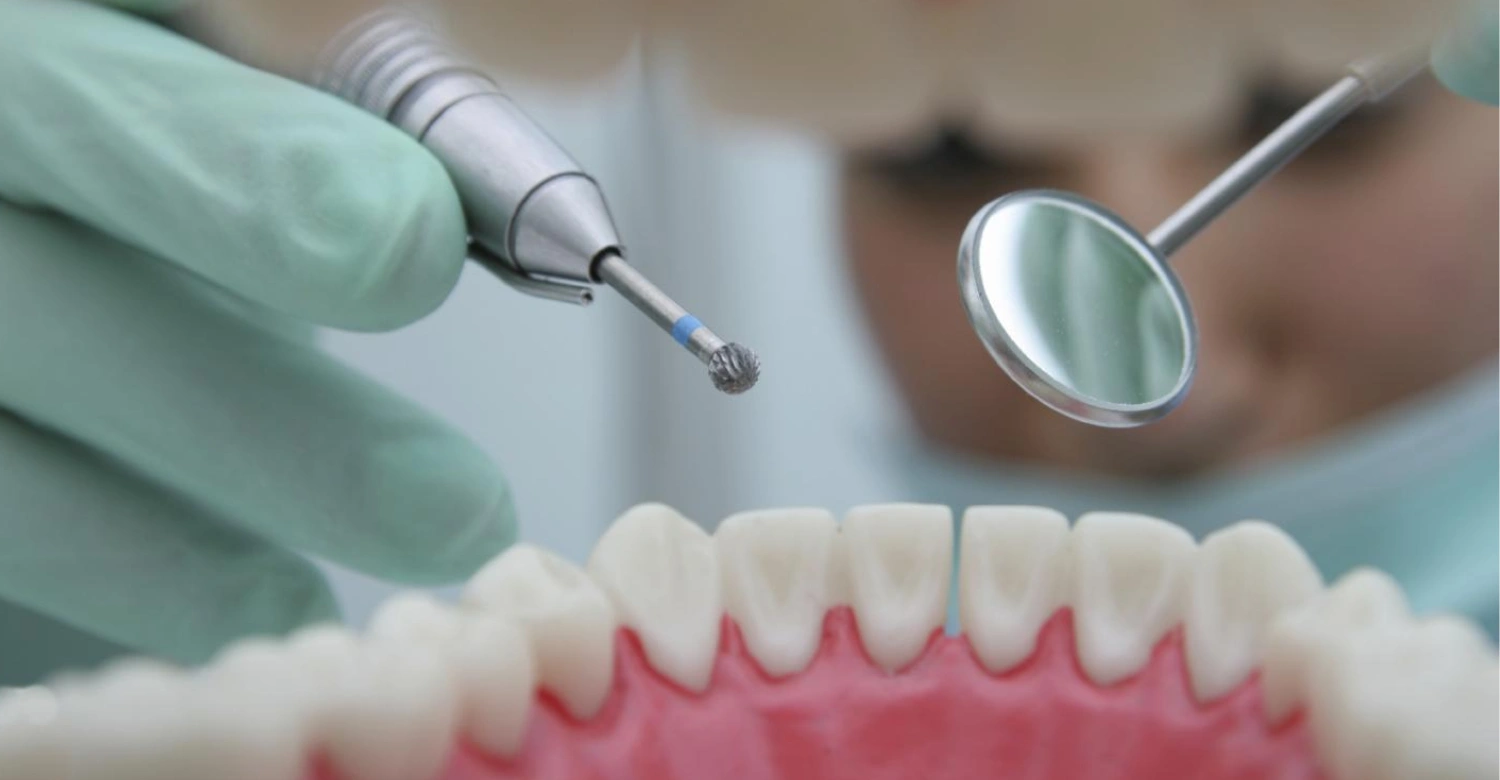
Note : All images used are for editorial and illustrative purposes only and may not originate from the original news provider or associated company.
Read more about roof of mouth rough
What We Notice Clinically About Early Tooth Decay
Clinicians often see important differences between early lesions and established cavities. Early lesions may be subtle and completely asymptomatic. Their appearance can vary depending on activity and location. Some lesions are visible, while others occur between teeth. A lesion may also remain stable rather than progressing continuously. This makes diagnosis more complex than simply looking for a hole. Dentists consider texture, color, location, radiographic appearance, and patient risk. Monitoring changes over time can provide additional information. Digital technology may improve visualization and documentation. The clinical objective is to identify disease early enough for Drill Free Tooth Decay Treatment when appropriate. Vitrin Clinic uses individualized assessment to connect diagnostic findings with treatment planning.
Why Early Diagnosis Matters
Early diagnosis can prevent minor mineral changes from becoming major structural defects. Noncavitated lesions provide more opportunities for Drill Free Tooth Decay Treatment. Once a cavity forms, restorative treatment becomes more likely. Early assessment can also identify multiple risk factors simultaneously. This allows prevention to begin before additional teeth become affected. WHO data highlight the continuing global burden of untreated dental caries. Regular examinations are therefore valuable even when patients feel no pain. Dental caries can progress silently for significant periods. A preventive visit can identify problems before they become emergencies. Early diagnosis ultimately supports tooth preservation and potentially simpler treatment.
How Dentists Determine If Decay Can Be Treated Without Drilling
Dentists combine clinical examination with diagnostic information. They assess whether the surface is intact or cavitated. Lesion location and depth also influence treatment decisions. Patient caries risk helps determine how aggressively the disease should be controlled. Radiographs may reveal lesions hidden between teeth. Digital imaging can support documentation and treatment planning. The dentist may also evaluate plaque, diet, saliva, and previous caries experience. These findings are interpreted together rather than individually. A small visible spot does not automatically mean that no treatment is needed. Likewise, discoloration does not automatically indicate active decay. Diagnosis therefore provides the foundation for Drill Free Tooth Decay Treatment.
Visual Dental Examination
Visual examination remains a fundamental part of caries diagnosis. Dentists inspect tooth color, surface texture, plaque accumulation, and structural integrity. Drying the tooth can make early enamel changes easier to identify. Lesion activity may be estimated through surface characteristics and location. The dentist also looks for cavitation, food trapping, and areas that are difficult to clean. Magnification or enhanced lighting may improve examination. However, visual inspection alone may not detect every proximal lesion. Additional diagnostic methods may therefore be necessary. Patients should avoid interpreting a normal-looking surface as proof that no hidden disease exists. Professional examination remains more reliable than self-assessment.
Dental X-Rays
Dental radiographs can help identify decay that is difficult to see clinically. Bitewing radiographs are particularly useful for detecting proximal lesions. Imaging can also provide information about lesion depth. Radiographs do not replace clinical examination because they have limitations. Very early mineral changes may not always be visible. Interpretation should therefore consider clinical findings and risk factors. Modern digital radiography can provide efficient image acquisition and documentation. Radiation exposure should always be justified according to clinical need. CBCT is generally reserved for specific diagnostic situations rather than routine caries screening. The dentist chooses imaging based on the question that needs answering.
Digital Diagnostic Technology
Digital technology can improve documentation, visualization, and treatment planning. Digital X-rays provide rapidly available diagnostic images. Intraoral scanners can create detailed digital records of tooth surfaces. These records may support monitoring and communication. Advanced systems can also help dentists compare changes during follow-up. CBCT can provide three-dimensional information when clinically indicated. However, technology does not replace clinical judgment. A digital image must still be interpreted within the patient's overall risk profile. Vitrin Clinic integrates digital diagnostics into personalized treatment planning when appropriate. The purpose is to improve decision-making rather than use technology simply because it is available.
Clinical Note: Not Every Dark Spot Is a Cavity
Dark areas on teeth can have several causes. Staining, pigmentation, old restorations, developmental changes, and inactive lesions can all alter tooth color. A dark fissure does not automatically mean that drilling is necessary. Dentists evaluate surface hardness, cavitation, lesion activity, and radiographic findings. They also consider whether the area can be cleaned effectively. Treating every dark mark could lead to unnecessary removal of healthy tissue. Ignoring every dark mark could allow active disease to progress. Diagnosis therefore requires balance. Patients should avoid self-diagnosing cavities from photographs or mirrors. Professional examination provides a more reliable basis for treatment decisions.
Explore the causes of jaw hurts on one side when chewing
Drill Free Tooth Decay Treatment at Vitrin Clinic
Vitrin Clinic approaches conservative dental care through diagnosis, treatment planning, and personalized clinical assessment. The first objective is determining the condition of the tooth. The team can evaluate whether a lesion appears early, noncavitated, cavitated, or advanced. Treatment recommendations depend on these findings. Digital diagnostics can support clinical evaluation when indicated. Preventive strategies may be discussed when the tooth remains suitable for Drill Free Tooth Decay Treatment. Restorative treatment may be recommended when structural loss requires reconstruction. The treatment plan should therefore reflect disease severity rather than a patient's preference for one technique. International patients can also receive coordinated treatment planning before their dental journey. Vitrin Clinic's broader digital workflow supports communication between diagnosis and treatment.
How Vitrin Clinic Evaluates Tooth Decay
Evaluation begins with a clinical dental examination. The dentist reviews visible changes, symptoms, existing restorations, and caries risk. Imaging may be recommended when disease cannot be fully assessed visually. The team considers lesion location and potential progression. Patient habits can also influence treatment recommendations. Diet, oral hygiene, fluoride exposure, and dry mouth are relevant factors. The goal is to determine whether Drill Free Tooth Decay Treatment is appropriate. If restoration is necessary, the treatment plan should still aim to preserve healthy structure. Vitrin Clinic can combine clinical examination with digital diagnostic tools. This supports a structured approach for patients seeking modern dental care in Istanbul.
Digital Diagnostics and Personalized Treatment Planning
Digital diagnostics can provide additional information during dental assessment. Digital X-rays can help visualize areas hidden from direct inspection. Intraoral scanning can document tooth surfaces and support treatment planning. These tools can improve communication between clinicians and patients. They may also help create organized digital records for treatment progression. However, each diagnostic tool should have a clinical purpose. Vitrin Clinic uses digital workflows to support individualized planning. Treatment recommendations are based on the complete diagnostic picture. This approach can be particularly useful for international patients coordinating multiple dental procedures. Personalized planning helps ensure that conservative options are considered before more invasive treatment when clinically appropriate.
Digital X-Rays and CBCT When Needed
Digital X-rays can help identify proximal or deeper lesions that are difficult to assess visually. Bitewing imaging is often useful when evaluating interproximal caries. CBCT provides three-dimensional information but is not routinely required for ordinary caries detection. Its use should depend on the diagnostic question and clinical indication. Vitrin Clinic can incorporate digital imaging when the dentist considers it appropriate. The objective is accurate diagnosis rather than unnecessary imaging. Patients should ask why a particular scan is recommended. Understanding the purpose of imaging can make treatment planning more transparent. Diagnostic technology should always complement clinical examination rather than replace it.
Digital Scanning and Treatment Planning
Intraoral scanning creates a digital representation of dental structures. It can help document tooth surfaces and existing dental conditions. Digital records may support treatment planning and patient communication. Scans can also contribute to restorative and cosmetic workflows when treatment becomes necessary. Vitrin Clinic uses modern digital technology as part of its broader treatment-planning process. The value of scanning depends on how clinicians interpret and use the information. A digital scan cannot independently diagnose every type of dental disease. It works best alongside clinical examination and appropriate imaging. This integrated approach supports more organized and individualized dental care.
Dr. Rifat Alsaman's Clinical Perspective
Dr. Rifat Alsaman emphasizes that conservative dentistry begins with accurate diagnosis. Patients should not be promised Drill Free Tooth Decay Treatment before the tooth is examined. Some lesions can be managed without conventional restoration, while others require intervention. The distinction depends on disease stage and structural integrity. Preserving healthy tooth structure remains an important principle of modern dentistry. However, preservation should never compromise disease control. At Vitrin Clinic, treatment planning considers the patient's clinical findings and long-term needs. Digital diagnostics can support this process when appropriate. The final recommendation should balance comfort, preservation, effectiveness, and prognosis. This patient-centered approach helps avoid both unnecessary treatment and harmful delays.
Learn more about melioidosis and oral health
Why Choose Vitrin Clinic for Modern Cavity Treatment?
Vitrin Clinic combines clinical assessment with modern digital dental workflows. The emphasis is on personalized treatment rather than one universal procedure. Patients can receive diagnostic evaluation before a treatment pathway is selected. This can help determine whether conservative or restorative care is appropriate. International patients can also benefit from coordinated treatment planning. Digital records can support communication before and during treatment. The clinic's broader dental services allow treatment plans to adapt when additional procedures are needed. Modern equipment can support efficient clinical workflows. However, technology remains a tool rather than a substitute for clinical expertise. Treatment decisions should always be based on diagnosis and individual needs.
Personalized Treatment Based on Decay Severity
Not every lesion requires the same treatment. Early enamel changes may require preventive management and monitoring. Noncavitated lesions may be candidates for minimally invasive procedures. Cavitated lesions may require restoration. Deeper disease may require more extensive treatment depending on pulp involvement. Vitrin Clinic can structure treatment plans around these clinical differences. This helps avoid a one-size-fits-all approach. Personalized care also considers patient risk factors. Diet, hygiene, previous caries, and saliva can influence long-term outcomes. The final plan should therefore address both the existing lesion and future disease risk.
Modern Dental Technology
Modern dentistry uses digital imaging, scanning, and computer-assisted treatment planning. These technologies can improve documentation and communication. Digital X-rays can provide rapid diagnostic information. Intraoral scanners can capture detailed digital dental records. CBCT can provide three-dimensional information for selected clinical indications. Vitrin Clinic integrates digital technology into its broader dental workflow. Technology can be particularly valuable for international patients who need coordinated treatment. Nevertheless, diagnostic technology should be used selectively. The quality of care depends on combining technology with clinical expertise. Digital tools support decisions rather than making those decisions independently.
Preserving Healthy Tooth Structure
Natural tooth structure has significant biological and mechanical value. Removing unnecessary enamel can weaken the remaining structure. Minimally invasive dentistry therefore emphasizes Drill Free Tooth Decay Treatment whenever appropriate. Early caries management can sometimes avoid conventional restorative preparation. Sealants, fluoride, and resin infiltration are examples of conservative approaches. However, preserving structure does not mean leaving active disease untreated. If structural damage has progressed, restoration may be necessary. Vitrin Clinic considers preservation within the context of disease control. The best treatment is the least invasive approach that safely manages the condition.
Support for International Patients
International patients often need clear planning before traveling for dental treatment. Vitrin Clinic can organize digital records and treatment discussions around the patient's clinical needs. Diagnostic information can help establish a more structured treatment pathway. Patients can discuss expected procedures before arriving in Istanbul. This can make the overall dental journey easier to organize. Treatment planning may also identify situations where Drill Free Tooth Decay Treatment is unsuitable. International dental care should prioritize safety and realistic expectations. Patients should understand that final treatment decisions may change after in-person examination. A professional assessment remains essential before definitive care.
Discover more about chewing pens and teeth
Tips for Patients Considering Drill-Free Cavity Treatment
Patients interested in Drill Free Tooth Decay Treatment should begin with professional diagnosis. Avoiding drilling should not become the only treatment goal. The priority is controlling disease while preserving healthy tissue. Ask whether the lesion is noncavitated or cavitated. Ask how deep the lesion appears and whether it is active. Discuss fluoride, sealants, resin infiltration, or monitoring when appropriate. Maintain consistent oral hygiene and fluoride exposure. Reduce frequent sugar consumption and address dry mouth. Attend recommended follow-up examinations. If a filling is eventually required, early treatment may reduce the amount of tissue that needs restoration. Conservative dentistry works best when patients and clinicians work together.
Schedule an Early Dental Examination
Early examinations provide an opportunity to identify disease before symptoms appear. Many early carious lesions are painless. Waiting for pain can allow structural damage to progress. A dentist can evaluate suspicious areas and determine whether intervention is necessary. Early diagnosis can also identify risk factors affecting multiple teeth. Patients with previous decay may benefit from more frequent monitoring. Professional examinations are especially important when symptoms such as sensitivity or food trapping develop. An early appointment does not automatically mean treatment. It may simply provide reassurance and preventive guidance. The earlier a problem is identified, the greater the potential for Drill Free Tooth Decay Treatment.
Do Not Treat Cavities Without Professional Guidance
Online remedies cannot reliably determine whether a tooth has a cavity. Home fluoride use can support prevention but cannot diagnose lesion depth. Whitening products should not be used as cavity treatments. Abrasive substances may damage enamel without controlling caries. Delaying professional assessment can allow disease to progress. A dentist can distinguish active decay from staining or other conditions. Professional guidance is particularly important when pain, swelling, or persistent sensitivity occurs. Drill Free Tooth Decay Treatment requires diagnosis rather than guesswork. Patients should therefore use home care as part of professional management, not as a substitute for examination.
Ask About Conservative Treatment Options
Patients have the right to ask about treatment alternatives. Ask whether the lesion is cavitated. Ask whether fluoride or remineralization strategies could be appropriate. For selected lesions, ask whether sealants or resin infiltration are options. Ask how the dentist plans to monitor the lesion. Also ask what would indicate that restoration has become necessary. These questions create a clearer understanding of treatment decisions. Conservative options are not appropriate for every lesion. However, discussing them can help prevent unnecessary intervention. A good treatment plan should explain both the recommended option and the reasons behind it.
Use Fluoride-Based Oral Care
Fluoride toothpaste remains a central part of caries prevention. Regular brushing provides repeated fluoride exposure to enamel. Cochrane evidence supports fluoride toothpaste for reducing caries compared with non-fluoride toothpaste. Patients at higher risk may require additional fluoride measures. These should be selected by a dental professional. Children need age-specific guidance because swallowing excessive fluoride can affect developing teeth. Patients should also maintain interdental cleaning. Fluoride works best when combined with effective plaque removal. It should not be viewed as a replacement for professional treatment when a cavity is already established.
Maintain Regular Dental Checkups
Regular checkups allow dentists to monitor existing lesions and identify new changes. Monitoring is particularly important after Drill Free Tooth Decay Treatment. A stable lesion may require continued prevention rather than restoration. A progressing lesion may require treatment escalation. Follow-up also allows the dentist to reassess caries risk. Changes in diet, medication, oral hygiene, or saliva can alter disease activity. Regular examinations can therefore support long-term prevention. The appropriate interval varies between patients. High-risk individuals may need closer monitoring than low-risk patients. Consistent follow-up is an important part of minimally invasive dentistry.
Explore more about tick borne diseases and oral health
When Is Traditional Cavity Treatment Necessary?
Traditional restorative treatment becomes necessary when tooth structure has been lost beyond what conservative methods can manage. Cavitation can create areas that are difficult to clean. Larger defects may compromise tooth strength and function. Deep decay can threaten the dental pulp. Symptoms such as spontaneous pain may indicate advanced disease. The dentist evaluates these factors before recommending restoration. Conventional treatment should not be viewed as a failure of preventive dentistry. Sometimes restoration is the most conservative option remaining. The objective remains to remove diseased tissue appropriately and preserve sound structure. Delaying necessary treatment can increase complexity and risk.
When Decay Has Progressed Beyond the Enamel
Decay extending into dentin requires careful assessment. Dentin is more vulnerable to rapid progression than enamel. Drill Free Tooth Decay Treatment decision depends on lesion depth and cavitation. Some noncavitated dentinal lesions may be monitored or managed conservatively. Others require restorative intervention. Radiographic findings can help estimate lesion extent. The dentist also considers whether the lesion can remain cleanable. Patient carries risk influences the likelihood of progression. Therefore, dentin involvement does not automatically dictate one procedure. Clinical context determines the safest treatment. Early detection remains valuable because deeper lesions generally provide fewer conservative options.
When Tooth Structure Has Been Lost
Lost tooth structure cannot simply be restored through fluoride exposure. Remineralization can strengthen remaining minerals but cannot rebuild a missing wall. A cavity may therefore require restorative material to restore shape and function. The amount of preparation should still be minimized when possible. Modern restorative dentistry increasingly emphasizes tissue preservation. The dentist may select materials and techniques based on lesion size and location. Delaying restoration can allow the cavity to enlarge. Larger defects may eventually require more complex procedures. Prompt professional assessment can therefore help prevent unnecessary progression.
When Decay Is Deep
Deep decay requires careful treatment planning because the pulp may be threatened. Symptoms can include prolonged sensitivity, spontaneous pain, or pain triggered by temperature. However, severe disease can sometimes remain asymptomatic. Radiographs can help assess the depth of a lesion. The dentist may use additional tests to evaluate pulp status. Treatment can range from conservative restoration to more extensive therapy. The exact choice depends on the tooth's condition. Deep lesions should never be managed solely through home remedies. Professional assessment becomes especially important when pain or swelling develops.
Symptoms of Advanced Tooth Decay
Advanced decay may cause persistent toothache or sensitivity. Pain can occur during eating, drinking, or exposure to hot and cold temperatures. Some patients experience spontaneous pain without an obvious trigger. Food may become trapped inside a cavity. A visible hole or broken tooth structure may also appear. Swelling can indicate infection and requires prompt dental attention. Bad taste or drainage can occur when an abscess develops. Symptoms vary considerably between individuals. Therefore, symptom severity does not always reflect lesion depth. Any persistent dental symptom deserves professional assessment.
Why Delaying Treatment Can Make Decay Worse
Active decay can continue destroying mineral and structural tissue. A small lesion may become a larger cavity over time. Deeper progression can increase sensitivity and treatment complexity. If disease reaches the pulp, root canal treatment may become necessary. Infection can also spread beyond the tooth. Early treatment can therefore reduce the risk of advanced complications. Avoiding drilling should not mean avoiding dental care. Drill Free Tooth Decay Treatment depends on treating disease at the earliest suitable stage. Patients who are concerned about restorative treatment should discuss minimally invasive options promptly. Early professional advice provides more choices than delayed intervention.
Dr. Rifat Alsaman's Opinion on When Fillings Are Needed
Dr. Rifat Alsaman emphasizes that fillings should be recommended according to clinical necessity. A visible spot alone does not automatically justify restoration. However, a true cavity with structural breakdown may require restorative treatment. The objective is to control disease while preserving as much sound tooth structure as possible. Treatment decisions should consider lesion activity, depth, cleansability, and progression risk. If Drill Free Tooth Decay Treatment cannot predictably control the disease, restoration may be safer. Patients should receive a clear explanation of why a filling is recommended. At Vitrin Clinic, personalized treatment planning supports this decision-making process. The priority remains long-term oral health rather than avoiding a particular instrument.
Learn more about H5N1 bird flu and oral health
Conclusion: Is Drill Free Tooth Decay Treatment Right for You?
Conservative caries management can be highly valuable when decay is detected early. Noncavitated lesions may respond to fluoride, remineralization, sealants, or resin infiltration. The suitability of each option depends on clinical diagnosis. Established cavities with structural loss may require restorative treatment. Therefore, the phrase "drill-free" should never replace proper dental assessment. Modern dentistry increasingly focuses on preserving healthy tooth structure and controlling disease early. Evidence supports nonrestorative approaches for selected carious lesions. Fluoride remains an important component of prevention and early management. Vitrin Clinic can evaluate individual dental conditions using clinical examination and appropriate digital diagnostics. The best treatment is the one that safely controls disease while preserving natural tooth structure.
Drill-Free Tooth Decay Treatment at Vitrin Clinic
For patients considering a Hollywood Smile at Vitrin Clinic, addressing existing tooth decay is an important part of preparing the teeth for cosmetic treatment. When suitable, Drill-Free Tooth Decay Treatment can provide a conservative approach for managing certain early cavities before moving forward with cosmetic smile enhancement. Once the teeth and gums are healthy and ready for treatment, patients can explore Vitrin Clinic’s Hollywood Smile treatment to improve the overall appearance, shape, and harmony of their smile. A personalized assessment helps determine the most appropriate sequence of treatments for each patient.
Reference
FAQs
Dr. Rifat Alsaman has more than 5 years of clinical experience in dentistry and currently serves as the Head of the Medical Team at Vitrin Clinic. He is dedicated to providing exceptional patient care, overseeing treatment planning, and ensuring the highest clinical standards across the team. His expertise, attention to detail, and commitment to continuous professional development have helped countless patients achieve healthier, more confident smiles.


