General Dentistry
September 14, 2026
What Are the Muscles Used for Chewing? Anatomy, Function & Movement
 Dr. Rifat Alsaman
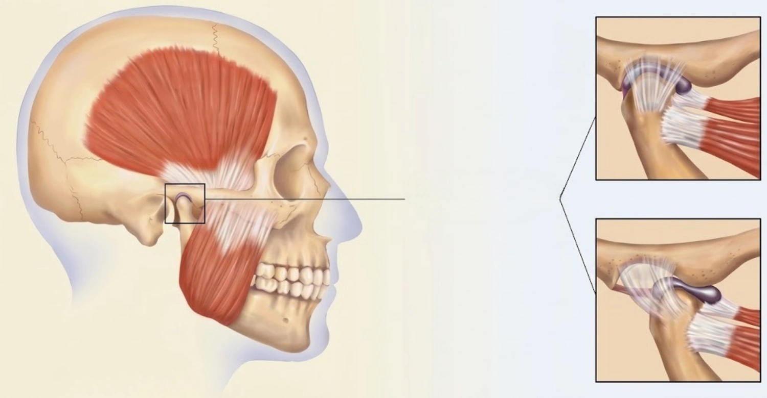
The muscles used for chewing are a coordinated group of muscles that move the lower jaw during biting, crushing, grinding, and food preparation. The four primary muscles are the masseter, temporalis, medial pterygoid, and lateral pterygoid. Together, they control important mandibular movements, including elevation, depression, protrusion, retraction, and side-to-side motion. The masseter, temporalis, and medial pterygoid mainly contribute to closing the jaw, while the lateral pterygoid helps with forward and lateral movements. These muscles work with the teeth, temporomandibular joints, tongue, and nervous system to create efficient mastication. The mandibular division of the trigeminal nerve provides their primary motor supply. Sensory feedback also helps regulate chewing force according to food texture and resistance. Understanding this anatomy can help explain jaw movement, chewing efficiency, muscle fatigue, and some causes of jaw discomfort. Vitrin Clinic considers oral function alongside dental structures when planning appropriate restorative and implant treatments.
What Are the Muscles Used for Chewing?
These muscles allow the lower jaw to move during biting, crushing, grinding, and food preparation. Four primary muscles control most mandibular movements. These include the masseter, temporalis, medial pterygoid, and lateral pterygoid. Each muscle performs different actions while working with the others. Their combined movements allow elevation, depression, protrusion, retraction, and side-to-side movement. The trigeminal nerve provides the primary motor supply to these muscles. Chewing also depends on sensory feedback from teeth, muscles, joints, and oral tissues. This feedback helps adjust force according to food texture and resistance. Vitrin Clinic considers these functional relationships when evaluating complex restorative and implant cases.
The Four Main Muscles of Mastication
The four primary muscles of mastication are the masseter, temporalis, medial pterygoid, and lateral pterygoid. Together, these muscles control most movements of the mandible. The masseter provides powerful jaw elevation and assists forward movement. The temporalis elevates and retracts the mandible. The medial pterygoid assists elevation, protrusion, and grinding movements. The lateral pterygoid contributes to opening, protrusion, and side-to-side movements. This muscle group therefore functions as a coordinated system rather than independent structures. Their activity changes throughout every chewing cycle. This coordination allows people to adapt chewing movements to different foods. Accessory muscles also contribute to jaw opening and stabilization when required.
Masseter Muscle
The masseter is a thick muscle located along the outer surface of the mandible. It extends between the zygomatic arch and mandibular region. Its primary action is elevating the mandible during jaw closure. The masseter also contributes to mandibular protrusion and stabilization. Its strong contraction supports forceful biting when food requires greater pressure. The muscles used for chewing depend on coordinated activation, so the masseter does not work alone. Temporalis and medial pterygoid activity supports effective mandibular closure. The masseter can also become highly active during clenching. Persistent clenching may contribute to muscular fatigue or tenderness. Clinicians can assess this muscle through external palpation. Its accessibility makes it particularly useful during examinations involving jaw discomfort.
Temporalis Muscle
The temporalis is a broad, fan-shaped muscle covering much of the temporal region. It extends toward the coronoid process of the mandible. Its anterior and middle fibers mainly elevate the mandible. Its posterior fibers contribute to mandibular retraction. The temporalis therefore supports both jaw closure and controlled positioning. The jaw muscles require precise timing during these movements. Temporalis activity can also help stabilize the mandible during tooth contact. Prolonged clenching may increase temporalis activity and contribute to discomfort. Some patients notice tenderness around the temple after waking. Others experience symptoms during prolonged concentration. Clinical evaluation can help determine whether muscular activity contributes to those symptoms.
Medial Pterygoid Muscle
The medial pterygoid is located deep within the facial skeleton. It has superficial and deep portions with different anatomical origins. The muscle attaches to the medial surface of the mandibular region. Its primary actions include mandibular elevation and protrusion. It also assists side-to-side movement during grinding. The muscles used for chewing rely on the medial pterygoid for controlled grinding movements. Unilateral activation can help produce lateral mandibular movement. This action helps position food between opposing tooth surfaces. The muscle also works closely with the masseter during jaw closure. Its deep location makes direct examination more challenging. Nevertheless, its functional contribution remains essential for normal mastication.
Lateral Pterygoid Muscle
The lateral pterygoid is positioned deep within the infratemporal region. It contains superior and inferior heads with different anatomical attachments. Bilateral contraction helps move the mandible forward. Unilateral contraction contributes to side-to-side mandibular movement. The inferior head also contributes to jaw opening through forward condylar movement. These jaw muscles depend on this muscle for controlled mandibular positioning. Its relationship with the temporomandibular joint makes it clinically important. The superior and inferior heads can demonstrate different activity patterns. This complexity allows the mandible to perform several coordinated movements. Problems affecting this region can sometimes influence chewing comfort and jaw mobility.
Clinical Note: Why These Muscles Matter for Jaw Function
Healthy chewing requires coordinated muscle activity rather than maximum muscular strength. The masseter, temporalis, and pterygoid muscles must work with the teeth and temporomandibular joints. The chewing muscles can become overloaded through repeated clenching or grinding. Temporomandibular disorders can also involve jaw muscles and nearby joint structures. NIDCR reports that TMD affects about 5% of adults in the United States. The organization also notes that prevalence varies between populations and diagnostic methods. Symptoms can include jaw pain, facial discomfort, and difficulty moving the jaw. Vitrin Clinic considers functional symptoms alongside dental findings during appropriate clinical evaluations. This approach can be particularly useful during extensive restorative treatment.
Find out more about opioids and dental pain
What Does Each Chewing Muscle Do?
Each primary muscle performs specific actions during mandibular movement. However, their functions overlap throughout the chewing cycle. The muscles used for chewing continuously adjust their activity according to food resistance. Harder foods can require stronger and more controlled muscle activation. Softer foods usually require less mechanical force. The nervous system coordinates these changes through sensory feedback. This process allows chewing to remain efficient while food characteristics change. Muscle coordination also helps maintain stable tooth contact. Understanding individual muscle actions makes jaw symptoms easier to interpret. It also helps explain why chewing involves more than simple opening and closing. Each movement depends on coordinated activity across several anatomical structures.
Masseter Muscle
The masseter is one of the primary muscles responsible for powerful mandibular elevation. It contracts when the lower jaw moves toward the upper jaw. This group of muscles works together during this closing movement. The masseter also contributes to forward mandibular movement. Its activity can increase when chewing resistant or firm foods. Strong contraction helps stabilize the mandible during forceful biting. However, continuous contraction can increase muscular workload. Clenching and grinding can therefore place additional demands on the masseter. Some patients may experience tenderness near the jaw angle. Dental professionals can assess this area during a clinical examination. Muscle findings should always be interpreted alongside dental and joint findings.
Location and Anatomy
The masseter occupies the lateral aspect of the mandibular ramus. It originates primarily from the zygomatic arch and nearby structures. Its fibers extend toward the lateral mandibular surface and angle. The muscles used for chewing have different orientations that create different mandibular movements. The masseter contains superficial and deeper fiber layers. These fibers contribute to elevation and stabilization. The muscle is easily felt when a person gently clenches the teeth. This makes it useful during external clinical assessment. Its position also explains why jaw-angle tenderness can involve muscular tissue. Anatomy varies between individuals, so examination remains important.
Main Function
The main function of the masseter is mandibular elevation. This movement brings the lower teeth toward the upper teeth. These mastication muscles coordinate this action with the temporalis and medial pterygoid. The masseter can also assist mandibular protrusion. Its strong contraction helps generate force during biting. The required force depends on food consistency and resistance. Sensory feedback helps the nervous system regulate this force. Excessive activation can occur during clenching or grinding. Persistent symptoms should be assessed rather than assumed to result from muscle activity alone.
Temporalis Muscle
The temporalis contributes significantly to mandibular elevation and retraction. Its anterior fibers are particularly involved in elevation. Posterior fibers contribute more strongly to backward mandibular movement. The muscles responsible for chewing rely on this division of function for controlled jaw positioning. The temporalis also helps stabilize the mandible during tooth contact. Excessive activation can accompany clenching or sustained jaw tension. Some people may notice temple discomfort after prolonged jaw activity. Others may experience fatigue without significant pain. Clinical assessment can help identify whether muscle activity contributes to symptoms.
Location and Anatomy
The temporalis covers a large area of the temporal region. It originates from the temporal fossa and associated fascia. Its fibers converge toward a tendon beneath the zygomatic arch. The tendon attaches to the mandibular coronoid process. The muscles used for chewing have different attachment patterns that influence their mechanical roles. The fan-shaped temporalis allows several fiber groups to contribute to mandibular movement. Its posterior fibers provide an important retraction component. This anatomy supports controlled jaw positioning throughout chewing.
Main Function
The temporalis primarily elevates and retracts the mandible. Elevation helps close the mouth during biting and chewing. Retraction moves the mandible backward after forward movement. These four muscles coordinate these actions with the pterygoid muscles. Temporalis activity can also stabilize the jaw during tooth contact. Persistent clenching may increase its workload. This can contribute to tenderness around the temple in some patients. Symptoms should be evaluated according to their duration and severity.
Medial Pterygoid Muscle
The medial pterygoid contributes to mandibular elevation and protrusion. It also assists lateral grinding movements. The muscles used for chewing depend on this muscle when the mandible moves laterally. Its action complements the masseter during forceful jaw closure. Unilateral activation contributes to lateral mandibular movement. This movement helps food pass across different tooth surfaces. The muscle lies deep within the facial region. Its position makes direct palpation more difficult than masseter assessment. Nevertheless, its function remains important during normal mastication.
Location and Anatomy
The medial pterygoid lies on the inner aspect of the mandibular ramus. It has superficial and deep heads. These portions originate from structures associated with the maxilla and sphenoid region. The muscle inserts near the medial mandibular angle. The masticatory muscles have anatomical relationships that determine their individual mechanical roles. The medial pterygoid works closely with the masseter. Together, they create a functional muscular sling around the mandibular angle. This arrangement contributes to effective jaw elevation and stabilization.
Main Function
The medial pterygoid elevates and protrudes the mandible. It also contributes to lateral grinding movements. These muscles use this movement to process food across opposing teeth. Alternating activation supports controlled side-to-side jaw motion. The muscle also works with the lateral pterygoid during mandibular movement. Its activity changes according to the chewing phase. Efficient grinding therefore requires coordinated rather than isolated contraction.
Lateral Pterygoid Muscle
The lateral pterygoid plays an important role in forward and lateral mandibular movement. It contains superior and inferior heads. Bilateral activation contributes to mandibular protrusion. Unilateral activation produces lateral movement. The muscles used for chewing depend on this movement for efficient food positioning. The muscle also interacts closely with the temporomandibular joint. Its anatomical attachment includes structures associated with the joint capsule and articular disc. This relationship gives the muscle particular clinical importance. Abnormal function may influence mandibular movement and chewing comfort.
Location and Anatomy
The lateral pterygoid lies deep in the infratemporal region. Its superior head originates from the greater wing of the sphenoid. Its inferior head originates from the lateral pterygoid plate. The muscle attaches toward the mandibular condyle and joint structures. This muscle group therefore connects several structures involved in mandibular movement. Its deep position makes clinical assessment different from superficial muscle examination. Its relationship with the joint helps explain its role during opening and protrusion.
Main Function
The lateral pterygoid contributes to protrusion and lateral mandibular movement. It also participates in jaw opening through forward condylar translation. The muscles used for chewing require this coordinated movement for efficient food positioning. Bilateral activity moves the mandible forward. Unilateral activity creates controlled lateral movement. Its superior and inferior heads can perform different roles. These coordinated actions allow the jaw to transition smoothly between chewing phases.
Discover the connection between heartburn and jaw pain
Which Muscle in an Animal's Head Is Used for Chewing?
Animals have specialized jaw muscles adapted to their diets and feeding behaviors. Mammals generally possess muscles corresponding to major human masticatory structures. However, muscle proportions and jaw mechanics vary considerably between species. The jaw muscles reflect evolutionary adaptations to different food types. Herbivores often require extensive grinding movements. Carnivores commonly emphasize powerful jaw closure and cutting. Omnivores combine mechanical strategies for different foods. Comparative anatomy demonstrates how diet influences skull structure, teeth, and muscle organization. These differences make animal chewing systems valuable for understanding mammalian anatomy.
Chewing Muscles in Mammals
Mammalian chewing systems generally contain homologous muscles with species-specific adaptations. Their jaws can differ substantially in movement patterns. Some species emphasize vertical movement during feeding. Others require extensive lateral movement for grinding. The muscles used for chewing therefore vary in size and functional emphasis. Tooth shape also influences the required jaw movement. Incisors, canines, premolars, and molars perform different mechanical tasks. Muscle activity adapts accordingly. These relationships demonstrate how feeding behavior shapes the entire masticatory system.
How Diet Influences Jaw Muscles
Diet determines many mechanical requirements placed on the jaw. Fibrous foods require repeated grinding and controlled lateral movement. Tough foods may require substantial bite force. Softer foods usually require less mechanical processing. These jaw muscles adapt their activity according to these demands. Animal studies show clear relationships between diet and craniofacial anatomy. Jaw shape, tooth morphology, and muscle development can differ between feeding groups. Human chewing also adapts according to food texture. Sensory feedback helps regulate force and movement during each chewing cycle.
Herbivores vs. Carnivores
Herbivores often require prolonged grinding because plant tissues can be mechanically resistant. Their jaw movements may include substantial lateral components. Carnivores commonly emphasize vertical closure for cutting and tearing. Their tooth shapes reflect these feeding requirements. The chewing muscles therefore operate within different mechanical systems. Omnivores combine elements of both patterns. Humans can process many food types through adaptable mandibular movements. This flexibility is supported by coordinated muscles, teeth, joints, and sensory feedback.
Human and Animal Chewing Muscles
Humans share the four primary masticatory muscles with many other mammals. However, human jaw structure reflects evolutionary, dietary, and functional changes. The muscles used for chewing coordinate with teeth and temporomandibular joints. Human chewing includes vertical crushing and lateral grinding. Brainstem circuits generate rhythmic movement while sensory feedback modifies the pattern. Animal species demonstrate different versions of these same principles. Their jaw mechanics reflect their diets and feeding strategies. Comparative anatomy therefore helps explain why chewing systems differ between species.
Learn about myotonic dystrophy and oral health
How Do the Muscles Used for Chewing Work Together?
This group of muscles operates as a coordinated neuromuscular system. Different muscles contract during different phases of each chewing cycle. Jaw elevation requires coordinated activity from several closing muscles. Opening involves the lateral pterygoid and accessory muscles. Protrusion and lateral movement require carefully timed activation. Sensory receptors provide continuous feedback about tooth contact and food resistance. This allows the nervous system to adjust force and movement continuously. Chewing is therefore rhythmic but highly adaptable. The same basic pattern can change according to food texture, age, and individual characteristics.
Jaw Elevation
Jaw elevation moves the mandible upward toward the maxilla. The masseter, temporalis, and medial pterygoid contribute strongly to this movement. The muscles used for chewing coordinate their contraction to produce controlled jaw closure. The amount of force depends on food resistance. Harder foods can require stronger activation. Softer foods usually require less force. Sensory feedback helps prevent unnecessary loading. This allows chewing to remain efficient across different food textures.
Jaw Depression
Jaw depression moves the mandible downward and opens the mouth. The lateral pterygoid contributes to this movement through forward condylar translation. Accessory muscles also assist mandibular opening. These mastication muscles therefore interact with additional muscles during opening. Gravity also contributes when the jaw moves downward. Normal opening prepares the mouth for food intake. It also creates space for the next chewing cycle.
Jaw Protrusion and Retraction
Protrusion moves the mandible forward relative to the upper jaw. The lateral pterygoid contributes strongly to this action. The medial pterygoid and masseter can also assist forward movement. Retraction moves the mandible backward. Posterior temporalis fibers contribute significantly to this movement. The muscles responsible for chewing coordinate these opposing actions throughout mastication. Forward movement can help position food between teeth. Backward movement helps return the mandible toward its starting position.
Side-to-Side Jaw Movement
Side-to-side movement is essential for grinding food. It depends heavily on coordinated pterygoid muscle activity. Unilateral activation produces movement toward the opposite side. The muscles used for chewing use this movement to distribute food across different tooth surfaces. Grinding becomes especially important when processing fibrous or resistant foods. Alternating movements help reduce food particle size. The tongue simultaneously repositions food within the oral cavity. This creates a coordinated chewing sequence.
Clinical Note: Muscle Coordination During Mastication
Chewing is controlled by rhythmic neural activity and continuous sensory feedback. Research identifies a brainstem central pattern generator involved in this rhythm. These four muscles receive coordinated motor signals throughout each cycle. Sensory receptors monitor tooth contact, muscle stretch, and food properties. The nervous system then adjusts movement according to incoming information. This explains why chewing changes when food becomes harder or softer. It also explains why jaw movement is not completely repetitive. Clinical symptoms can occur when muscles, joints, teeth, or neural control become disrupted.
Find out how to get feeling back in your mouth after the dentist
What Is the Process of Chewing?
Chewing transforms food into smaller particles suitable for swallowing. The process combines teeth, jaw muscles, tongue movement, saliva, and sensory feedback. The muscles used for chewing repeatedly move the mandible through coordinated cycles. Each cycle can change according to food texture and resistance. Hard foods generally require stronger mechanical processing. Softer foods often require fewer chewing cycles. Sensory feedback helps adjust movement throughout the process. The tongue continuously repositions food between chewing surfaces. Saliva also helps lubricate and organize the developing food bolus. Efficient mastication therefore involves several systems working simultaneously.
Biting and Jaw Closure
Biting begins when food is positioned between the teeth. The jaw then closes through coordinated muscle contraction. The masseter, temporalis, and medial pterygoid contribute significantly to closure. The masticatory muscles regulate force according to food resistance. Stronger foods require greater mechanical force. Excessive force is unnecessary for softer foods. Sensory feedback helps control this adjustment. Proper tooth contact also influences the distribution of chewing forces. The first bite begins the process of reducing food into smaller pieces.
Crushing and Grinding Food
After initial biting, repeated jaw movements break food into smaller particles. Vertical movements crush food between opposing teeth. Lateral movements help grind food across the chewing surfaces. These muscles coordinate these directional changes. Pterygoid activity contributes significantly to lateral jaw movement. The tongue continuously moves food back toward appropriate tooth surfaces. This repeated process gradually creates smaller and more manageable particles. The required number of cycles varies according to food properties.
Preparing Food for Swallowing
Chewing continues until food becomes sufficiently processed for swallowing. Saliva moistens the particles and contributes to bolus formation. The tongue gathers the processed material into a cohesive mass. The muscles used for chewing support the repeated movements needed for adequate food breakdown. Sensory feedback helps determine when additional chewing is necessary. Once the bolus reaches an appropriate consistency, swallowing can begin. Efficient preparation reduces the mechanical demands placed on later swallowing stages.
How Teeth and Jaw Muscles Work Together
Teeth provide hard surfaces that mechanically break down food. Jaw muscles generate the forces needed to move those surfaces together. This muscle group adjusts force according to sensory information from oral tissues. Periodontal receptors can provide information about tooth loading. The tongue then repositions food between the teeth. This interaction creates a highly coordinated mechanical system. Damaged or missing teeth can alter how forces are distributed. Restorative treatment may therefore consider both tooth structure and functional movement.
Explore how tongue posture affects teeth
What Controls the Muscles Used for Chewing?
The nervous system controls chewing through motor signals and sensory feedback. The trigeminal nerve provides the primary motor supply to the four main muscles. Brainstem networks generate rhythmic chewing patterns. Higher brain regions can modify these patterns according to voluntary actions. Sensory receptors provide information about food, teeth, muscles, and joints. The muscles used for chewing respond to these continuously changing signals. This creates adaptable rather than fixed jaw movements. Research describes mastication as a distributed neural process involving several interconnected systems.
The Trigeminal Nerve and Mastication
The trigeminal nerve is cranial nerve five. Its mandibular division carries the primary motor supply for mastication. The jaw muscles receive motor signals through branches of this division. The same nerve also carries important sensory information from oral structures. This creates a close connection between movement and sensation. The nervous system can therefore modify muscle activity according to incoming sensory information. This mechanism supports controlled biting and chewing.
Mandibular Division of the Trigeminal Nerve
The mandibular division is called V3. It is the only trigeminal division containing substantial motor fibers. V3 supplies the masseter, temporalis, medial pterygoid, and lateral pterygoid. These jaw muscles therefore share an important neural pathway. V3 also carries sensory information from several oral and facial structures. This combination supports both motor control and sensory monitoring. Damage to relevant neural pathways can affect jaw movement or sensation.
Motor Control of Mastication
Motor signals activate specific muscles according to the desired jaw movement. Closing requires coordinated activation of several elevator muscles. Opening involves different muscles and mandibular translation. Lateral movement requires asymmetric muscle activation. The muscles used for chewing therefore receive precisely timed signals. Brainstem circuits contribute to rhythmic movement generation. Higher brain centers can modify these movements according to voluntary control. Sensory feedback then fine-tunes ongoing muscle activity.
Brain and Neuromuscular Control
Chewing involves brainstem networks, cortical input, and sensory feedback. A central pattern generator produces the basic rhythmic pattern. The chewing muscles then receive coordinated activation during different movement phases. Sensory feedback modifies the pattern according to food properties. Higher brain regions can also influence chewing behavior. This distributed system allows automatic movement while preserving adaptability. Modern research continues investigating the detailed neural mechanisms behind mastication.
Chewing Rhythm
Chewing follows a repeating sequence of opening, closing, and food-processing movements. Brainstem circuits generate the basic rhythmic pattern. The muscles used for chewing activate according to specific phases within this cycle. Food properties can modify chewing speed and force. Voluntary control can also interrupt or change the rhythm. This makes chewing automatic while remaining flexible. The rhythm is therefore not identical during every meal.
Sensory Feedback
Sensory feedback comes from teeth, muscles, joints, and oral tissues. Periodontal mechanoreceptors provide information about tooth loading. Muscle receptors provide information about muscle length and movement. This group of muscles adjusts its activity according to this information. Harder food can trigger changes in chewing force. Unexpected tooth contact can also modify movement. This feedback system protects against inappropriate force and improves chewing efficiency.
Bite Force Regulation
Bite force must match the mechanical resistance of food. Excessive force can increase unnecessary loading on teeth and supporting structures. Insufficient force may not adequately process resistant food. The muscles used for chewing adjust force through sensory and neural mechanisms. Food texture provides important information for this adjustment. Research describes sensory feedback as a major component of adaptive mastication. This regulation allows efficient chewing across different foods.
Note : All images used are for editorial and illustrative purposes only and may not originate from the original news provider or associated company.
Discover more about craniofacial disorders and dental health
What We Notice Clinically
Jaw-muscle symptoms can develop when normal muscular activity becomes excessive or poorly coordinated. Common complaints include tenderness, fatigue, stiffness, and facial discomfort. These mastication muscles may become overloaded through clenching or grinding. Temporomandibular disorders can also involve the masticatory muscles. NIDCR reports that TMD affects about 5% of U.S. adults. Prevalence varies according to diagnostic methods and studied populations. Persistent symptoms should receive professional evaluation. Vitrin Clinic considers symptoms together with dental and functional findings when appropriate.
Signs of Overactive or Imbalanced Jaw Muscles
Overactive jaw muscles can produce tenderness or fatigue. Some patients notice symptoms during chewing. Others experience discomfort after waking or during stressful periods. The muscles responsible for chewing can remain active during unconscious clenching. This creates a workload different from normal rhythmic mastication. Symptoms can also originate from temporomandibular joints or dental structures. Therefore, muscle symptoms should not automatically be treated as isolated muscular problems. A comprehensive examination can help identify contributing factors.
Jaw Muscle Tenderness
Jaw muscle tenderness may occur after prolonged clenching or chewing. The masseter and temporalis can often be assessed through external palpation. The muscles used for chewing may feel sensitive when excessive activity occurs. However, tenderness does not identify the cause by itself. Dental, joint, and neurological factors can produce similar symptoms. Persistent tenderness should therefore receive professional evaluation. Appropriate diagnosis can help guide conservative or restorative treatment.
Jaw Fatigue
Jaw fatigue can feel like heaviness, stiffness, or reduced chewing endurance. It may appear after prolonged chewing or repeated clenching. These four muscles can become tired when activity remains elevated. Temporary fatigue may resolve after rest. Persistent fatigue deserves clinical assessment. A dentist can evaluate teeth, joints, muscles, and functional patterns. Treatment depends on the underlying cause.
Facial Discomfort
Facial discomfort may occur around the cheek, temple, jaw angle, or joint region. TMD can produce pain affecting muscles and nearby structures. The muscles used for chewing may contribute to symptoms when they become overactive. However, facial pain has many possible causes. Persistent or worsening symptoms should receive appropriate professional evaluation. Clinical history helps determine which structures require closer examination.
Clenching and Bruxism
Clenching involves forceful or sustained tooth contact outside normal chewing. Bruxism includes repeated jaw-muscle activity involving clenching or grinding. The masticatory muscles can experience increased workload during these behaviors. Repeated activity may contribute to muscle fatigue and tooth wear. Symptoms vary considerably between individuals. Assessment should consider timing, frequency, dental findings, and reported discomfort. Treatment should address contributing factors rather than symptoms alone.
How Clenching Affects the Jaw Muscles
Clenching keeps jaw-closing muscles active for longer periods. Normal chewing instead alternates activity with periods of relaxation. These muscles may become fatigued when contraction continues unnecessarily. Persistent clenching can also increase mechanical loading on teeth. Patients may notice morning tenderness or facial fatigue. Reducing unnecessary daytime clenching may help some individuals. Persistent symptoms still require professional assessment.
Masseter and Temporal Overactivity
The masseter and temporalis can become highly active during clenching. Their sustained contraction may contribute to tenderness or facial fatigue. The muscles used for chewing can also become more prominent in some people with chronic activity. Muscle size alone cannot diagnose a functional disorder. Clinical assessment should consider symptoms, habits, tooth findings, and jaw movement. A professional evaluation can determine whether muscular overactivity appears clinically significant.
Dr. Rifat Alsaman's Clinical Opinion
Dr. Rifat Alsaman, Head of the Medical Team at Vitrin Clinic and cosmetic dentist, emphasizes functional assessment before complex restorative treatment. This muscle group should be considered alongside teeth, joints, and bite relationships. A restoration should support practical oral function as well as appearance. This principle becomes especially important during extensive rehabilitation. Digital diagnostic tools can help visualize dental structures before treatment. However, technology should support clinical judgment rather than replace it. Each patient's anatomy, symptoms, and treatment objectives require individual consideration.
Learn how to get rid of numbness after the dentist
What Can Cause Pain in the Muscles Used for Chewing?
Jaw-muscle pain can have several possible causes. Common contributors include clenching, grinding, stress, and temporomandibular disorders. Dental problems can also change normal chewing patterns. The muscles used for chewing may then experience altered or increased workload. NIDCR identifies TMDs as a common cause of jaw pain. Some symptoms resolve without extensive treatment. Others can become persistent and require professional management. Accurate diagnosis is important because similar symptoms can originate from different structures.
Teeth Grinding and Clenching
Grinding and clenching increase muscular activity beyond normal chewing requirements. Repeated episodes can place additional stress on jaw muscles. The jaw muscles may become tired or tender after repeated activity. Tooth wear can also occur with persistent grinding. Some people grind during sleep without awareness. Others clench during concentration or stressful situations. Dental evaluation can identify signs associated with these behaviors. Management depends on the individual clinical situation.
Temporomandibular Disorders
Temporomandibular disorders affect the jaw joints, muscles, or associated structures. Symptoms can include pain, restricted movement, and difficulty chewing. These jaw muscles may contribute to symptoms in muscular TMD presentations. NIDCR reports that TMD affects approximately 5% of U.S. adults. TMD prevalence can differ depending on diagnostic criteria. Persistent symptoms should be assessed by an appropriately trained professional. Diagnosis should consider both muscular and joint-related findings.
Stress and Muscle Tension
Stress can influence jaw tension and unconscious clenching. Sustained contraction increases muscular workload over time. The muscles used for chewing may therefore become fatigued or tender. Some patients notice symptoms during concentration or emotionally demanding periods. Relaxation and awareness strategies may help reduce unnecessary tension. Persistent symptoms should not automatically be attributed to stress. Professional assessment remains important when discomfort continues.
Dental and Bite-Related Factors
Damaged or missing teeth can change chewing patterns. Patients may unconsciously favor one side during eating. The chewing muscles may then receive uneven functional demands. However, current evidence does not support the idea that a bad bite alone causes TMD. Dental problems can still affect chewing comfort and function. A clinical examination can determine whether restorative issues contribute to symptoms.
When Should Jaw Muscle Pain Be Evaluated?
Jaw pain deserves evaluation when it persists, returns frequently, or affects eating. Difficulty opening the mouth can also require professional assessment. The muscles used for chewing may not be the only source of symptoms. Joint disorders, dental problems, and other conditions can produce similar complaints. NIDCR recommends professional assessment for persistent TMD-related symptoms. Severe swelling, trauma, or sudden functional changes require prompt attention. Early evaluation can help identify the underlying cause.
Explore the link between atherosclerosis and periodontal disease
Tips for Patients
Healthy jaw function depends on comfortable movement and stable oral conditions. Simple habits can reduce unnecessary mechanical demands. This group of muscles normally alternates activity during regular mastication. Excessive gum chewing can increase workload. Daytime clenching can also maintain unnecessary muscle tension. Good oral hygiene protects teeth involved in food processing. Patients with persistent symptoms should seek professional evaluation. Vitrin Clinic can assess dental structures and develop individualized treatment plans when appropriate.
How to Reduce Jaw Muscle Strain
Reducing unnecessary jaw activity may help decrease muscular workload. Patients can avoid prolonged gum chewing when symptoms appear. Softer foods may temporarily reduce mechanical demands during acute discomfort. These mastication muscles need normal periods of relaxation between functional activities. Awareness of daytime clenching can also be helpful. However, persistent symptoms should not be managed indefinitely without diagnosis. Professional assessment can identify the underlying cause and appropriate treatment approach.
Avoid Excessive Chewing
Long periods of gum chewing can increase jaw-muscle activity. Very chewy foods can create similar mechanical demands. The muscles used for chewing may become fatigued when activity continues for extended periods. Patients experiencing symptoms can temporarily reduce these activities. Normal eating can usually continue according to comfort. Persistent pain requires professional evaluation rather than prolonged self-management.
Be Aware of Daytime Clenching
Daytime clenching can occur without conscious awareness. People may clench during concentration, driving, computer work, or stressful situations. The muscles responsible for chewing remain active during these periods. Periodic reminders can help patients notice unnecessary tooth contact. Keeping the jaw relaxed may reduce excessive muscular activity. Persistent symptoms should still receive professional assessment.
Maintain Good Oral Health
Healthy teeth support effective food processing and comfortable chewing. Brushing and interdental cleaning help reduce dental disease. The muscles used for chewing depend on healthy teeth for normal mechanical function. Regular examinations can identify damaged teeth or restorations. Early treatment may prevent dental problems from affecting chewing patterns. Good oral hygiene also supports long-term restorative outcomes.
When to See a Dentist
A dental evaluation is appropriate when jaw pain persists or repeatedly returns. Difficulty chewing or opening the mouth also warrants assessment. These four muscles can produce symptoms that overlap with joint disorders. Tooth sensitivity, fractures, and sudden bite changes should also receive attention. Severe symptoms or trauma may require prompt care. A dentist can determine whether further diagnostic assessment is appropriate.
Discover the veneers Turkey package and Istanbul price
How Vitrin Clinic Evaluates Jaw Function
Vitrin Clinic uses digital technologies to support comprehensive dental assessment. These technologies can provide detailed information about teeth and supporting structures. The muscles used for chewing form part of the wider functional system. Clinical examination remains essential when assessing jaw movement or symptoms. Digital records can support diagnosis, communication, and treatment planning. Vitrin Clinic combines clinical findings with appropriate digital workflows for suitable patients. The goal is individualized treatment rather than a standardized approach.
Digital Dental Examination
A digital dental examination can document tooth condition and existing restorations. Clinicians can assess potential functional concerns alongside oral health findings. The masticatory muscles should be considered when patients report jaw discomfort. Clinical examination can identify tenderness, movement limitations, and dental problems. Digital records can support treatment planning and communication. Findings should always be interpreted according to individual symptoms and anatomy.
CBCT and Digital X-Ray Assessment
CBCT provides three-dimensional imaging of selected dental and maxillofacial structures. Digital X-rays provide additional information about teeth and supporting tissues. These muscles cannot be diagnosed through imaging alone. However, imaging can reveal structural factors relevant to treatment planning. CBCT is particularly valuable for selected implant and complex dental cases. Imaging should be prescribed according to clinical need. Vitrin Clinic can incorporate appropriate imaging into individualized treatment workflows.
3D Intraoral Scanning
A 3D intraoral scanner creates digital records of dental surfaces. These records can support restorative planning and digital workflows. The muscles used for chewing interact with teeth during normal function. Therefore, accurate dental information can contribute to broader functional planning. Digital scans can help visualize tooth position and restoration requirements. Clinical examination remains necessary because scans do not replace functional assessment.
Personalized Digital Treatment Planning
Digital information can be combined into an individualized treatment plan. Planning may consider teeth, bone, bite relationships, and restorative objectives. This muscle group forms part of the functional environment surrounding restorations. Complex cases may require multiple diagnostic information sources. Vitrin Clinic uses digital workflows to support treatment planning for appropriate patients. The final plan depends on clinical findings and individual treatment goals.
Dr. Rifat Alsaman's Clinical Perspective
Dr. Rifat Alsaman emphasizes that restorative dentistry should balance aesthetics with practical oral function. The muscles used for chewing influence how the mandible moves against restored teeth. Tooth position and restoration design can therefore influence functional relationships. Digital tools can help visualize dental structures before treatment begins. Clinical judgment remains essential when interpreting these findings. Individual anatomy and symptoms should determine the final treatment approach.
Learn more about the cost of a tooth filling
Restoring Oral Function at Vitrin Clinic
Restoring missing or damaged teeth can improve oral function in appropriately selected patients. Treatment may involve implants, crowns, bridges, or comprehensive rehabilitation. The jaw muscles interact with restored teeth during normal function. Implant-supported rehabilitation has demonstrated improvements in several measures of oral function. Research has reported improvements in bite force and masticatory performance after rehabilitation. Treatment suitability depends on bone, gums, teeth, systemic factors, and individual goals. Vitrin Clinic develops treatment plans according to clinical findings. Functional assessment can therefore complement aesthetic planning.
Dental Implants
Dental implants replace missing tooth roots and support prosthetic restorations. Proper planning considers bone availability, implant position, and restorative requirements. These jaw muscles apply forces to implant-supported restorations during function. Implant rehabilitation can improve oral function in suitable patients. However, treatment outcomes depend on individual clinical factors. Imaging and comprehensive examination are important before implant placement. Vitrin Clinic uses digital planning tools to support appropriate implant cases.
Crowns and Restorative Dentistry
Crowns can restore damaged teeth when sufficient supporting structure remains. Proper restoration design should support comfortable function and appropriate tooth contact. The muscles used for chewing interact with restored surfaces during every functional cycle. Treatment selection depends on tooth condition and remaining structure. Vitrin Clinic provides restorative options based on individual assessment. Complex restorative cases may benefit from digital planning. Functional considerations remain important alongside aesthetic objectives.
Full-Mouth Rehabilitation
Full-mouth rehabilitation addresses multiple dental problems through coordinated treatment. It may involve implants, crowns, bridges, or other restorative procedures. The chewing muscles operate across the entire restored dental system. Treatment planning therefore requires attention to multiple functional relationships. Digital workflows can help coordinate different treatment stages. Vitrin Clinic develops individualized plans based on dental findings and patient objectives. Complex rehabilitation should always begin with comprehensive diagnosis.
Creating a Personalized Treatment Plan
Every patient has different anatomical and functional requirements. A personalized plan can consider teeth, gums, bone, bite, and jaw movement. The muscles used for chewing provide an important functional context for restorative treatment. Imaging and digital scanning can provide supporting information. Clinical examination determines how those findings should be interpreted. Vitrin Clinic can combine these diagnostic elements into an individualized treatment pathway. The final approach depends on clinical suitability and patient goals.
Find out about hemochromatosis and dental extractions
Conclusion About the Muscles Used for Chewing
The human chewing system depends on four primary muscles working together. These include the masseter, temporalis, medial pterygoid, and lateral pterygoid. This group of muscles creates elevation, depression, protrusion, retraction, and lateral movement. The trigeminal nerve provides their primary motor supply. Brainstem networks generate rhythmic chewing patterns. Sensory feedback continuously modifies these movements according to food properties. Jaw pain, fatigue, or restricted movement can have several possible causes. Persistent symptoms should receive professional assessment. Vitrin Clinic considers functional and structural factors during appropriate dental evaluations. A personalized assessment remains essential before selecting complex restorative or implant treatment.
Explore hypogammaglobulinemia and gum disease
Reference
FAQs
Dr. Rifat Alsaman has more than 5 years of clinical experience in dentistry and currently serves as the Head of the Medical Team at Vitrin Clinic. He is dedicated to providing exceptional patient care, overseeing treatment planning, and ensuring the highest clinical standards across the team. His expertise, attention to detail, and commitment to continuous professional development have helped countless patients achieve healthier, more confident smiles.


