General Dentistry
September 23, 2026
What Are Osteopetrosis and Tooth Eruption Problems?
 Dr. Rifat Alsaman
Osteopetrosis and tooth eruption problems are closely linked, because the bone cells that fail in this rare disorder are the same cells teeth need to break through the jaw. Osteopetrosis, often called "marble bone disease," makes bones abnormally dense. One of its earliest signs can appear in the mouth, as baby or permanent teeth that arrive very late or never erupt at all. Understanding osteopetrosis and tooth eruption problems helps families recognize warning signs early, and a dentist is often the first professional to notice that something is wrong. This guide explains how the condition affects osteopetrosis oral health, which dental abnormalities may develop, how problems are diagnosed, and how Vitrin Clinic supports patients with careful, individualized dental care.
What Is Osteopetrosis and How Does It Affect Oral Health?
Osteopetrosis is a rare inherited bone disorder in which bones become too dense because bone-resorbing cells called osteoclasts do not work properly. In the mouth, this leads to osteopetrosis and tooth eruption problems, abnormal tooth development, weak enamel, and a higher risk of decay and serious jaw infection.
Understanding Osteopetrosis
Osteopetrosis is a group of rare hereditary bone disorders with markedly increased bone density. It is caused by defective bone resorption. The autosomal recessive form affects about 1 in 250,000 births, and the autosomal dominant form affects about 1 in 20,000. Higher rates have been reported in parts of Costa Rica, the Middle East, Sweden and Russia.
Osteopetrosis and Oral Health
Osteopetrosis oral health concerns begin with the jawbone. When osteoclasts cannot remodel bone normally, the jaws stay dense and hard, which affects how teeth form, erupt and stay healthy. As a result, osteopetrosis and tooth eruption problems often appear together with decay, missing teeth and a higher risk of jaw infection.
Why Osteopetrosis and Tooth Eruption Problems Are Linked
Osteoclasts remove the bone covering a developing tooth crown and dissolve the roots of baby teeth so they can shed. They also help form the roots of permanent teeth. When these cells fail, both eruption and tooth formation are affected, so problems rarely involve just one tooth.
Clinical Note
Osteopetrosis has three clinical forms: a severe infantile ("malignant") recessive form, a milder intermediate recessive form, and an adult dominant form. Severe infantile disease is the most common presentation of TCIRG1-related osteopetrosis and can be fatal early in life without successful treatment. Dental manifestations vary with the type and severity of the condition.
Learn more about tongue bumps and possible causes
Can Osteopetrosis Cause Delayed Tooth Eruption?
Yes. Osteopetrosis and delayed tooth eruption are strongly connected. Teeth depend on osteoclasts in the jaw to clear a path through the alveolar bone into the mouth, and these cells do not work well in osteopetrosis. This is why absent or severely delayed eruption is a characteristic feature of osteopetrosis and tooth eruption problems.
Osteopetrosis and Delayed Tooth Eruption
Eruption is an active process. A tooth does not simply push its way upward. Osteoclasts must resorb the bone above each tooth to open an eruption pathway. In osteopetrosis, this bone remains in place, so baby and permanent teeth may stay trapped in the jaw far beyond their expected eruption age.
How Abnormal Bone Density Can Affect Tooth Eruption
Animal research supports this link. In osteopetrotic mice (c-src knockout and op/op strains), the incisors never erupt, and the difference can be recognized about 10–12 days after birth. Researchers concluded that the eruption failure results directly from defective osteoclastic bone resorption. This confirms that osteopetrosis and tooth eruption problems stem from the disease itself.
Delayed Tooth Eruption Causes
In osteopetrosis, delayed tooth eruption causes usually act together. Dense bone, disturbed tooth development and blocked eruption pathways can all contribute at the same time. Other dental or general health factors may also be involved. For this reason, every late tooth needs a full evaluation before its cause is assumed.
Dense Jawbone
The jaws in osteopetrosis are unusually thick and hard. Because the bone is not resorbed normally, it forms a physical barrier over developing teeth. Even a well-formed tooth may be unable to move into the mouth, which makes dense bone the central factor behind late or failed eruption.
Abnormal Tooth Development
Researchers suggest that dense bone gradually restricts the nutrient supply to developing tooth germs. This may lead to malformed crowns, short or absent roots and other structural changes. Abnormal development and blocked eruption often occur together, so both need to be considered when teeth are late.
Limited Eruption Pathways
Normally, bone above a tooth is removed ahead of it, creating a clear channel into the mouth. In osteopetrosis, this channel may never form. Baby teeth may also fail to shed on time because their roots are not resorbed, and this blocks the permanent teeth waiting underneath.
Other Dental or Systemic Factors
Not every late tooth is caused by osteopetrosis. Crowding, retained baby teeth, cysts, hormonal conditions and other syndromes can also delay eruption. The dental team must weigh these possibilities, especially when a child also has vision, hearing or general health concerns that point toward a systemic condition.
Tips for the Patients
Maintain regular dental examinations during childhood and adolescence.
Monitor changes in tooth eruption and development.
Seek a dental evaluation when permanent teeth do not erupt within the expected timeframe.
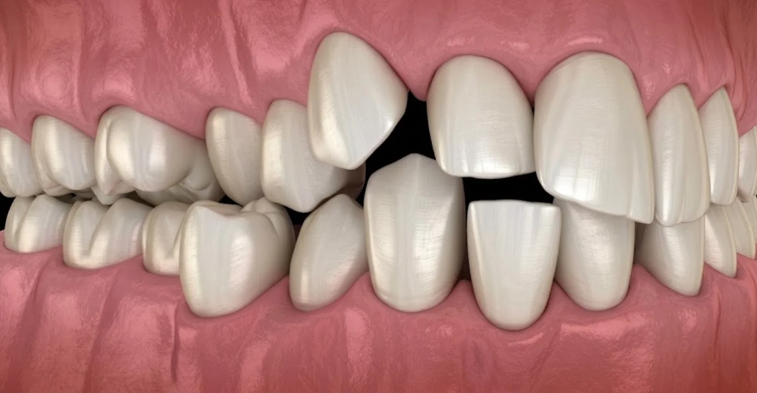
Note : All images used are for editorial and illustrative purposes only and may not originate from the original news provider or associated company.
Discover more about tongue-tie and its effects
What Dental Problems Can Osteopetrosis Cause?
Osteopetrosis dental problems include delayed or failed eruption, impacted teeth, congenitally missing teeth, malformed crowns and roots, weak enamel, severe decay and jaw infection. Together, osteopetrosis and tooth eruption problems can affect chewing, appearance and overall health, so every affected child and adult needs early and ongoing dental monitoring.
Osteopetrosis Dental Problems
Reported osteopetrosis dental problems extend well beyond late teeth. Published case reports describe oligodontia (several congenitally missing teeth), conical crowns, short or absent roots, enamel hypoplasia and abnormal pulp chambers. These findings show that the disease affects how teeth form, and it also affects when they appear.
Osteopetrosis Teeth Problems
Many osteopetrosis teeth problems involve the tooth structure itself. Studies of enamel and dentin show a reduced calcium-to-phosphorus ratio, which suggests altered mineral metabolism and weaker crystal formation. Weaker teeth are more vulnerable to damage and decay, so daily oral care and regular professional checkups become especially important.
Osteopetrosis Tooth Abnormalities
Osteopetrosis tooth abnormalities may affect the crown, root or pulp. Crowns can appear cone-shaped, roots may be short or missing, and pulp chambers may look unusual on X-rays. These changes can resemble other conditions, so they should be interpreted alongside the patient's eruption pattern and medical history.
Delayed Tooth Eruption
Delayed eruption is the most characteristic dental sign. Teeth may appear years late or remain completely buried in the jaw. Osteoclasts are needed to open the eruption path, so late teeth are often the first visible clue that the underlying bone remodeling process is not working normally.
Abnormal Tooth Development
Dense bone may limit nourishment to developing teeth. This can lead to conical crowns, enamel hypoplasia, short or absent roots and abnormal pulp chambers. Many of these changes cannot be seen in the mouth and are detected only on dental X-rays taken during routine or targeted examinations.
Impacted Teeth
Teeth that form but cannot break through the bone become impacted. In osteopetrosis, several baby and permanent teeth may be impacted at the same time. When a child has multiple impacted teeth, further investigation is warranted rather than simply waiting for the teeth to appear.
Increased Risk of Dental Infection
Weak, decay-prone enamel allows bacteria to reach the tooth pulp more easily. In osteopetrosis, dental infection can spread into the jaw and cause osteomyelitis, a serious bone infection. For this reason, infection prevention is one of the most important goals of dental care in these patients.
Clinical Note
The presence and severity of dental abnormalities differ widely between patients. One person may have only a few late teeth, while another has multiple impacted, malformed and missing teeth. Each patient should be assessed individually with a careful clinical examination, appropriate imaging and a review of their medical history.
Explore the causes and treatment options for a swollen tongue
Why Does Osteopetrosis Cause Impacted Teeth?
Osteopetrosis and impacted teeth are linked because osteoclasts cannot remove the bone covering developing teeth. Without this resorption, teeth remain trapped in the jaw even when they are fully formed. Impaction is one of the clearest osteopetrosis and tooth eruption problems, and it often affects several baby and permanent teeth.
Osteopetrosis and Impacted Teeth
In one reported case, an 8-year-old girl with multiple impacted baby and permanent teeth went undiagnosed for years, even though she had visual, hearing and dental problems. Her case shows why osteopetrosis and tooth eruption problems deserve attention, because impacted teeth can signal an underlying bone disorder.
How Increased Bone Density Can Interfere With Tooth Eruption
Healthy eruption depends on a balance between bone removal and tooth movement. In osteopetrosis, bone density increases because resorption is defective, so the jaw does not give way ahead of the tooth. The tooth stays embedded in dense bone, which is the direct reason impaction is so common.
Signs of an Impacted Tooth
Impacted teeth are not always painful, so they are easy to miss. Warning signs include gaps where teeth should be, baby teeth that stay too long, swelling, and crooked or drifting neighboring teeth. Any of these signs calls for a dental examination and, usually, an X-ray.
A Missing Tooth That Does Not Erupt
A space that stays empty long after the expected age may hide a tooth still inside the bone. A dental X-ray can show whether the tooth is present but trapped, malformed, or truly absent. Each of these findings leads to a different management plan.
Swelling or Discomfort
Swelling, tenderness or pain around an unerupted tooth can signal pressure or infection. In osteopetrosis, jaw infections can become serious and difficult to control, so swelling should be examined promptly by a dental professional. Patients should not wait to see whether it settles on its own.
Abnormal Tooth Position
Impacted teeth may develop tilted, rotated or far from their normal location. They can also press against the roots of neighboring teeth. Dental imaging helps map the exact position of each tooth, which is essential for monitoring and for planning any treatment safely.
Crowding
When some teeth erupt and others stay buried, the dental arch may become uneven, with crowding or drifting teeth. Crowded teeth are harder to clean, which raises the risk of decay. This is a particular concern because enamel in osteopetrosis is already weaker than normal.
Tips for the Patients
Regular dental imaging may be recommended when tooth eruption is delayed.
Report swelling, pain, or unexplained missing teeth to a dental professional.
Explore the possible causes of itchy gums
Can Osteopetrosis Cause Missing Teeth?
Yes. Osteopetrosis and missing teeth are linked in two ways. Some teeth never form at all (oligodontia), while others form but remain hidden in the bone. Distinguishing between these two situations is essential in osteopetrosis and tooth eruption problems, because the right treatment depends on it.
Osteopetrosis and Missing Teeth
Congenitally missing teeth have been reported in osteopetrosis, possibly because dense bone restricts the nutrient supply to developing tooth germs. Delayed eruption, crown anomalies and missing teeth can still occur after hematopoietic stem cell transplantation (HSCT), and their severity may depend on the child's age at transplant.
Delayed Eruption vs. Congenitally Missing Teeth
A tooth that is late is different from a tooth that never formed. X-rays reveal whether a tooth germ exists beneath the gum. This distinction is central to dental planning, because a buried tooth needs monitoring, while an absent tooth needs long-term replacement planning.
Why a Missing Tooth Should Be Evaluated
A gap in the smile can have several explanations, and each one leads to a different plan. Evaluation identifies whether the tooth is impacted, malformed, lost to infection, or genuinely absent. Without this assessment, treatment decisions may be premature or unsuitable for the patient.
Impacted Permanent Teeth
Permanent teeth may be fully formed but unable to break through dense bone. On an X-ray they appear inside the jaw, often beneath retained baby teeth. These teeth need careful monitoring, because their position and condition guide all later dental decisions.
Developmental Abnormalities
Some teeth form with conical crowns, short or absent roots, or enamel hypoplasia. A severely malformed tooth may not function normally even if it erupts. Identifying these abnormalities early helps the dental team plan realistic, long-term care for the patient.
Previous Dental Infection
Teeth weakened by decay may become infected and eventually be lost. In osteopetrosis, pulp necrosis and tooth extraction are key triggers for jaw osteomyelitis. A missing tooth with a history of infection therefore needs careful assessment of the surrounding bone.
Tooth Agenesis or Other Causes
Tooth agenesis means a tooth never developed. It can occur in osteopetrosis, and it can also occur in the general population or with other conditions. A full history and imaging help determine whether an absent tooth is related to the bone disorder or to another cause.
Discover what you should know about big teeth
Is Osteopetrosis Associated With Dental Caries?
Yes. Osteopetrosis and dental caries are associated because enamel and dentin show a reduced calcium-to-phosphorus ratio, suggesting weaker crystal formation. This makes patients highly susceptible to decay. Among osteopetrosis and tooth eruption problems, caries is especially important because it can open the way to serious jaw infection.
Osteopetrosis and Dental Caries
Weaker tooth structure means that acids from bacteria can damage enamel more easily. Once decay reaches the pulp, the tooth may become necrotic. In osteopetrosis, this sequence is dangerous because pulp necrosis is a key trigger for osteomyelitis of the jaw.
Why Dental Caries Can Become a Concern
In most people, a cavity is a local problem. In osteopetrosis, decay can lead to abscesses and to osteomyelitis, particularly of the mandible. Extractions also carry a higher risk, so preventing cavities is far safer than treating their complications.
Factors That May Increase the Risk of Tooth Decay
Several factors can combine to raise caries risk, including weak enamel, crowded or partially erupted teeth, difficulty cleaning, and existing infection. Identifying the specific factors for each patient allows the dental team to target prevention more effectively.
Enamel or Tooth Development Abnormalities
Enamel hypoplasia and altered mineral content leave teeth less resistant to acid. Thin or defective enamel can decay faster and more deeply than healthy enamel, so fluoride and close monitoring are especially valuable for these teeth.
Difficulty Maintaining Oral Hygiene
Irregular tooth positions, crowding and partially erupted teeth create areas that are hard to reach with a toothbrush. Plaque builds up in these spots and increases the risk of decay. Professional cleanings and tailored hygiene instructions help patients manage these difficult areas.
Delayed Tooth Eruption
Teeth that erupt slowly or only partially may leave gum flaps and pockets where food and bacteria collect. Baby teeth that stay longer than normal also remain exposed to decay for more years, which adds to the overall caries risk.
Dental Infection
Existing infection can spread and damage nearby teeth and bone. Untreated decay in one tooth may lead to abscesses affecting the surrounding area. Early treatment of small problems limits their spread and reduces the need for higher-risk procedures later.
Clinical Note
Osteopetrosis should not automatically be considered the direct cause of every cavity. Diet, oral hygiene, fluoride exposure and tooth position all play a part. Identifying individual risk factors for dental caries allows the dental team to build a prevention plan suited to each patient.
Tips for the Patients
Brush twice daily with fluoride toothpaste.
Learn how dental and periodontal charting worksClean between the teeth regularly.
Reduce frequent exposure to sugary foods and drinks.
Attend routine dental checkups.
What Are the Main Osteopetrosis Dental Complications?
The main osteopetrosis dental complications are impacted teeth, crowding, severe decay, gum problems and dental infection. The most serious is osteomyelitis of the jaw, especially the mandible. Many osteopetrosis and tooth eruption problems are connected, because decay and abscesses can progress to this dangerous bone infection.
Osteopetrosis Dental Complications
Osteomyelitis is the main complication of autosomal dominant osteopetrosis type II, and it is linked to caries and abscesses. In one series, 13% of patients had osteomyelitis of the maxilla or mandible. Pulp necrosis and tooth extraction are key triggers, so prevention is essential.
Oral Infections and Dental Health
Oral infections that would be minor in healthy bone can become serious in osteopetrosis. Dense jawbone may struggle to fight the spread of infection. Prompt treatment of decay, abscesses and gum inflammation protects both the teeth and the jaw.
Possible Complications of Delayed or Abnormal Tooth Eruption
Delayed or abnormal eruption sets off a chain of problems. Buried teeth, uneven arches and hard-to-clean areas all increase dental risk. Recognizing how these eruption problems lead to later complications helps families understand why early monitoring matters so much.
Impacted Teeth
Impacted teeth may stay silent for years or cause pressure, swelling and damage to neighboring roots. Their management must be planned carefully, because any surgical removal in osteopetrosis carries an increased risk of infection and requires special precautions.
Crowding
Uneven eruption can leave teeth crowded or drifting into empty spaces. Crowding affects appearance and bite, and it creates plaque traps. Over time, these areas become common sites for decay and gum inflammation.
Tooth Decay
Reduced mineral quality in enamel and dentin leaves teeth highly susceptible to caries. Decay can progress quickly to the pulp. Because pulp necrosis is a trigger for osteomyelitis, early detection and treatment of cavities is a key safety measure.
Gum Problems
Plaque around crowded or partially erupted teeth can cause gum inflammation. Inflamed gums bleed easily and may allow bacteria to spread more deeply. Professional cleanings and good home care help keep the gums healthy and reduce the overall infection risk.
Dental Infections
Abscesses from decayed or necrotic teeth can spread into the surrounding bone. In osteopetrosis, this may develop into osteomyelitis, especially of the mandible. Any swelling, pain or discharge should be assessed promptly by a dental professional.
Clinical Note
Treatment decisions depend on the patient's age, dental development, symptoms and overall oral condition. Antibiotic prophylaxis is recommended before any surgical procedure. The dental team should weigh the risks of each intervention carefully and coordinate with the patient's medical team when needed.
Find out more about osteopetrosis and tooth eruption problems
How Are Osteopetrosis and Tooth Eruption Problems Diagnosed?
Diagnosis combines a clinical dental examination, a review of eruption patterns and dental imaging, usually a panoramic X-ray. Experts recommend this X-ray whenever teeth erupt late for a child's age. Recognizing osteopetrosis and tooth eruption problems may also lead to referral to a pediatrician when systemic disease is suspected.
Dental Examination
The dentist checks which teeth are present, their shape, the condition of the enamel, signs of decay and the health of the gums. Any swelling or signs of infection are noted. This examination is often the first step toward recognizing an underlying bone disorder.
Reviewing Tooth Eruption Patterns
The dental team compares the child's erupted teeth with the expected eruption timeline for their age. Multiple late or missing teeth, retained baby teeth, or teeth appearing in an unusual order are important clues, and they need further investigation.
Dental Imaging for Impacted or Delayed Teeth
A panoramic X-ray shows all developing teeth and the surrounding bone in a single image. In osteopetrosis, it can reveal impacted teeth, missing tooth germs, root abnormalities and unusually dense jawbone that cannot be seen during a visual examination.
Clinical Examination
The clinical examination records the number, shape and condition of the visible teeth, along with gum health and any swelling. It also documents any medical history of vision, hearing or bone problems that could point toward a systemic condition.
Dental X-Rays
X-rays confirm whether unerupted teeth are present and show their stage of development. They also reveal short or absent roots, abnormal pulp chambers and hidden decay. Imaging is essential because many of these findings are invisible in the mouth.
Evaluation of Tooth Position
Imaging shows whether impacted teeth are tilted, rotated or close to important structures. Knowing each tooth's exact position helps the dental team decide whether to monitor it, guide its eruption, or plan any intervention with appropriate precautions.
Assessment of Jaw and Bone Structure
Unusually dense, uniform jawbone on an X-ray can raise suspicion of osteopetrosis. When bone findings are combined with multiple impacted teeth or systemic symptoms, referral for a medical evaluation is recommended, since the dentist may be the first to notice the disease.
Tips for the Patients
Good records make changes easier to spot over time:
Keep regular dental records during tooth development.
Follow recommended dental imaging and monitoring.
Discuss any unusual eruption pattern with the dental team.
Understand the connection between Epstein-Barr virus and oral health
How Can Patients Manage Osteopetrosis and Oral Health?
Managing osteopetrosis oral health focuses on prevention and close monitoring. Recommended care includes frequent follow-up to guide eruption, professional cleanings, fluoride to prevent decay, and antibiotic prophylaxis before any surgical procedure. Consistent care reduces the impact of osteopetrosis and tooth eruption problems and lowers the risk of jaw infection.
Preventive Dental Care
Prevention is the foundation of care. Regular professional cleanings, fluoride applications and personalized hygiene advice help protect weaker enamel. By preventing decay, patients can avoid many extractions and surgical procedures that carry a higher infection risk in osteopetrosis.
Monitoring Osteopetrosis and Tooth Eruption Problems
Frequent follow-up visits allow the dental team to track eruption and guide it where possible. Regular monitoring of osteopetrosis and tooth eruption problems helps identify impacted, malformed or missing teeth early and allows the care plan to change as the child grows.
Protecting Teeth From Decay and Infection
Fluoride, sealants where suitable, limited sugar intake and prompt treatment of small cavities all protect teeth from decay. Early care keeps decay from reaching the pulp, which is important because pulp necrosis can trigger osteomyelitis of the jaw.
Managing Impacted or Abnormally Erupting Teeth
Impacted teeth are usually monitored carefully and managed according to their position and the patient's overall condition. When any surgical procedure is needed, antibiotic prophylaxis is recommended beforehand. Decisions should always balance the expected benefit against the risk of infection.
What We Notice Clinically
The medical team at Vitrin Clinic notices that long-term monitoring makes the greatest difference for patients with osteopetrosis and tooth eruption problems. Regular visits allow small issues to be detected early, before decay or infection progresses. Preventive care is always safer and more comfortable than treating serious complications later.
Tips for the Patients
These habits help protect teeth and jawbone over the long term:
Maintain consistent oral hygiene.
Do not delay dental examinations when permanent teeth fail to erupt.
Ask about appropriate imaging when teeth appear missing or impacted.
Explore erythromelalgia and premature tooth lossSeek prompt dental care for pain, swelling, or signs of infection.
How Can Vitrin Clinic Help With Osteopetrosis and Tooth Eruption Problems?
Vitrin Clinic provides careful dental assessment and individualized care for osteopetrosis dental problems. The team evaluates eruption, tooth development, decay and gum health, and then builds a preventive, personalized plan. Our aim is to manage osteopetrosis and tooth eruption problems safely while protecting the teeth and the jaw.
Comprehensive Dental Assessment at Vitrin Clinic
Vitrin Clinic evaluates tooth eruption, tooth development, dental caries, gum health and other oral concerns through a detailed clinical examination and appropriate dental imaging. Each assessment is individualized and based on the patient's age, dental condition and medical history.
Personalized Treatment Planning at Vitrin Clinic
Based on the examination findings, Vitrin Clinic creates an individualized dental treatment plan. This may include preventive care such as cleanings and fluoride, restorative treatment for decay, and other dental treatments when indicated. Safety precautions are planned for any procedure that could raise infection risk.
What We Notice Clinically
Dr. Rifat Alsaman, Head of the Medical Team at Vitrin Clinic and a cosmetic dentist, notes that the most important step comes before any treatment begins. The team first determines whether a tooth is delayed, impacted, developmentally abnormal or genuinely missing. With osteopetrosis and tooth eruption problems, this distinction shapes every later decision and helps avoid unnecessary or risky procedures.
Clinical Note
Patients with osteopetrosis require an individualized dental assessment. No one should assume that every eruption problem has the same cause. Dense bone, abnormal development, infection and unrelated dental factors can all be involved, and each one needs a different approach.
Discover the relationship between mitochondrial disease and oral health
Resource:
https://pubmed.ncbi.nlm.nih.gov/15721137/
FAQs
Dr. Rifat Alsaman has more than 5 years of clinical experience in dentistry and currently serves as the Head of the Medical Team at Vitrin Clinic. He is dedicated to providing exceptional patient care, overseeing treatment planning, and ensuring the highest clinical standards across the team. His expertise, attention to detail, and commitment to continuous professional development have helped countless patients achieve healthier, more confident smiles.


.webp&w=3840&q=75)


